

 PDF
PDF ePub
ePub Citation
 Print
Print


Abstract
Cerebral aspergillosis accounts for about 10% of all the cases of invasive aspergillosis. The brain is the only infected site in less than 10% of cases. The patients at high risk for of aspergillosis are immunocompromised patients such as those in a neutropenic state after chemotherapy, AIDS and etc. We experienced a case of cerebral aspergillosis in a patient with acute leukemia that was in complete remission. The patient visited our hospital's ER due to nasal bleeding, and then he was quickly diagnosed as having acute promyelocytic leukemia. After the first induction chemotherapy, he achieved a complete remission. Loss of consciousness developed on day 31 after chemotherapy. High signal intensity in the right temporooccipital lobe and multiple nodular lesions in both cerebral hemisphereswere observed on the brain MRI. Stereotaxic biopsy showed septate aspergillus hyphae in the brain specimen. Despite of the use of amphotericin B deoxycholate, the patient died of recurrent grand mal seizure and multiple organ failure.
Go to : 
REFERENCES
1). Hori A, Kami M, Kishi Y, Machida U, Matsumura T, Kashima T. Clinical significance of extra-pulmonary involvement of invasive aspergillosis: a retrospective autopsy-based study of 107 patients. J Hosp infect. 2002; 50:175–82.

2). Denning DW. Invasive aspergillosis. Clin Infect Dis. 1998; 26:781–805.
3). Lin S, Schranz J, Teutsch SM. Aspergillosis case-fatality rate: systemic review of the literature. Clin Infect Dis. 2001; 32:358–66.
4). Rubin RH, Young LS, van Furth R. Clinical approach to infection in the compromised host. 4th ed.New York: Kluwer Academic/Plenum Publishers;2006. p. 193–6.
5). Kim YI, Han SR, Kim BK, Chung TI, Rye SY. Two cases of invasive aspergillosis of sino-nasal origin. J Korean Neuro Assoc. 2000; 18:368–71.
6). Kim JD, Kim EJ, Park SJ, Cho CW, Youn SM. Two cases of cerebral aspergillosis following cranial operation. J Korean Neurosurg Soc. 2000; 29:1094–7.
7). Karim M, Alam M, Shah Aa, Ahmed R, Sheikh H. Chronic invasive aspergillosis in apparently immunocompetent hosts. Clin Infect Dis. 1997; 24:723–33.
8). Sharma Rr. Gurusinghe NT, Lynch PG. Cerebral infarction due to aspergillosis arteritis following glioma surgery. Br J Neurosurg. 1992; 6:485–90.
9). Kim SS, Jeong JI, Cha JK, Kim SH, Kim JW, Kim HD. A case of intracerebral aspergillus abscess presenting as cerebral infarction. J Korean Neurol Assoc. 1998; 16:87–90.
10). Lyon RW, Andriole VT. Fungal infections of the CNS. Neurol Clin. 1986; 4:159–70.
11). Kei Y, David AS, Ana R, et al. Imaging findings in intracranial aspergillosis. Acad Radiol. 2002; 9:163–71.
12). Coleman JM, Hogg GG, Rosenfeld JV, Waters KD. Invasive central nervous system aspergillosis: cure with liposomal amphotericin B, itraconazole, and radical surgery-case report and review of the literature. Neurosurg. 1995; 36:858–63.
13). De Lastorus V, Lefort A, Zappa M, Dufour V, Belmatoug N, Fantin B. Two cases of cerebral aspergillosis successfully treated with voriconazole. Eur J Clin Microbiol Infect Dis. 2003; 22:297–9.
14). Lim JY, Choi JO, Lee KS, Chae SH, Joo MJ. MR imaging of cerebral aspergillosis in an infant with normal-immunity: a case report. J Korean Radiol Soc. 2000; 42:605–8.
15). Lee TK, Choi JH, Choi CW, et al. A case of cerebral aspergillosis in a patients with rheumatoid arthritis. J Korean Rheuma Assoc. 2003; 10:438–41.
16). Woo YS, Kim MJ, Jang JH, et al. Successful treatment of cerebral aspergillosis with liposomal amphotericin B and minimal invasive surgery in a patient who received renal transplant 10 year ago. Korean J Nephrol. 2001; 20:702–6.
17). Lee J, Cho B, Lee HJ, Kim SY, Chung NG, Kim HK. Successful treatment of cerebral aspergillosis in a child with acute lymphoblastic leukemia. Korean J Pediatr Hematol-Oncol. 2001; 7:121–8.
Go to :
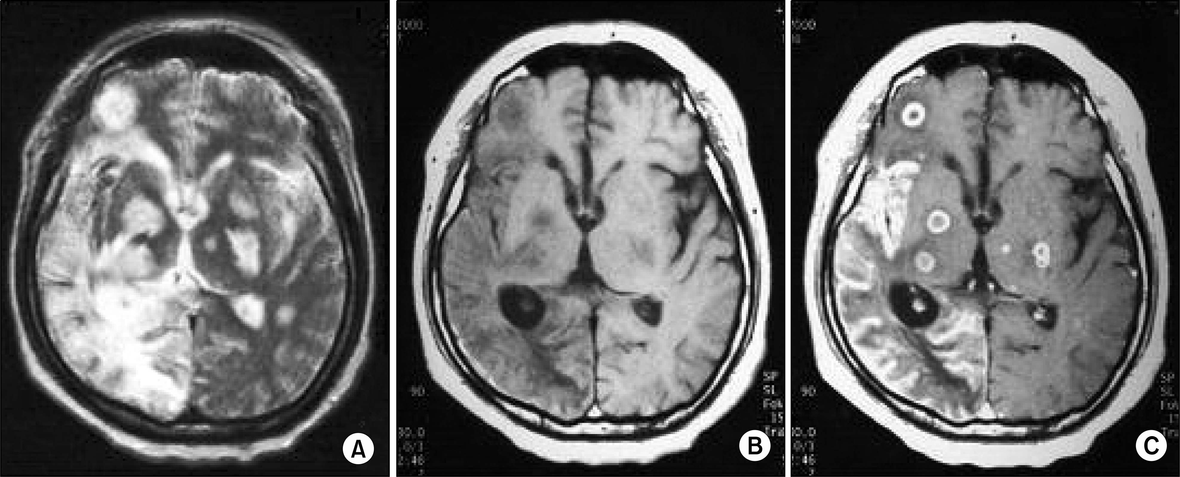 | Fig. 1.(A) Axial T2-weighted image shows confluent high signal intensity in the right temporal and occipital regions. Multiple nodular high signal intensity lesions are scattered in both cerebral hemispheres. (B) Axial T1-weighted image shows hyposignal intensity in the corresponding areas of T2 abnormality. Thin high signal along the surface of the right temporo-occpital region may indicate hemorrhage. (C) Axial T1-weighted image with Gadolinium enhancement demonstrates diffuse gyral enhancement of the right temporal and occipital lobes and multiple ring enhancing nodules in both cerebral hemispheres. C onsistent with subacute infarction and multiple brain abscesses. |

 XML Download