Abstract
A benign cementoblastoma, which is another name for a true cementoma, is a rare neoplasm that develops from odontogenic ectomesenchyme. It is characterized by a mineralized mass attached to the apex of the root produced by neoplastic cementoblasts. More than 75% of cases arise in the mandible, with 90% of them manifesting in the molar and premolar regions. This neoplasm occurs most commonly in children and young adults, with males being affected slightly more than females. Radiographically, the tumor is observed as a well-defined radiopaque mass that is fused to a tooth root and is surrounded by a radiolucent rim. The treatment of benign cementoblastoma consists of removal of the lesion and extraction of the affected tooth. This report presents an unusual case of benign cementoblastoma in a 31-year-old female, presenting as a densely mineralized mass seen at the apex of the impacted right mandibular canine tooth on radiographs.
Cementoblastoma is a relatively rare odontogenic benign neoplasm characterized by a calcified cementum-like deposit attached to the apex of the root and produced by neoplastic cementoblasts. It represents a very small proportion of all odontogenic tumors, with a percentage less than 1%1. The World Health Organization first named this neoplasm "benign cementoblastoma" and also "true cementoma" in their 1971 classification. This terminology was altered in 2005, and the benign prefix was dropped because there is no malignant neoplasm originating from cementum tissue23.
Cementoblastomas usually affect patients in the second or third decade of life4. Although males are affected slightly more, there is no significant sex predilection14. The neoplasm exhibits a slow but limitless growth pattern, and the mandible is involved more often than the maxilla15. Typically, the lesion is seen on the posterior region of the mandible and commonly involves a first molar14. The lesions vary in size between 0.5 and 5.5 cm4. Differential diagnosis should be made with condensing osteitis, osteoblastoma, odontoma, cementoossifying fibroma, periapical cemental dysplasia, and hypercementosis6.
The treatment of a cementoblastoma consists of excision of the tumor, extraction of the effected tooth, and osseous curettage. Apical resection and root canal treatment of the affected tooth may be preferred as an alternative treatment that increases the risk of recurrence14.
In this report, a case of cementoblastoma with involvement of an impacted mandibular canine tooth is presented due to the extremely rare tooth and site preference for this neoplasm.
A 31-year-old female was referred to our clinic for an asymptomatic lesion in the mandible that was noticed incidentally on panoramic radiograph during a dental examination performed by her dentist. The patient's medical history was unremarkable, and there was no reported history of maxillofacial trauma.
The clinical examination revealed a barely detectable, hard, non-tender area of swelling on the buccal side of the right mandibular canine area and a persistent deciduous right mandibular canine tooth. A radiopaque mass that had a close relationship with the root apex of the impacted mandibular right canine tooth was detected on the panoramic radiograph. The mass was surrounded by a thin radiolucent rim. Cone-beam computed tomography (CBCT) was determined to be appropriate for further radiological examination of the mass (Galileos Comfort Plus CBCT unit; Sirona Dental Systems Inc., Bensheim, Germany). A homogenous hyperdense deposit with a size of 9.5×9.5×6.5 mm was seen in the mentioned area of the CBCT images. There was also a slight expansion on the mandibular buccal side. Significant thinning on the adjacent buccal cortical bone was detected, but there was no cortical perforation. The oblique sagittal images showed that the deposit was continuous with the root apex of the impacted mandibular right canine tooth, and there was a well-defined hypodense border between the deposit and surrounding bone.(Fig. 1)
Clinical and radiological examination led to the diagnosis of cementoblastoma, and surgical excision of the mass with the involved tooth was planned. The lesion was removed surgically with extraction of the involved canine tooth through an intraoral approach under general anesthesia. The surgical specimen was sent for histopathologic examination (Fig. 2), which revealed dense, mineralized, cementum-like material and vascular soft tissue areas that consisted of cementoblasts.(Fig. 3) Based on these findings, the diagnosis of cementoblastoma was confirmed. The patient was monitored for 6 months without any postoperative symptoms or local recurrence.
Cementoblastoma most commonly occurs on the posterior region of the mandible and is often attached to the root of an erupted permanent premolar or first molar14. On rare occasions, an impacted tooth, multiple teeth, or a deciduous tooth may be affected as well1578. To our knowledge, there have been 8 reported cases of cementoblastoma that developed on anterior jaws in the literature910111213141516, with only two of them located in the mandible910. One case was related to an impacted permanent incisor9, while the other case was related to a deciduous incisor10. This present case is unique due to its occurrence in the anterior mandible and involvement of an impacted mandibular canine tooth.
Pain, tenderness, and swelling are present in most patients with cementoblastoma, although the lesion can be asymptomatic. The involved tooth is vital and often exhibits pain146. In the present case, the patient was asymptomatic, and the lesion was discovered incidentally on a panoramic radiograph.
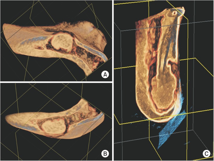
Radiographically, cementoblastoma appears as a well-defined circumscribed radiopaque or mixed mineralized mass fused to one or more tooth roots of the involved tooth and has a radiolucent rim1. Though rate, some tumors, especially recurrent ones, may be completely radiolucent due to reduced mineralization317. Usually, radiological examination is sufficient to diagnose the lesion. The tumor may cause various amounts of external resorption, bony expansion, displacement of adjacent teeth, and in some cases, cortical perforation, jaw deformity, and pathologic fracture146. CBCT images show the radiopaque mass attached to the roots of the involved tooth and provide detailed information about the presence of buccal and/or lingual perforations. In the present case, cementoblastoma was observed on CBCT as a well-defined radiopaque mass fused to the root of the mandibular canine without cortical perforation.(Fig. 4)
Simple excision of the lesion and extraction of the involved Kartooth are sufficient treatments1. A 37.1% recurrence rate has been recently reported, especially for cases with aggressive features4. In the present case, there was no recurrence more than 6 months after the surgical procedure.
Histologically, the tumor tissue consists of acellular and irregular layers of a calcified cementum-like mass attached to the root of the tooth, with trabecular calcified bone-like areas and cementoblasts observed in the calcified matrix and fibrovascular stroma. The periphery of the tumor is less mineralized and includes cementoblasts. The histopathologic presentation of cementoblastoma closely resembles that of osteoblastoma, but can be distinguished by fusion of the tumor with the involved tooth118.
During the differential diagnosis of cementoblastoma, there are some clinical and radiographic details that have to be considered for each similar disease. Condensing osteitis is an inflammatory sclerotic bone change of the periradicular region with dental inflammatory stimulus, as is not found in cementoblastoma. The periradicular sclerotic area resembles cementoblastoma, but the absence of a radiolucent rim and tooth-mass continuity with an irregular and poorly defined shape of the sclerotic area and inflammatory pulpal changes with associated symptoms are remarkable for condensing osteitis. Hypercementosis of the supraerupted or inflamed tooth may resemble an emerging cementoblastoma. Continuity of the periodontal ligament around the enlarged root is characteristic of hypercementosis. Osteoblastoma has very similar clinical and radiographic findings but tooth-mass continuity does not exist. Generally, osteoblastomas have more irregular mass radiopacity than cementoblastomas. Also most osteoblastomas have osteolytic borders and cause persistent pain19. Additionally, osteoblastoma may have more aggressive clinical features than cementoblastoma. Odontomas have a radiolucent rim like cementoblastomas, but the mass in the capsule consists of rudimentary tooth-like deposits or an irregularly shaped deposit that exhibits radiographic opacity similar to teeth. Cemento-ossifying fibromas have the same site preference as cementoblastomas. Unlike cementoblastomas, bilateral expansion and mixed radiolucency with sclerotic borders are seen with this fibrous-osseous lesion19. Periapical cemental dysplasia is usually seen on the anterior mandible with the involvement of multiple teeth, which is not seen with cementoblastomas. The mature stage of this disease may resemble cementoblastoma minus the tooth-mass continuity.
In conclusion, cementoblastoma usually involves an erupted permanent tooth and most commonly occurs in the posterior region of the mandible. This case report presents a unique example with an extremely rare tooth and site preference for this neoplasm. In addition, the involvement of the impacted tooth at this mentioned site is another rarity. Other than these mentioned differences, the observed clinical, radiological, and histopathological features and prognosis of the neoplasm were similar to all of the other cases of cementoblastoma found in the literature.
Acknowledgements
The study was presented as a poster presentation in the 17th International Congress on Oral Pathology and Medicine at May 25-30, 2014 held in Istanbul, Turkey.
References
1. Neville BW, Damm DD, Allen CM, Bouquot JE. Oral and maxillofacial pathology. 2nd ed. Philadelphia: W.B. Saunders;2002.
2. Barnes L, Eveson JW, Reichart P, Sidransky D. World Health Organization classification of tumors pathology & genetics head and neck tumors. Lyon: IARC Press;2005.
3. Noffke CE, Raubenheimer EJ, MacDonald D. Fibro-osseous disease: harmonizing terminology with biology. Oral Surg Oral Med Oral Pathol Oral Radiol. 2012; 114:388–392. PMID: 22862981.
4. Brannon RB, Fowler CB, Carpenter WM, Corio RL. Cementoblastoma: an innocuous neoplasm? A clinicopathologic study of 44 cases and review of the literature with special emphasis on recurrence. Oral Surg Oral Med Oral Pathol Oral Radiol Endod. 2002; 93:311–320. PMID: 11925541.
5. Pynn BR, Sands TD, Bradley G. Benign cementoblastoma: a case report. J Can Dent Assoc. 2001; 67:260–262. PMID: 11398388.
6. White SC, Pharoah MJ. Oral radiology: principles and interpretation. 6th ed. St. Louis: Mosby;2009.
7. Monti LM, Souza AM, Soubhia AM, Jorge WA, Anichinno M, Da Fonseca GL. Cementoblastoma: a case report in deciduous tooth. Oral Maxillofac Surg. 2013; 17:145–149. PMID: 22855308.
8. Ohki K, Kumamoto H, Nitta Y, Nagasaka H, Kawamura H, Ooya K. Benign cementoblastoma involving multiple maxillary teeth: report of a case with a review of the literature. Oral Surg Oral Med Oral Pathol Oral Radiol Endod. 2004; 97:53–58. PMID: 14716256.
9. Karaçal N, Agdoğan Ö, Livaoğlu M, Uraloğlu M, Özel B. Giant cementoblastoma of the impacted mandibular incisor. J Craniofac Surg. 2011; 22:e26–e27. PMID: 22134313.
10. Papageorge MB, Cataldo E, Nghiem FT. Cementoblastoma involving multiple deciduous teeth. Oral Surg Oral Med Oral Pathol. 1987; 63:602–605. PMID: 3473380.
11. Hirai E, Yamamoto K, Kounoe T, Kondo Y, Yonemasu H, Kurokawa H. Benign cementoblastoma of the anterior maxilla. J Oral Maxillofac Surg. 2010; 68:671–674. PMID: 20171487.
12. Costa FW, Pereira KM, Magalhães Dias M, da Costa Miguel MC, de Paula Miranda MA, Studart Soares EC. Maxillary cementoblastoma in a child. J Craniofac Surg. 2011; 22:1910–1913. PMID: 21959464.
13. Slimani F, Elbouihi M, Oukerroum A, Lazreqh H, Mahtar M, Karkouri M, et al. Maxillary cementoblastoma. A case report. Rev Med Brux. 2009; 30:185–188. PMID: 19642490.
14. Keyes G, Hilderbrand K. Successful surgical endodontics for benign cementoblastoma. J Endod. 1987; 13:566–569. PMID: 3482232.
15. Brocheriou C, Guilbert F, Matar A, Champion P, Couly G. Benign cementoblastoma of jaws: report of 6 cases and review of the literature (author's transl). Arch Anat Cytol Pathol. 1979; 27:29–34. PMID: 453932.
16. Souza AS, Cardoso JA, Silva VP, Oliveira MC, Azoubel E, Farias JG. Cementoblastoma affecting the maxilla of a pediatric patient: a case report. Rev Port Estomatol Med Dent Cir Maxillofac. 2013; 54:43–47.
17. Eversole LR, Sabes WR, Dauchess VG. Benign cementoblastoma. Oral Surg Oral Med Oral Pathol. 1973; 36:824–830. PMID: 4524834.
18. Slootweg PJ. Cementoblastoma and osteoblastoma: a comparison of histologic features. J Oral Pathol Med. 1992; 21:385–389. PMID: 1432731.