Abstract
Traumatic bone cyst (TBC) occurs preferentially on the mandibular symphysis and body, but rarely on the mandibular condyle. When TBC occurs in the condylar area, it can usually be related with or misdiagnosed as a temporomandibular joint disorder. A 15-year-old female patient visited the Temporomandibular Joint Clinic with a 5-year history of pain and noise localized in the left temporomandibular joint. On imaging, a well demarked oval-shaped radiolucent lesion was observed on the left condyle head. The patient underwent cyst enucleation and repositioning of the bony window on the lateral cortex of the affected condyle head under the impression of subchondral cyst or TBC; however, no cystic membrane was found. The bone defect resolved and showed no recurrence on the serial radiographic postoperative follow-up for 43 months after surgery.
Traumatic bone cyst (TBC) is a very rare intraosseous lesion with the cause not clearly identified, although correlation with trauma has been reported, including the formation of an intraosseous empty cavity following medullary hemorrhage caused by a traumatic injury1.
This cyst occurs preferentially on the mandibular symphysis and mandibular body2 but rarely on the mandibular condyle 345. TBC usually occurs in people in their twenties6, and it is not strongly related to the vitality of the teeth. In many cases, a blood-colored exudate is discharged3. TBC is radiologically found as a unicystic radiolucent lesion with well defined margins. Histologically, it is not considered a genuine cyst because it is covered with a thin, loose connective tissue layer without a lining epithelium.
We report a rare case in which an operation was performed for a cystic lesion and repositioning of the bony window on the left mandibular condyle with serial postoperative follow-up radiographs, which was subsequently diagnosed as a TBC.
A 15-year-old female patient presented to the Temporomandibular Joint Clinic with complaints of pain in the left temporomandibular joint and joint noise when opening the mouth. These symptoms had started five years prior to the presentation. She denied any specific trauma involving the mandible and any abnormal habits such as clenching or bruxism. On the clinical examination, the maximum inter-incisal mouth opening measure was 57 mm without deviation during mandibular movements. However, the patient had left temporomandibular joint pain and crepitus with opening the mouth. The panoramic X-ray showed a radiolucent lesion with a relatively well defined margin on the left mandibular condyle and a narrow left temporomandibular joint space. (Fig. 1) Computed tomography showed a cystic lesion with a size of 14×8×9 mm on the left mandibular condyle, accompanied by an expansion of the cortical bone.(Fig. 2) Magnetic resonance imaging showed a high signal intensity lesion in the same location on the T2-weighted images.(Fig. 3) Based on these imaging studies, the main differential diagnosis was a TBC versus a subchondral cyst, and surgery was scheduled for the removal of the lesion.
For the operation, general anesthesia was administered by nasoendotracheal intubation, and the left condyle was exposed by a preauricular incision. A fibrous adhesion was found between the articular disc and the mandibular condyle and was released. There was no bony defect on the cortical surface of the mandibular condyle, but a bluish black region with a diameter of about 7 mm was found at the outer posterior region at about 10 mm inferior to the upper part of the condyle. Osteotomy was carried out in that region with a semi-circular corticotomy by fissure burr. A bony window was created by a greenstick fracture without removing the bone fragment. No lining epithelium was found in the exposed cystic lesion, and the surrounding bone wall of the lesion was relatively smooth. Curettage was performed over the entire inner bone wall; the bone window was relocated after checking for hemorrhage inside the empty defect.(Fig. 4) Based on the surgical findings and the radiological findings, the final diagnosis was TBC of the mandibular condyle.
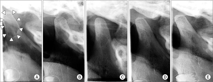
There were no specific postoperative complications such as abnormal hemorrhage, edema, facial nerve damage, or pathologic condyle fracture. A postoperative follow-up was conducted periodically for more than 4 years after the surgery, and the mouth opening increased from 20 mm on the sixth day to 30 mm in the first month, to 45 mm in the second month, and to 60 mm in the fifth month. The patient did not have any pain in the left temporomandibular joint with mouth opening during the follow-up examinations. The postoperative radiological images showed gradual bony deposition, and it was difficult to find the bony defect since 5 months follow-up on X-rays.(Fig. 5, 6, 7) There was no recurrence on the image study through 43 months of follow-up.
TBC has been reported as various names including simple bone cyst, solitary bone cyst, and hemorrhagic cyst5. It is a benign intraosseous lesion with the etiology not clearly identified123456, and its correlation with trauma has been controversial for quite some time. Beasley7 reported that only about 25% of TBC cases were related to trauma. When TBC occurs in the mandibular condyle, it might be related with temporomandibular joint disorders, and the patients often complain of joint pain, discomfort, or noise8. According to the traumahemorrhage theory of Olech et al.9, it is presumed that an intraosseous hematoma occurs as a result of mild trauma, not strong enough to cause a bone fracture. The hematoma is liquefied and becomes an intraosseous cystic lesion when it fails to undergo the organization and healing process9.
TBC of the mandible is usually an incidental finding on general radiography, but it is difficult to differentiate a TBC based only on the radiological studies because various types of cystic lesions occur in the mandible610. TBC is asymptomatic in most cases, but it can cause pain, tooth hypersensitivity, or expansion of the cortical bone in rare cases568. When it is detected in a tooth-bearing area or the body or symphysis of the mandible, differential diagnosis between odontogenic and non-odontogenic cysts is difficult11.
The general treatment for TBS is surgical exposure and curettage of the lesion, which also helps in the diagnosis38. Complete recovery can be achieved with surgical exposure performed for the diagnosis and by the hemorrhage inflicted by the curettage.
In our case, the TBC occurred on the mandibular condyle, different from the typical cases. The patient presented to the clinic with symptoms of joint noise in the temporomandibular joint and pain with mouth opening. The initial panoramic X-ray showed a radiolucent lesion on the left mandibular condyle. Magnetic resonance T2-weighted imaging showed a high signal intensity lesion in the same region, and computed tomography showed a partially thin-walled cystic lesion accompanied by expanded cortical bone.
In this case, based on the fact that the patient denied any history of macrotrauma, the presence of an adhesion between the left joint disc and the mandibular condyle, and the presence of temporomandibular joint noise and pain with mouth opening, it can be assumed that the TBC might have occurred as a result of microtrauma accumulated at the left temporomandibular joint or from trauma that the patient had not been able to recognize or was not aware of.
Because the lesion was close to the load-bearing articular surface, approach to the lesion was performed carefully, and the lateral cortical bone window was not removed to prevent soft tissue ingrowth after cyst removal. During the operation, no lining epithelium was found inside the cyst, and the inner bone wall was relatively smooth. The exudate or blood that is commonly found inside a TBC was not observed in this case12. However, since it is difficult to make an accurate judgment because of the suction and irrigation during the surgery, the surgical method to intraoperatively observe the inside of the cyst needs to be considered in the future. When cortex of the condyle head is exposed, needle aspiration would be useful to identify contents of the lesion13. If preoperative diagnosis is assumed to be a TBC, an intraoral endoscopic approach as a minimally invasive surgery would be useful for surgical exploration and cosmetic outcome1415. Minimally invasive surgery is recommended to prevent accidental or pathologic condyle fracture after surgery. When additional joint surgery such as disc repositioning, release of adhesions, as in this case, or correction of bony surface is required, a preauricular open joint approach would be more effective than endoscopic surgery.
Ameloblastoma and odontogenic myxoma are similar to TBC in that all are unilocular or multilocular radiolucent lesions and are prone to occur in the posterior region of the mandible in young people. However, these diseases were excluded from the differential diagnosis because the cyst in our case was found on the left mandibular condyle and was not considered an odontogenic cyst. The cyst in this case looked similar to a subchondral cyst in that it occurred at the joint. A subchondral cyst is, however, different from the cyst in our case because there are few reported cases of a subchondral cyst occurring in the temporomandibular joint8, a subchondral cyst occurs after skeletal maturity, is lined with a fibrous membrane, and is filled with gelatin16. Moreover, it is more probable that the cyst in this case was a TBC that occurred inside a bone, because a subchondral cyst occurs between the subchondral bone and the articular cartilage in the bone joint and expands to the inside of the joint. The cyst was finally diagnosed as a TBC based on the liquid filling the cyst, as demonstrated on the T2-weighted image12; the cortical bone expansion accompanying the unilocular cyst on computed tomography; and the surgical findings.
In this case, recurrence did not take place during more than 4 years of follow-up, and the postoperative radiography showed bony deposition in the previous cystic region and a relatively normal mandibular condyle shape.
References
1. Kaugars GE, Cale AE. Traumatic bone cyst. Oral Surg Oral Med Oral Pathol. 1987; 63:318–324. PMID: 3473360.

2. Martins-Filho PR, Santos Tde S, Araújo VL, Santos JS, Andrade ES, Silva LC. Traumatic bone cyst of the mandible: a review of 26 cases. Braz J Otorhinolaryngol. 2012; 78:16–21. PMID: 22499365.
3. Forssell K, Forssell H, Happonen RP, Neva M. Simple bone cyst. Review of the literature and analysis of 23 cases. Int J Oral Maxillofac Surg. 1988; 17:21–24. PMID: 3127484.
4. An SY, Lee JS, Benavides E, Aminlari A, McDonald NJ, Edwards PC, et al. Multiple simple bone cysts of the jaws: review of the literature and report of three cases. Oral Surg Oral Med Oral Pathol Oral Radiol. 2014; 117:e458–e469. PMID: 24842451.

5. Shigematsu H, Fujita K, Watanabe K. Atypical simple bone cyst of the mandible. A case report. Int J Oral Maxillofac Surg. 1994; 23:298–299. PMID: 7890974.

6. Oliveira DT, Hoshi AT, dos Reis OP. Clinical management of traumatic bone cyst in children: a case report. J Clin Pediatr Dent. 2000; 24:107–110. PMID: 11314316.
7. Beasley JD 3rd. Traumatic cyst of the jaws: report of 30 cases. J Am Dent Assoc. 1976; 92:145–152. PMID: 1060677.

8. Ogasawara T, Kitagawa Y, Ogawa T, Yamada T, Yamamoto S, Hayashi K. Simple bone cyst of the mandibular condyle with severe osteoarthritis: report of a case. J Oral Pathol Med. 1999; 28:377–380. PMID: 10478964.

9. Olech E, Sicher H, Weinmann JP. Traumatic mandibular bone cysts. Oral Surg Oral Med Oral Pathol. 1951; 4:1160–1172. PMID: 14882788.

10. Bechtel K, Soltis M. Traumatic bone cyst of the mandible in a 10-year-old boy. Pediatr Emerg Care. 2009; 25:96–97. PMID: 19225375.

11. Dincer O, Kose TE, Cankaya AB, Aybar B. Traumatic bone cyst mimicking radicular cyst. BMJ Case Rep. 2012; DOI: 10.1136/bcr-2012-007316.

12. Tanaka H, Westesson PL, Emmings FG, Marashi AH. Simple bone cyst of the mandibular condyle: report a case. J Oral Maxillofac Surg. 1996; 54:1454–1458. PMID: 8957127.
13. Magliocca KR, Edwards SP, Helman JI. Traumatic bone cyst of the condylar region: report of 2 cases. J Oral Maxillofac Surg. 2007; 65:1247–1250. PMID: 17517316.

14. Hatakeyama D, Tamaoki N, Iida K, Yonemoto K, Kato K, Makita H, et al. Simple bone cyst of the mandibular condyle in a child: report of a case. J Oral Maxillofac Surg. 2012; 70:2118–2123. PMID: 22248797.

15. Saia G, Fusetti S, Emanuelli E, Ferronato G, Procopio O. Intraoral endoscopic enucleation of a solitary bone cyst of the mandibular condyle. Int J Oral Maxillofac Surg. 2012; 41:317–320. PMID: 22024140.

16. Huang TL, Ghosh L, Barmada R. Subchondral cyst of bone: a case report. Clin Orthop Relat Res. 1979; (144):212–214. PMID: 535226.
Fig. 1
Panoramic view at the first visit. A well-defined radiolucent lesion with an irregular cortical surface of the condyle head (arrow) is observed on the left mandibular condyle.
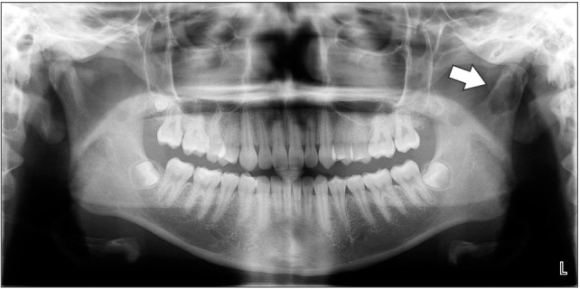
Fig. 2
Preoperative computed tomography. Trabecular pattern is not detected in the bone marrow space of the left condyle head (arrows) on axial (A), coronal (B), or sagittal view (C).
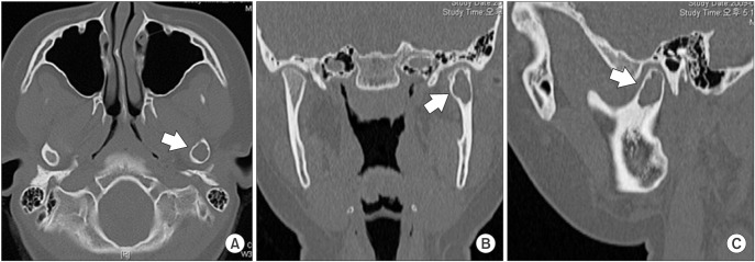
Fig. 3
Preoperative sagittal T1-weighted (A) and sagittal (B) and coronal (C) T2-weighted magnetic resonance images of the left temporomandibular joint at closed mouth position. Bone marrow space of the condyle head shows high signal intensity (arrows).
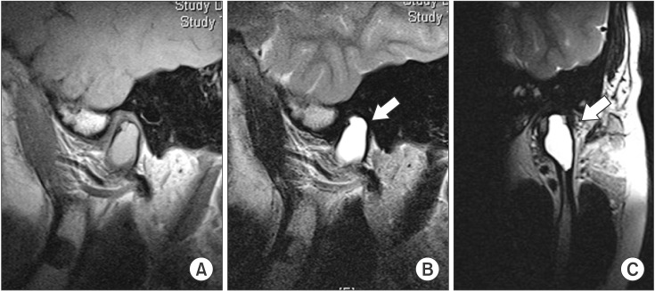
Fig. 4
Operative findings. (A) The head of the left condyle (arrow) was exposed. (B) After creation of a bony window with an initial semicircular corticotomy. (C) Putting the bony wall aside, no cystic membrane was found in the cavity. (D) Repositioning of the bony wall (asterisk) just before closure.
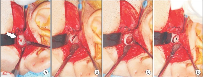
Fig. 5
Serial evaluation of the left condylar cystic lesion (arrow) on the panoramic X-rays. Preoperative image (A), and images at 2 months (B), 5 months (C), 9 months (D), and 43 months (E) after surgery.


 PDF
PDF ePub
ePub Citation
 Print
Print



 XML Download