Abstract
The uvulopalatal flap (UPF) technique is a modification of uvulopalatopharyngoplasty (UPPP) for the surgical treatment of obstructive sleep apnea. In the UPF technique, an uvulopalatal flap is fabricated and sutured to the residual mucosa of the soft palate to expand the anteroposterior dimensions of the oropharyngeal inlet. In the extended uvulopalatal flap (EUPF) technique, an incision at the tonsillar fossa is added to the classical UPF technique followed by the removal of mucosa and submucosal adipose tissue for additional expansion of the lateral dimension. The EUPF technique is more conservative and reversible than UPPP. Therefore, complications, such as velopharyngeal insufficiency, dysphagia, dryness, nasopharyngeal stenosis and postoperative pain, are reduced. In the following case report, the patient was diagnosed with obstructive sleep apnea and treated with the EUPF technique. The patient's total respiratory disturbance events per hour (RDI) was decreased to 15.4, the O2 saturation during the sleep was increased, and the excessive daytime sleepiness had disappeared after the surgery without complications. The authors report this case with a review of the relevant literature.
Go to : 
References
1. Won CH, Li KK, Guilleminault C. Surgical treatment of obstructive sleep apnea: upper airway and maxillomandibular surgery. Proc Am Thorac Soc. 2008; 5:193–9.

2. Cho KS. Surgical management for obstructive sleep apnea. J Clin Otolaryngol Head Neck Surg. 2008; 19:168–76.

3. Rhee CS, Han DH. Surgical management of sleep-disordered breathing. Tuberc Respir Dis. 2009; 66:417–30.

4. Young T, Palta M, Dempsey J, Skatrud J, Weber S, Badr S. The occurrence of sleep-disordered breathing among middle-aged adults. N Engl J Med. 1993; 328:1230–5.

5. Huang SG, Li QY. Sleep Respiratory Disorder Study Group Respiratory Disease Branch Shanghai Medical Association. Prevalence of obstructive sleep apnea-hypopnea syndrome in Chinese adults aged over 30 yr in Shanghai. Zhonghua Jie He He Hu Xi Za Zhi. 2003; 26:268–72.
6. Grote L, Hedner J, Grunstein R, Kraiczi H. Therapy with nCPAP: incomplete elimination of sleep related breathing disorder. Eur Respir J. 2000; 16:921–7.

7. Fujita S, Conway W, Zorick F, Roth T. Surgical correction of anatomic azbnormalities in obstructive sleep apnea syndrome: uvulopalatopharyngoplasty. Otolaryngol Head Neck Surg. 1981; 89:923–34.
8. Walker-Engstro¨m ML, Tegelberg A, Wilhelmsson B, Ringqvist I. 4-year follow-up of treatment with dental appliance or uvulopalatopharyngoplasty in patients with obstructive sleep apnea: a randomized study. Chest. 2002; 121:739–46.
9. Lee YK, Myung H, Hwang SJ, Seo BM, Lee JH, Choung PH, et al. Clinical study of surgical treatments for snoring and obstructive sleep apnea. J Korean Assoc Oral Maxillofac Surg. 2008; 34:435–44.
10. Fairbanks DN. Uvulopalatopharyngoplasty complications and avoidance strategies. Otolaryngol Head Neck Surg. 1990; 102:239–45.

11. Haavisto L, Suonpa¨a¨ J. Complications of uvulopalatopharyngoplasty. Clin Otolaryngol Allied Sci. 1994; 19:243–7.

12. Li HY, Chen NH, Shu YH, Wang PC. Changes in quality of life and respiratory disturbance after extended uvulopalatal flap surgery in patients with obstructive sleep apnea. Arch Otolaryngol Head Neck Surg. 2004; 130:195–200.

13. Powell N, Riley R, Guilleminault C, Troell R. A reversible uvulopalatal flap for snoring and sleep apnea syndrome. Sleep. 1996; 19:593–9.

14. Li HY, Li KK, Chen NH, Wang PC. Modified uvulopalatopharyngoplasty: the extended uvulopalatal flap. Am J Otolaryngol. 2003; 24:311–6.

15. Johns MW. A new method for measuring daytime sleepiness: the Epworth sleepiness scale. Sleep. 1991; 14:540–5.

16. Friedman M, Ibrahim H, Joseph NJ. Staging of obstructive sleep apnea/hypopnea syndrome: a guide to appropriate treatment. Laryngoscope. 2004; 114:454–9.

17. Kim JH. Sleep apnea. Korean Society of Otorhinolayngology-Head and Neck Surgery. Otorhinolaryngology-Head and Neck Surgery. 1st ed.Seoul: Ilchokak;2002. p. 1103–14.
18. Li HY, Li KK, Chen NH, Wang CJ, Liao YF, Wang PC. Three-dimensional computed tomography and polysomnography findings after extended uvulopalatal flap surgery for obstructive sleep apnea. Am J Otolaryngol. 2005; 26:7–11.

19. Kim SW. Eun YG. Surgical therapy for obstructive sleep apnea. J Clin Otolaryngol Head Neck Surg. 2005; 16:193–206.
Go to : 
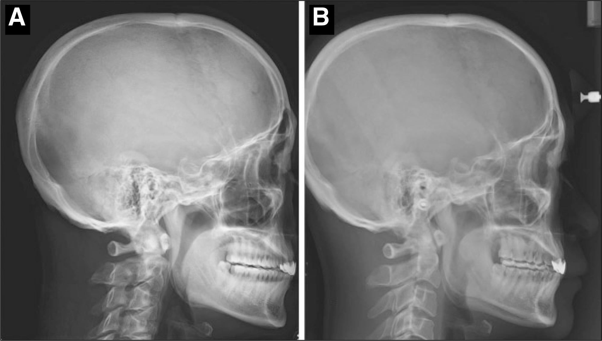
 | Fig. 2.Preoperative and postoperative lateral cephalogram. A. Preoperative lateral cephalogram. The narrow posterior airway space and long soft palate was observed. The length of the soft palate (posterior nasal spine – uvular tip) was 56 mm. B. Postoperative lateral cephalogram. The xray was taken at 3 weeks after the surgery. The shortening of soft palate was observed. The length of the soft palate was changed to 38 mm. |
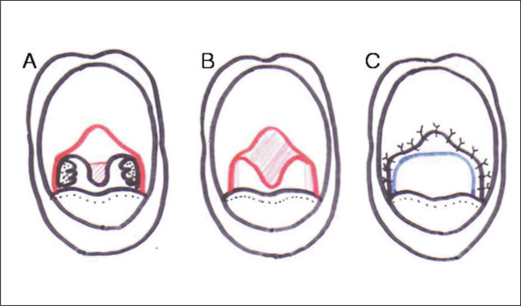
 | Fig. 3.A procedure of the extended uvulopalatal flap technique. A. The uvular and soft palate is retracted forward. B. The margin of the uvulopalatal flap is marked. C. The overlapping mucosa and submucosal adipose tissue are resected from the incision line to the uvular tip. D. The flap is folded and sutured to the residual mucosa of the soft palate. Also, the wound at tonsillar fossa for the removal of tonsils, mucosa and submucosal adipose tissue is closed. E. To achieve more effective expanding of posterior airway space, radiofrequency ablation of the tongue base is combined. F. At the postoperative one week, the shortening of soft palate and the expanding oropharyngeal inlet was observed. |
Table 1.
Classification of Friedman palate position16
Table 2.
Classification of tonsil size16
Table 3.
The modified Friedman staging system for patients with obstructive sleep apnea/hypopnea syndrome16


 PDF
PDF ePub
ePub Citation
 Print
Print


 XML Download