Abstract
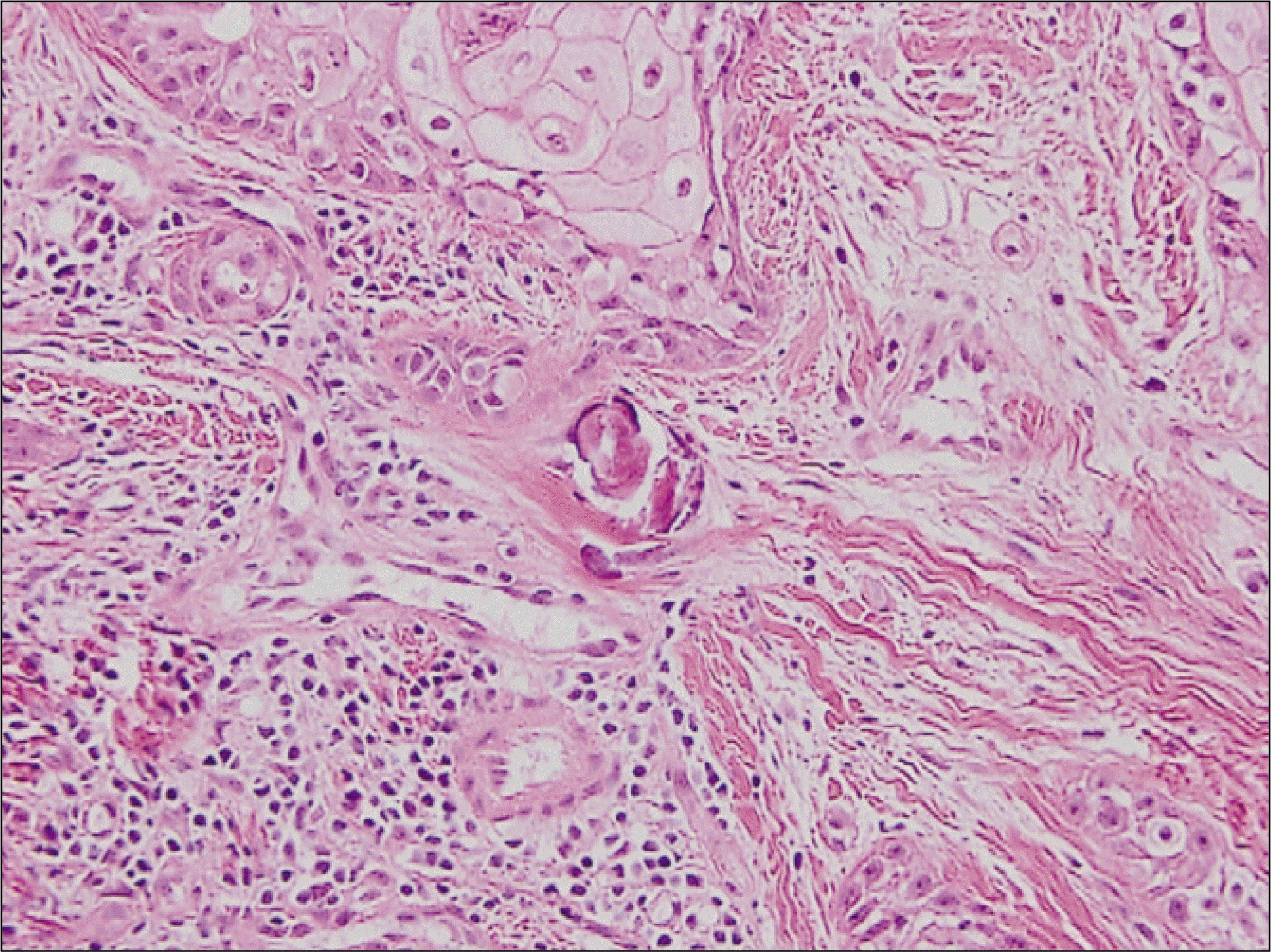
A calcifying epithelial odontogenic tumor (CEOT) was first described as a separate entity in 1955 by Pindborg, and has since been referred to as Pindborg tumor. CEOT is characterized by the presence of squamous-cell proliferation, calcification and amyloid deposits, and accounts for only 1% of all odontogenic tumors. CEOT is a benign, though occasional locally invasive, slow-growing neoplasm. It is located either intraosseously or extraosseously, and is usually associated with an unerupted permanent tooth.
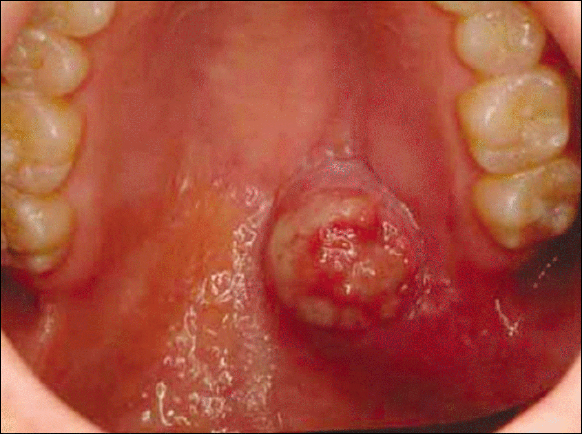
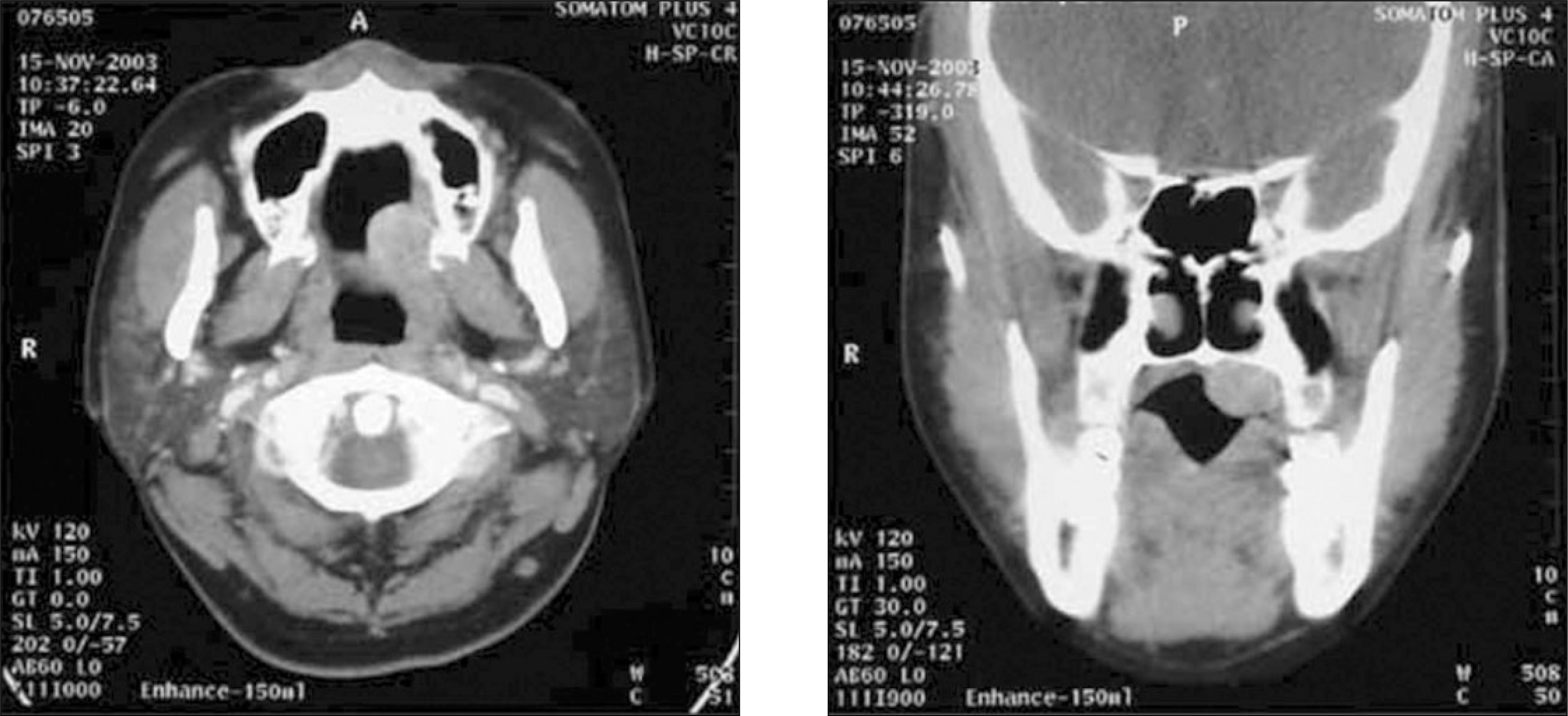
A 24 year-old female visited our clinic, presenting with a palatal swelling and intra-oral ulcer. After an incisional biopsy, the lesion was confirmed to be odontogenic tumor. A tumor resection and reconstruction surgery with tongue flap were performed.
References
2. Anavi Y, Kaplan I, Citir M, Calderon S. Clear-cell variant of calcifying epithelial odontogenic tumor: clinical and radiographic characteristics. Oral Surg Oral Med Oral Pathol Oral Radiol Endod. 2003; 95:332–9.

3. Korean Academy of Oral and Maxillofacial Radiology, ed. Oral and Maxillofacial Radiology. 2nd ed.Seoul: Yiwoo;1996.
4. Sapp JP, Eversole LR, Wysocki GP. Contemporary oral and maxillofacial pathology. St. Louis, Mo.: Mosby;1998.
5. Lim CY, Hong SP, Lee JI. Color atlas of oral pathology. 2nd ed.Seoul: Korea Medical Book;1999.
6. Maiorano E, Renne G, Tradati N, Viale G. Cytogical features of calcifying epithelial odontogenic tumor (Pindborg tumor) with abundant cementum-like material. Virchows Arch. 2003; 442:107–10.

7. Bouckaert MM, Raubenheimer EJ, Jacobs FJ. Calcifying epithelial odontogenic tumor with intracranial extension: report of a case and review of the literature. Oral Surg Oral Med Oral Pathol Oral Radiol Endod. 2000; 90:656–62.

8. Belmonte-Caro R, Torres-Lagares D, Mayorga-Jimenez F, Garcia-Perla Garcl ′ a A, Infante-Cossio P, Gutierrez-Perez JL. Calcifying epithelial odontogenic tumor (Pindborg tumor). Med Oral. 2002; 7:309–15.
9. Veness MJ, Morgan G, Collins AP, Walker DM. Calcifying epithelial odontogenic (Pindborg) tumor with malignant transformation and metastatic spread. Head Neck. 2001; 23:692–6.

10. Philipsen HP, Reichart PA. Calcifying epithelial odontogenic tumour: biological profile based on 181 cases from the literature. Oral Oncol. 2000; 36:17–26.

11. Martin-Duverneuil N, Roisin-Chausson MH, Behin A, Favre-Dauvergne E, Chiras J. Combined benign odontogenic tumors: CT and MR findings and histomorphologic evaluation. AJNR Am J Neuroradiol. 2001; 22:867–72.
12. Cheng YS, Wright JM, Walstad WR, Finn MD. Calcifying epithelial odontogenic tumor showing microscopic features of potential malignant behavior. Oral Surg Oral Med Oral Pathol Oral Radiol Endod. 2002; 93:287–95.

13. Cross JJ, Pilkington RJ, Antoun NM, Adlam DM. Value of computed tomography and magnetic resonance imaging in the treatment of a calcifying epithelial odontogenic (Pindborg) tumour. Br J Oral Maxillofac Surg. 2000; 38:154–7.

14. Crivelini MM, de Arau ′ jo VC, de Sousa SO, de Arau ′ jo NS. Cytokeratins in epithelia of odontogenic neoplasms. Oral Dis. 2003; 9:1–6.

15. Mesquita RA, Lotufo MA, Sugaya NN, de Arau ′ jo NS, de Arau ′ jo VC. Peripheral clear cell variant of calcifying epithelial odontogenic tumor: report of a case and immunohistochemical investigation. Oral Surg Oral Med Oral Pathol Oral Radiol Endod. 2003; 95:198–204.


 PDF
PDF ePub
ePub Citation
 Print
Print


 XML Download