

 PDF
PDF ePub
ePub Citation
 Print
Print


Abstract
Introduction
Endoscope-assisted open reduction and internal fixation (EAORIF) reduces the amount of facial scaring, but limitations, such as the possibility to convert to the open technique and the large learning curve, remain.
Materials and Methods
The medical records of 19 patients diagnosed as lateral overridden subcondyle fractures and treated with endoscope-assisted open reduction and internal fixation at Yonsei University Health System from December 2006 to August 2010 were reviewed.
Results
11 patients underwent temporary discomfort or pain such as limitation of mouth opening, temporomandibular joint discomfort, lip paresthesia or facial weakness, but the symptoms disappeared within 3 months. There was no severe long-term complication except 2 patients with refractures of operated subcondyles.
Go to : 
References
1. Lindahl L. Condylar fractures of the mandible. I. Classification and relation to age, occlusion, and concomitant injuries of teeth and teeth-supporting structures, and fractures of the mandibular body. Int J Oral Surg. 1977; 6:12–21.
2. Kellman RM. Endoscopic approach to subcondylar mandible fractures. Facial Plast Surg. 2004; 20:239–47.

3. Choi BH, Yi CK, Yoo JH. Clinical evaluation of 3 types of plate osteosynthesis for fixation of condylar neck fractures. J Oral Maxillofac Surg. 2001; 59:734–7. discussion 738.
4. Lee C, Mueller RV, Lee K, Mathes SJ. Endoscopic subcondylar fracture repair: functional, aesthetic, and radiographic outcomes. Plast Reconstr Surg. 1998; 102:1434–43. discussion 1444–5.
5. Kellman RM, Cienfuegos R. Endoscopic approaches to subcondylar fractures of the mandible. Facial Plast Surg. 2009; 25:23–8.
6. Ducic Y. Endoscopic treatment of subcondylar fractures. Laryngoscope. 2008; 118:1164–7.
7. Cho-Lee GY, Rodrl′guez Campo FJ, Gonza′lez Garcl′a R, Mun ̃oz Guerra MF, Sastre Pe′rez J, Naval Gl′as L. Endoscopically-assisted transoral approach for the treatment of subcondylar fractures of the mandible. Med Oral Patol Oral Cir Bucal. 2008; 13:E511–5.
8. Troulis MJ. Endoscopic open reduction and internal rigid fixation of subcondylar fractures. J Oral Maxillofac Surg. 2004; 62:1269–71.
9. Vural E. Treatment of adult subcondylar mandibular fractures: closed vs open vs endoscopic approach. Arch Otolaryngol Head Neck Surg. 2004; 130:1228–30.
10. Paeng JY, Ok YJ, Myoung H, Hwang SJ, Seo BM, Choi JY, et al. Endoscopic-assisted open reduction and internal fixation (EAORIF) for condylar fracture. J Korean Assoc Oral Maxillofac Surg. 2006; 32:474–81.
11. Schoen R, Fakler O, Metzger MC, Weyer N, Schmelzeisen R. Preliminary functional results of endoscope-assisted transoral treatment of displaced bilateral condylar mandible fractures. Int J Oral Maxillofac Surg. 2008; 37:111–6.
Go to :
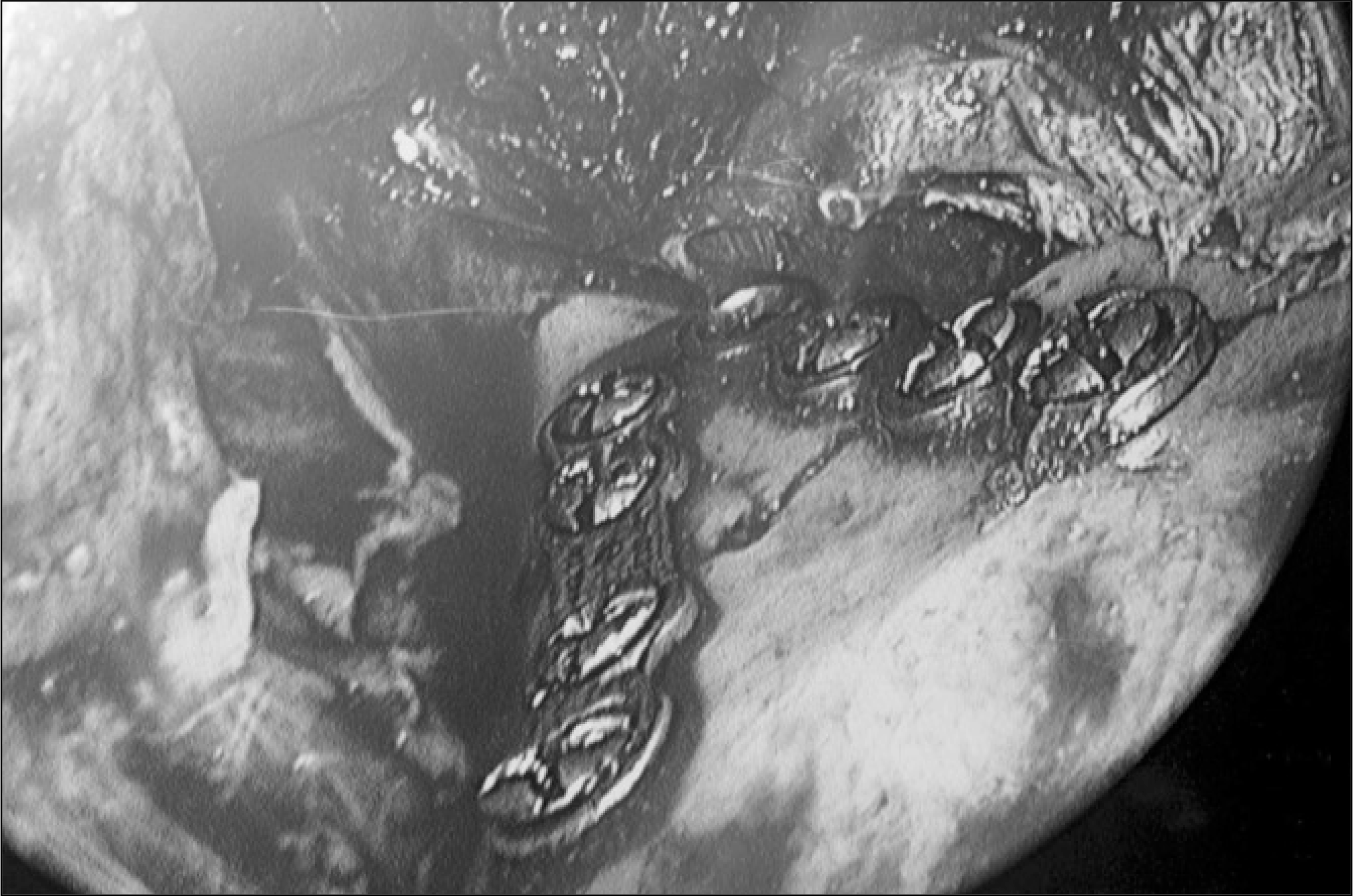 | Fig. 2.Endoscopic view after EAORIF of lateral-overridden subcondyle fracture.(EAORIF: endoscope-assisted open reduction and internal fixation) |
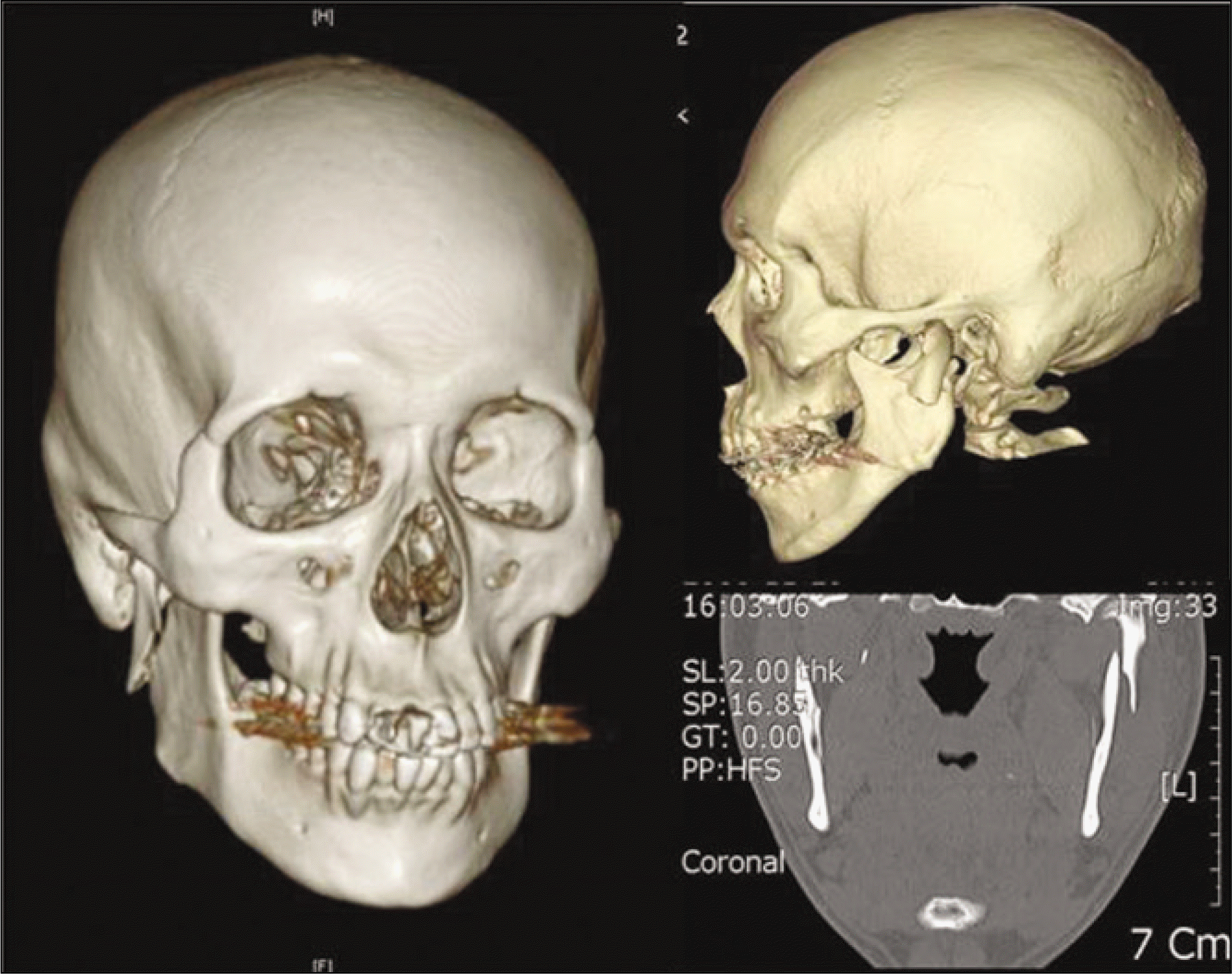
 | Fig. 3.A images of preoperative (left), postoperative (right) 3-dimensional computed tomography image of lateral-over-rided subcondyle fracture in a patient. |
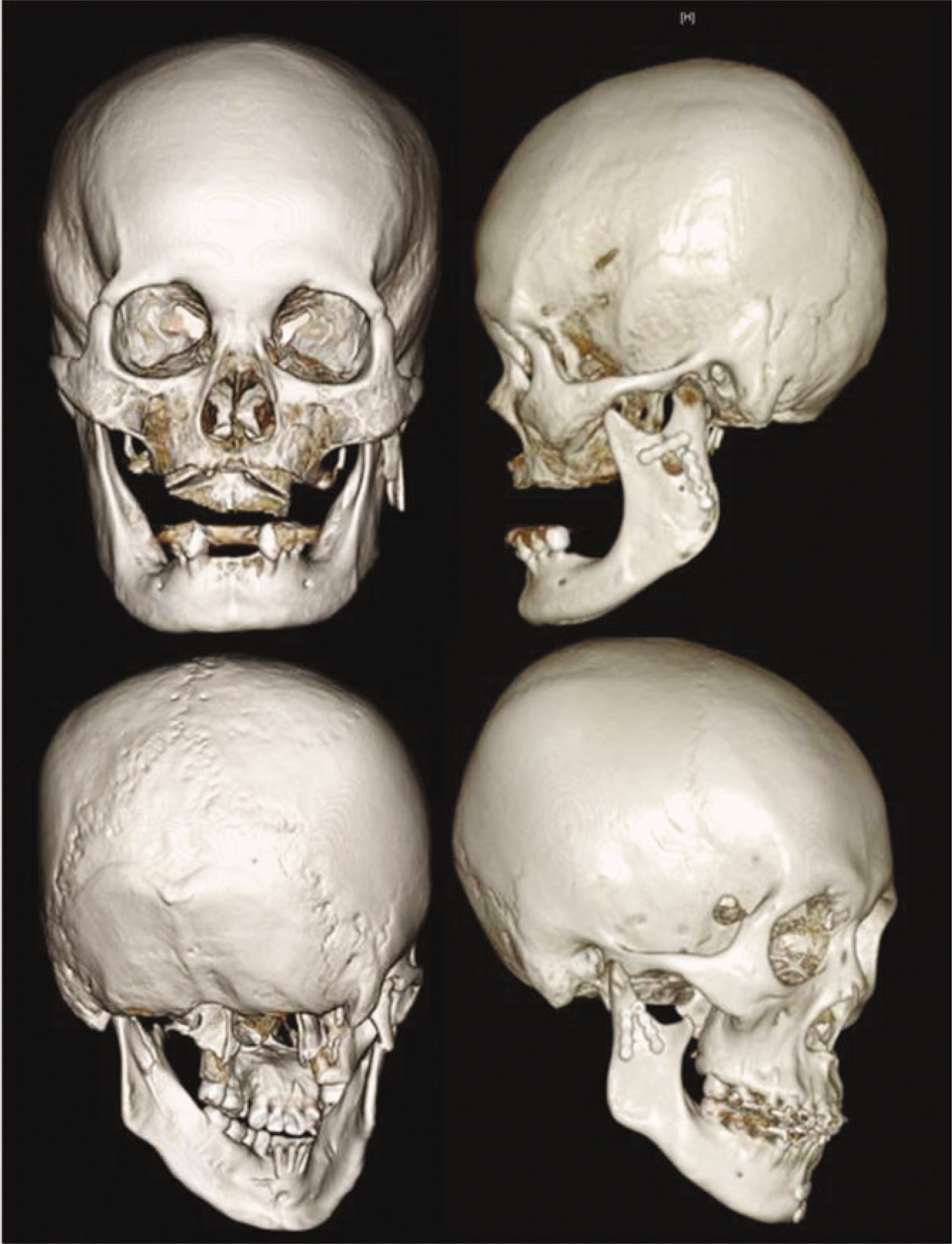
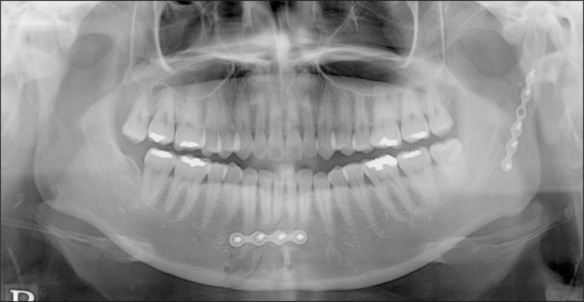
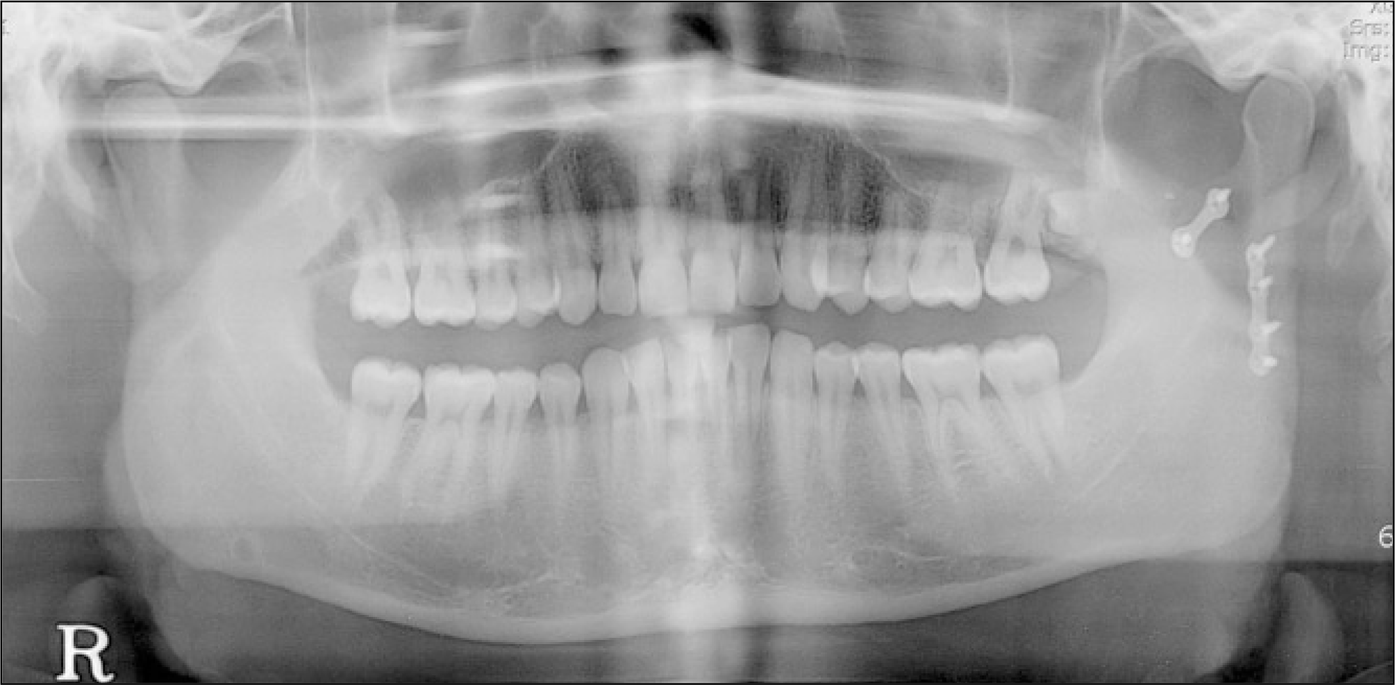
 | Fig. 4.Panoramic view of the patient who was diagnosed as subcondyle refracture 1 month after EAORIF. A. Before operation. B. Five days after operation. C. One month after operation.(EAORIF: endoscope-assisted open reduction and internal fixation) |
 | Fig. 5.EAORIF of subcondyle fracture was done with a 6-hole miniplate.(EAORIF: endoscope-assisted open reduction and internal fixation) |
 | Fig. 6.EAORIF was done with one 4-hole miniplate and one 2-hole miniplate.(EAORIF: endoscope-assisted open reduction and internal fixation) |
Table 1.
Results of EAORIF of lateral override subcondylar fracture (n=19)
| Patients | Age | Sex | Operated Sites | Number of Plates | Number of Screws | Periods of IMF | Temporary Complications1 | Long-term Complications | Operation Time (minutes) | Follow Up Periods (months) |
|---|---|---|---|---|---|---|---|---|---|---|
| 1 | 27 | M | Lt. Sc + P | 1 | 4 | 0 | TMJ pain | − | 125 | 8 |
| 2* | 36 | M | Rt. Sc | 1 | 4 | 14 | − | Premature contact, refracture | 106 | 14 |
| 3 | 59 | F | Rt. Sc + S | 1 | 3 | 14 | Lip paresthesia | − | 138 | 7 |
| 4 | 34 | F | Lt. Sc + P | 2 | 7 | 7 | Limited lateral movement | 218 | 9 | |
| 5 | 26 | M | Lt. Sc | 1 | 3 | 7 | Limited opening | Premature contact | 143 | 9 |
| 6 | 66 | F | Rt. Sc | 2 | 7 | 3 | − | Premature contact | 122 | 6 |
| 7 | 27 | F | Rt. Sc + P | 1 | 4 | 14 | Limited opening | − | 229 | 4 |
| 8 | 22 | M | Rt. Sc | 1 | 5 | 0 | − | Premature contact | 102 | 12 |
| 9 | 27 | F | Lt. Sc + S | 1 | 5 | 0 | Intermittent TMJ pain | − | 197 | 3 |
| 10 | 60 | M | Rt. Sc + P | 1 | 5 | 0 | TMJ pain, facial nerve weakness, chin paresthesia | 261 | 4 | |
| 11 | 29 | M | Lt. Sc + S | 2 | 6 | 10 | Infection | − | 171 | 5 |
| 12 | 32 | M | Rt. Sc | 2 | 6 | 0 | − | − | 150 | 7 |
| 13 | 37 | M | Lt. Sc + P | 2 | 5 | 0 | Limited opening | − | 56 | 4 |
| 14 | 24 | M | Lt. Sc +P | 2 | 6 | 0 | Intermittent TMJ pain limited opening | 144 | 6 | |
| 15 | 79 | F | Lt. Sc | 2 | 7 | 0 | − | − | 144 | 6 |
| 16 | 46 | F | Rt. Sc | 2 | 6 | 0 | − | refracture | 150 | 5 |
| 17 | 28 | M | Lt. Sc | 2 | 6 | 0 | − | − | 100 | 3 |
| 18 | 24 | M | Rt. Sc + P | 2 | 7 | 0 | − | − | 180 | 5 |
| 19 | 28 | M | Lt. Sc + S | 2 | 6 | 0 | − | − | 120 | 4 |
 XML Download