

 PDF
PDF ePub
ePub Citation
 Print
Print


Abstract
Synovial sarcoma (SS) is a malignant soft tissue tumor comprising 5–10% of all soft tissue sarcomas. This tumor normally occurs in the para-articular regions of the extremities but is rare in head and neck sites. SS is sometimes difficult to diagnose because it can mimic benign lesions both clinically and radiologically. This paper presents a rare case of a SS of the buccal space of a 25-year old man. The histology examination and immunohistochemistry of the mass led to a diagnosis of synovial sarcoma. The patient was treated primarily with a surgical resection, followed by radiotherapy and chemotherapy. The follow up examination 17-months after surgery showed no signs of tumor relapse or metastasis.
Fig. 1.
Pre-operative extraoral photo. Ji-Hoon Han et al: Synovial sarcoma in the buccal space: a case report. J Korean Assoc Oral Maxillofac Surg 2011
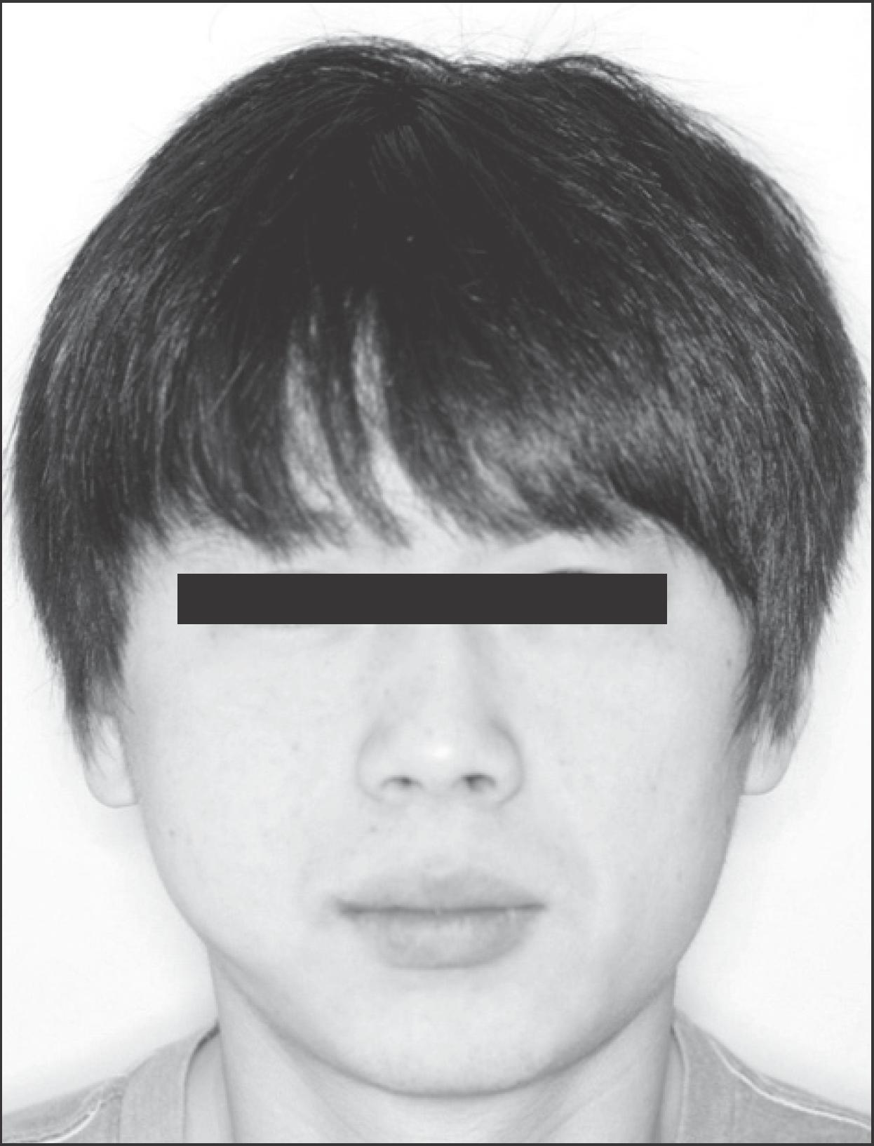
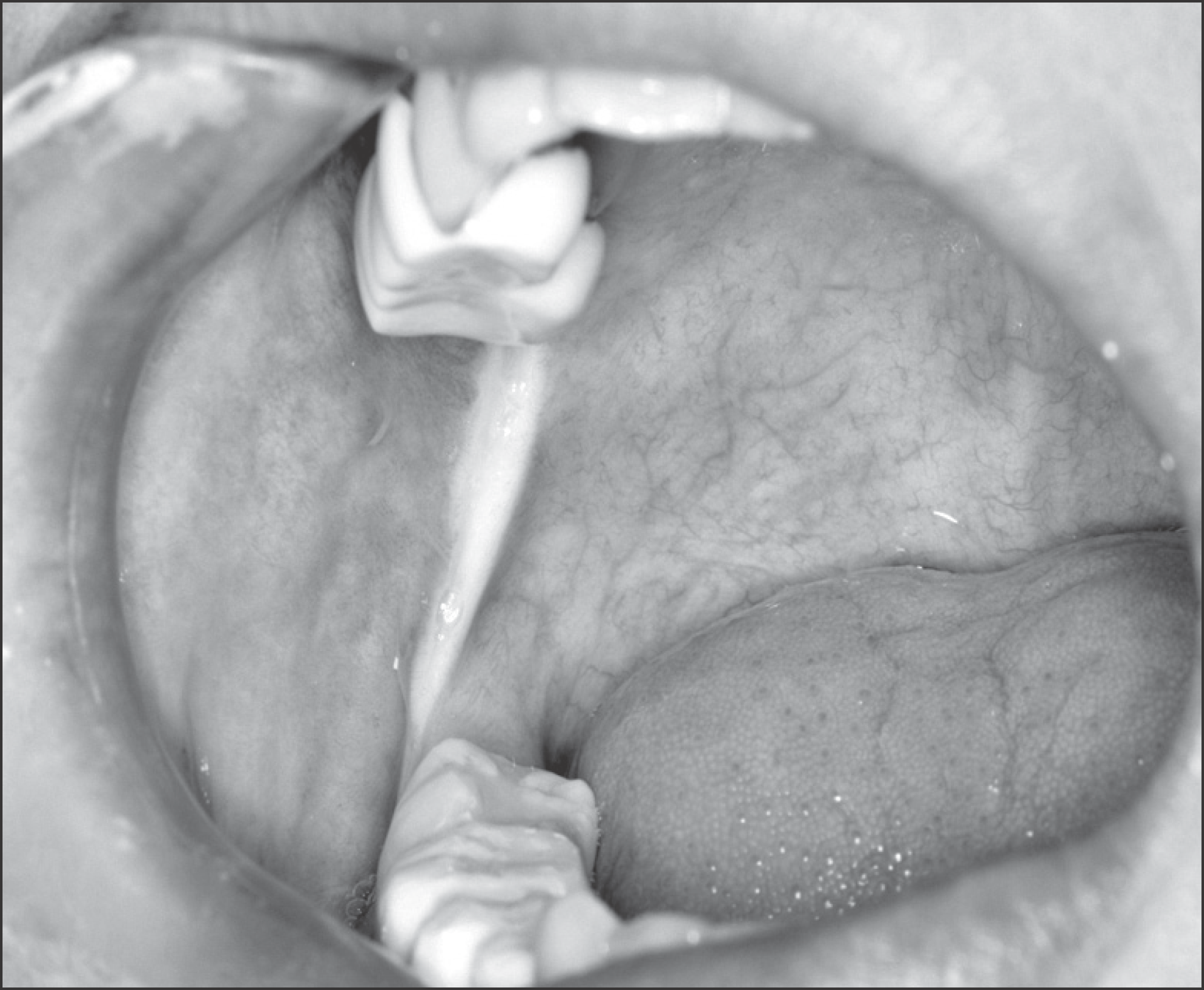
Fig. 2.
Pre-operative intraoral photo. Palpable elevated mass on right buccal mucosa and retromolar area. Ji-Hoon Han et al: Synovial sarcoma in the buccal space: a case report. J Korean Assoc Oral Maxillofac Surg 2011
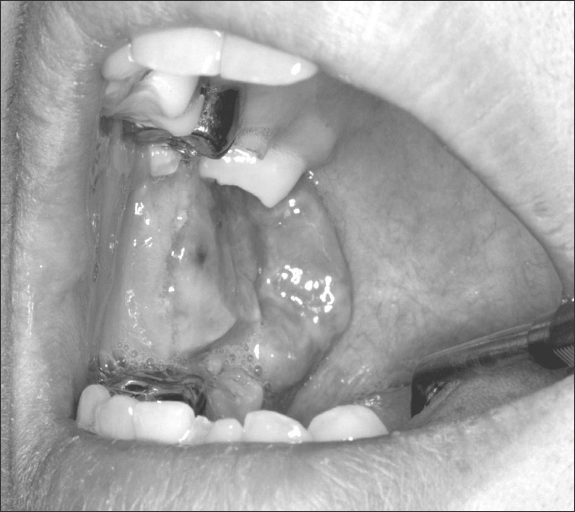
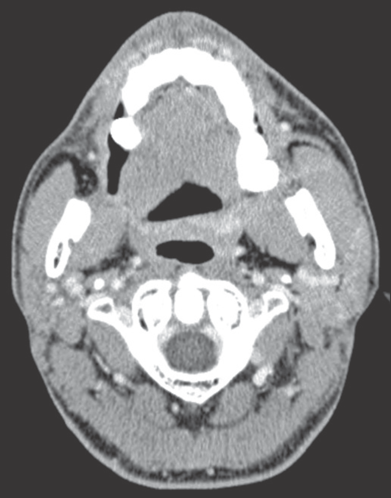
Fig. 3.
Pre-operative computed tomography. Round shaped relatively well-defined mild to moderately enhancing inhomogeneous mass (3.2 cm×2.8 cm, arrow) with internal high density foci in right buccal space without adjacent bony erosions. Ji-Hoon Han et al: Synovial sarcoma in the buccal space: a case report. J Korean Assoc Oral Maxillofac Surg 2011
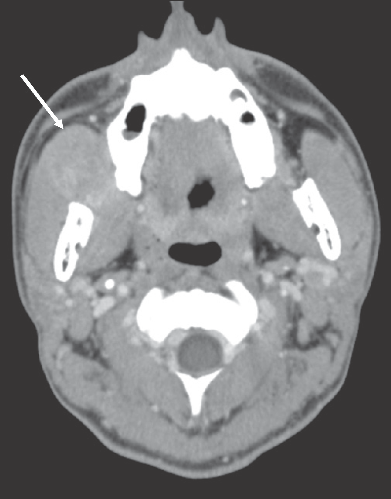
Fig. 4.
Pre-operative salivary gland sonography. Ovoid shaped well-defined heterogeneous mass (2.8 cm×2.5 cm×3.9 cm) with increased blood flow in right buccal space. Ji-Hoon Han et al: Synovial sarcoma in the buccal space: a case report. J Korean Assoc Oral Maxillofac Surg 2011
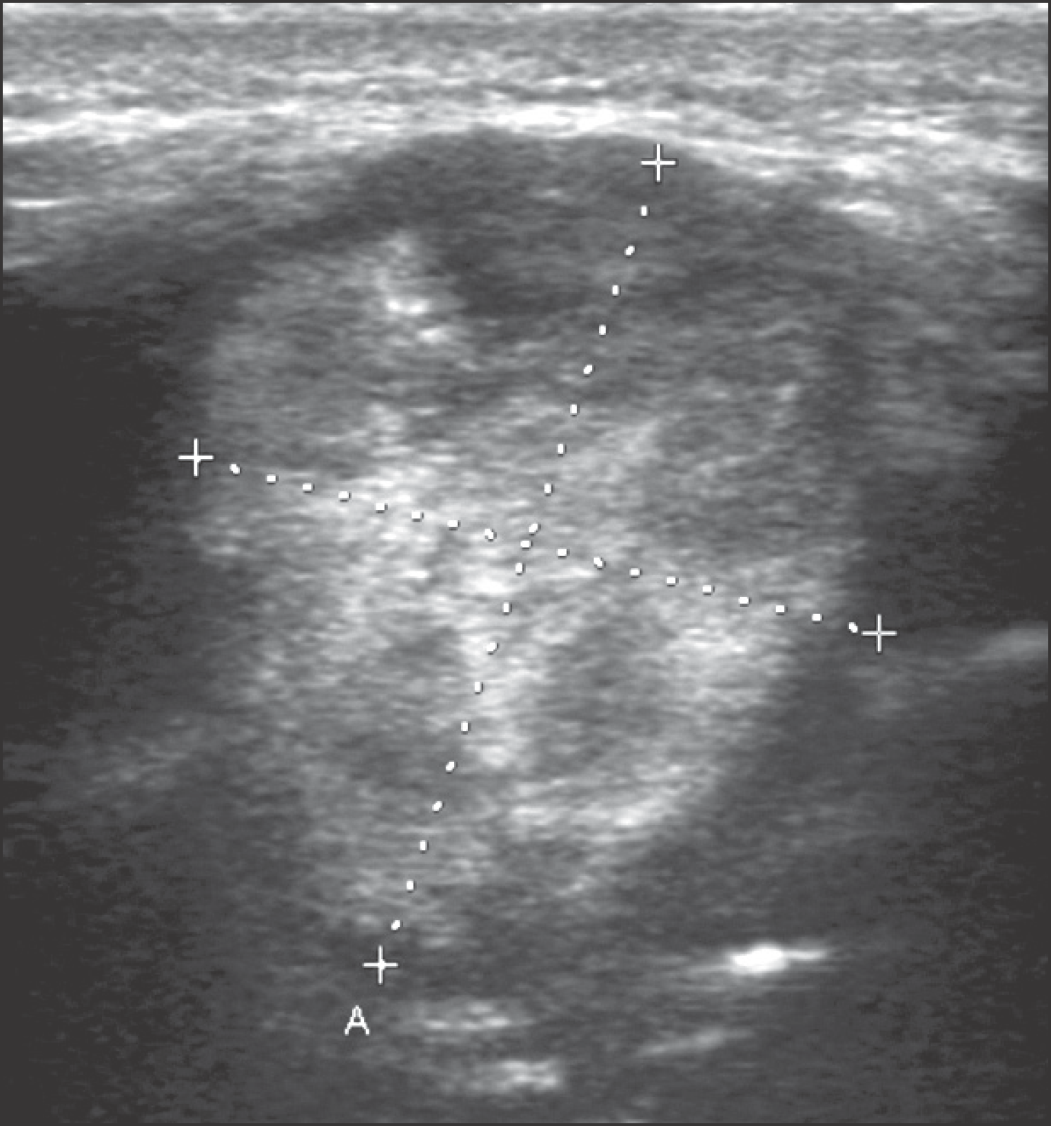
Fig. 5.
Gross finding. Well defined pinkish gray soft tissue (5 cm× 4 cm×3 cm). Ji-Hoon Han et al: Synovial sarcoma in the buccal space: a case report. J Korean Assoc Oral Maxillofac Surg 2011
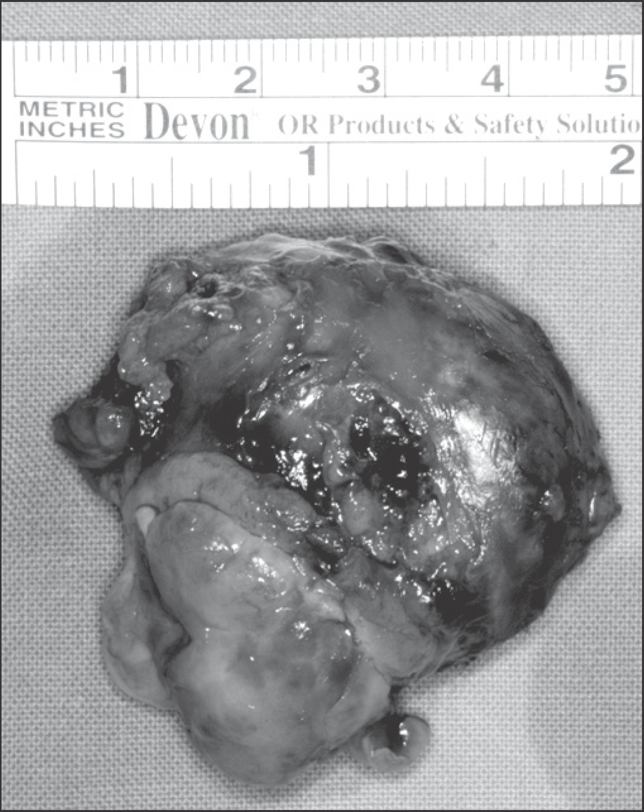
Fig. 7.
Post-operative intraoral photo (15-months after). Scar formation on right retromolar area. Ji-Hoon Han et al: Synovial sarcoma in the buccal space: a case report. J Korean Assoc Oral Maxillofac Surg 2011

Fig. 6.
Photomicrographs of synovial sarcoma in the buccal space. A. It reveals proliferation of spindle cells and the epithelial components forming gland-like structures (H&E staining, ×200). B. The spindle cells are stained for bcl-2 immunoreactivity (×200). C. The spindle cells are stained for CD99 immunoreactivity (×200). D. The epithelial components are stained for cytokeratin immunoreactivity (×200). Ji-Hoon Han et al: Synovial sarcoma in the buccal space: a case report. J Korean Assoc Oral Maxillofac Surg 2011
 XML Download