Abstract
Cemento-osseous dysplasia occurs in the tooth bearing areas of the jaws and is probably the most common fibroosseous manifestation. They are usually classified into three main groups according to their extent and radiographic appearance: periapical (surrounds the periapical region of teeth and are bilateral), focal (single lesion) and florid (scleroticsymmetrical masses) cemental-osseous dysplasias. Florid cemento-osseous dysplasia clearly appears to be a form of bone and cemental dysplasia that is limited to the jaws. Patients do not have laboratory or radiologic evidence of bone disease in other parts of the skeleton. For asymptomatic patients, the best management consists of regular recall examinations with prophylaxis and the reinforcement of good home hygiene care to control periodontal disease and prevent tooth loss. The treatment of symptomatic patients is more difficult. At this stage, there is an inflammatory component caused by the disease and the process is basically a chronic osteomyelitis involving dysplastic bone and cementum. Antibiotics might be suggested, but are not always effective. Two cases of florid cemento-osseous dysplasia diagnosed in two Korean females are reported with a review of the relevant literature.
Go to : 
References
1. Kramer IRH, Pindborg JJ, Shear M. Histologic typing of odontogenic tumors. WHO international Classification of Tumors. 2nd ed.Berlin: Springer-Verlag;1992. : 29.
2. Kawai T, Hiranuma H, Kishino M, Jikko A, Sakuda M. Cemento-osseous dysplasia of the jaws in 54 Japanese patients: A radiographic study. Oral Surg Oral Med Oral Pathol Oral Radiol Endod. 1999; 87:107–14.
4. Neville BW, Damm DD, Allen CM, Bouquot JE. Oral & Maxillofacial Pathology. 2nd ed.Philadelphia: WB Saunders Co.;1995. p. 449–51.
5. Singer SR, Mupparapu M, Rinaggio J. Florid cemento-osseous dysplasia and chronic diffuse osteomyelitis: Report of a simultaneous presentation and review of the literature. J Am Dent Assoc. 2005; 136:927–31.
6. Higuchi Y, Nakamura N, Tashiro H. Clinicopathologic study of cemento-osseous dysplasia producing cysts of the mandible: report of four cases. Oral Surg Oral Med Oral Pathol. 1988; 65:339–42.
7. Summerlin DJ, Tomich CE. Focal cemento osseous dysplasia: a clinicopathologic study of 221 cases. Oral Surg Oral Med Oral Pathol. 1994; 78:611–20.
8. Toffanin A, Benetti R, Manconi R. Familial florid cemento-osseous dysplasia: a case report. J Oral Maxillofac Surg. 2000; 58:1440–6.

9. Wolf J, Hietanen J, Sane J. Florid cemento-osseous dysplasia (gig-antiform cementoma) in a Caucasian woman. Br J Oral Maxillofac Surg. 1989; 27:46–52.

10. Coleman H, Altini M, Kieser J, Nissenbaum M. Familial florid cemento-osseous dysplasia: a case report and review of the literature. J Dent Assoc S Afr. 1996; 51:766–70.
11. Gonçalves M, Pispico R, Alves Fde A, Lugão CE, Gonçalves A. Clinical, radiographic, biochemical and histological findings of florid cemento-osseous dysplasia and report of a case. Braz Dent J. 2005; 16:247–50.

12. Groot RH, van Merkesteyn JP, Bras J. Diffuse sclerosing osteomyelitis and florid osseous dysplasia. Oral Surg Oral Med Oral Pathol Oral Radiol Endod. 1996; 81:333–42.

13. Johannsen A. Chronic sclerosing osteomyelitis of the mandible: radiographic differential diagnosis from fibrous dysplasia. Acta Radiol Diagn (Stockh). 1977; 18:360–8.
14. White SC, Pharoah MJ. Oral Radiology-Principles and Interpretation. St. Louis: Mosby;2000. p. 439–48.
15. Lee SK, Krebsbach PH, Matsuki Y, Nanci A, Yamada KM, Yamada Y. Ameloblastin expression in rat incisors and human tooth germs. Int J Dev Biol. 1996; 40:1141–50.
Go to : 
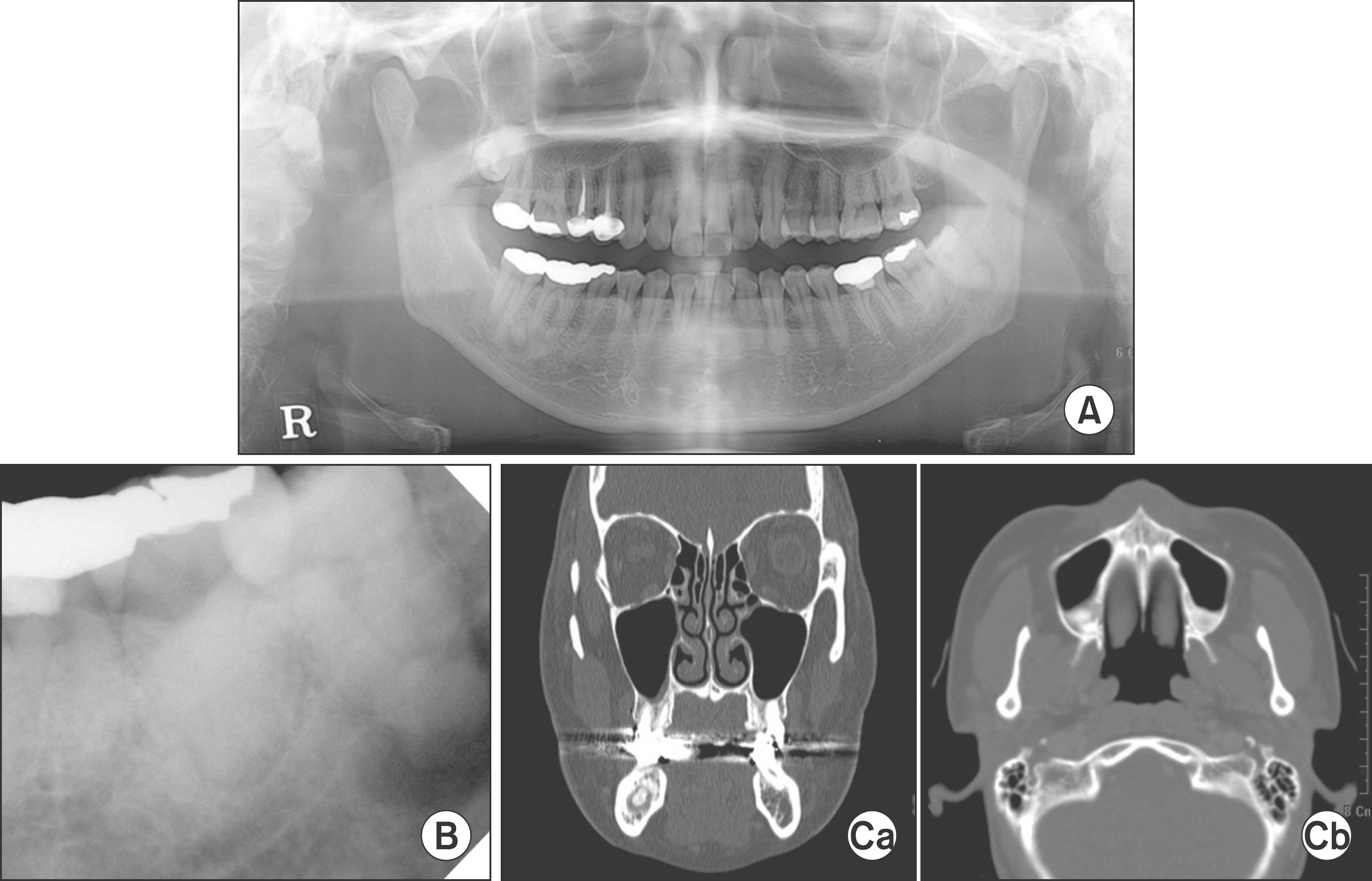 | Fig. 1.Pre-operation. Panoramic (A), periapical view (B), computer tomography findings (Ca, Cb) demonstrated multiple radiopaque/radiolucent admixed lesions on maxilla and mandible. Nam-Kyun Kim et al: Florid cemento-osseous dysplasia: a report of two cases. J Korean Assoc Oral Maxillofac Surg 2011
|
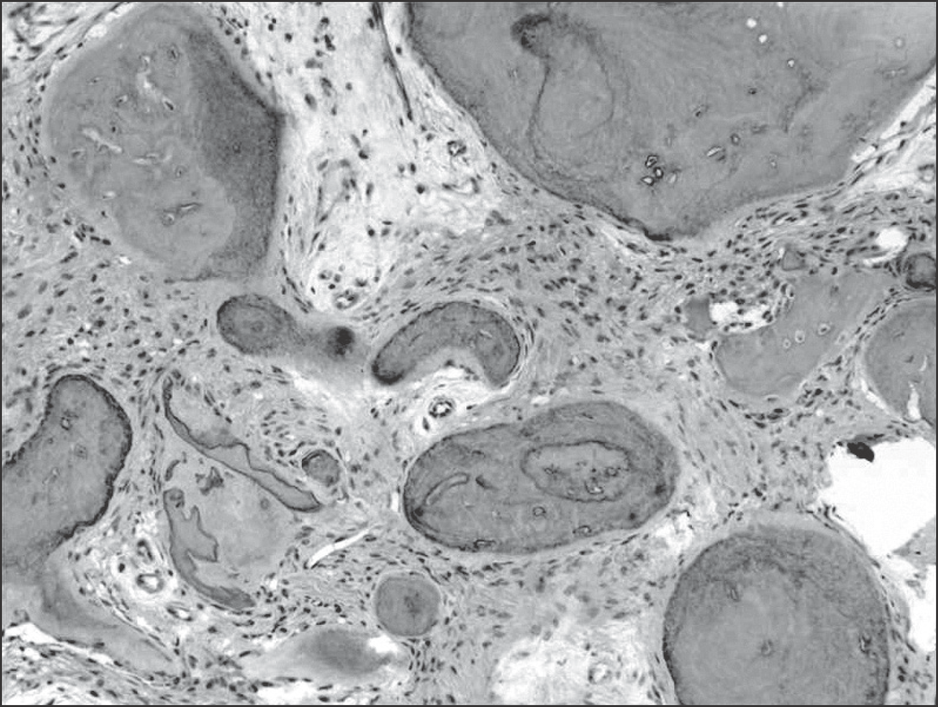
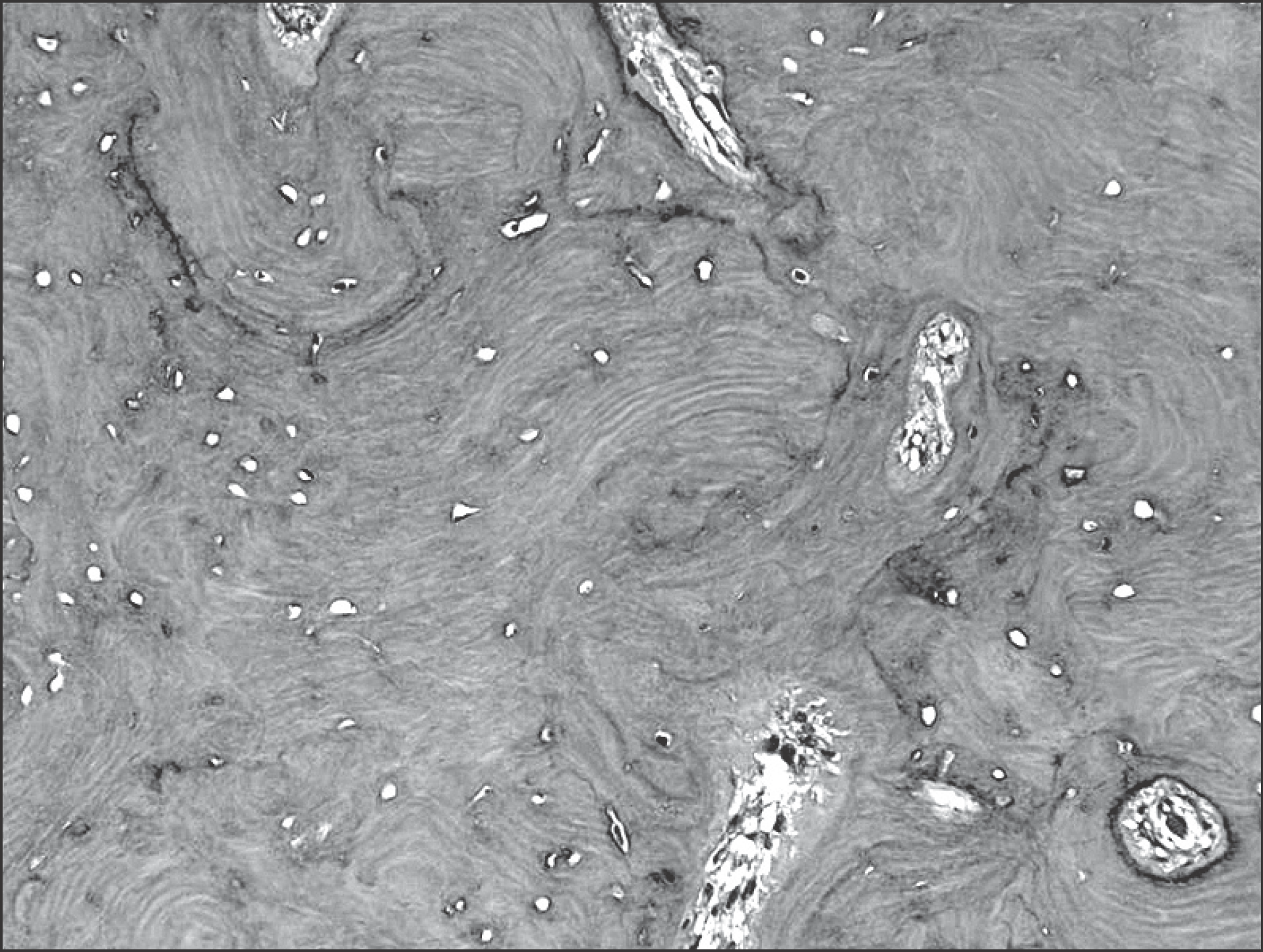 | Fig. 2.Microscopic features showed dense immature bony tissue with concentric calcification (H & E staining, x100). Nam-Kyun Kim et al: Florid cemento-osseous dysplasia: a report of two cases. J Korean Assoc Oral Maxillofac Surg 2011
|
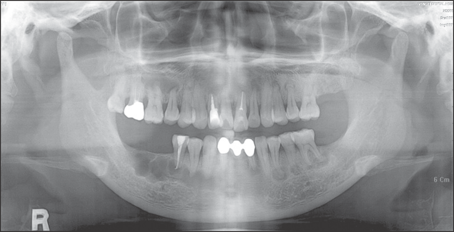
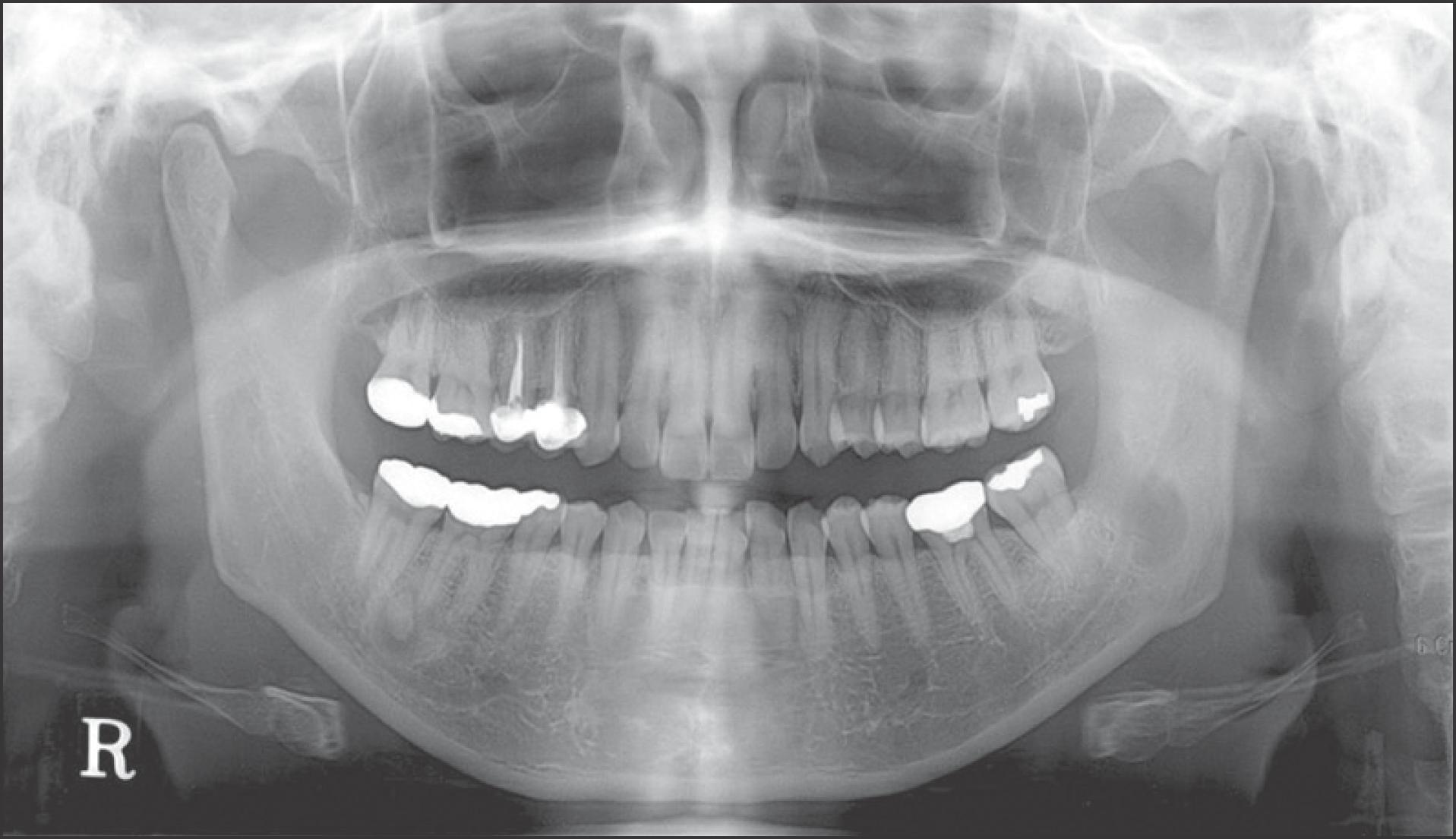 | Fig. 3.Post-operation. Panoramic view (2 months) showed good healing state. Nam-Kyun Kim et al: Florid cemento-osseous dysplasia: a report of two cases. J Korean Assoc Oral Maxillofac Surg 2011
|
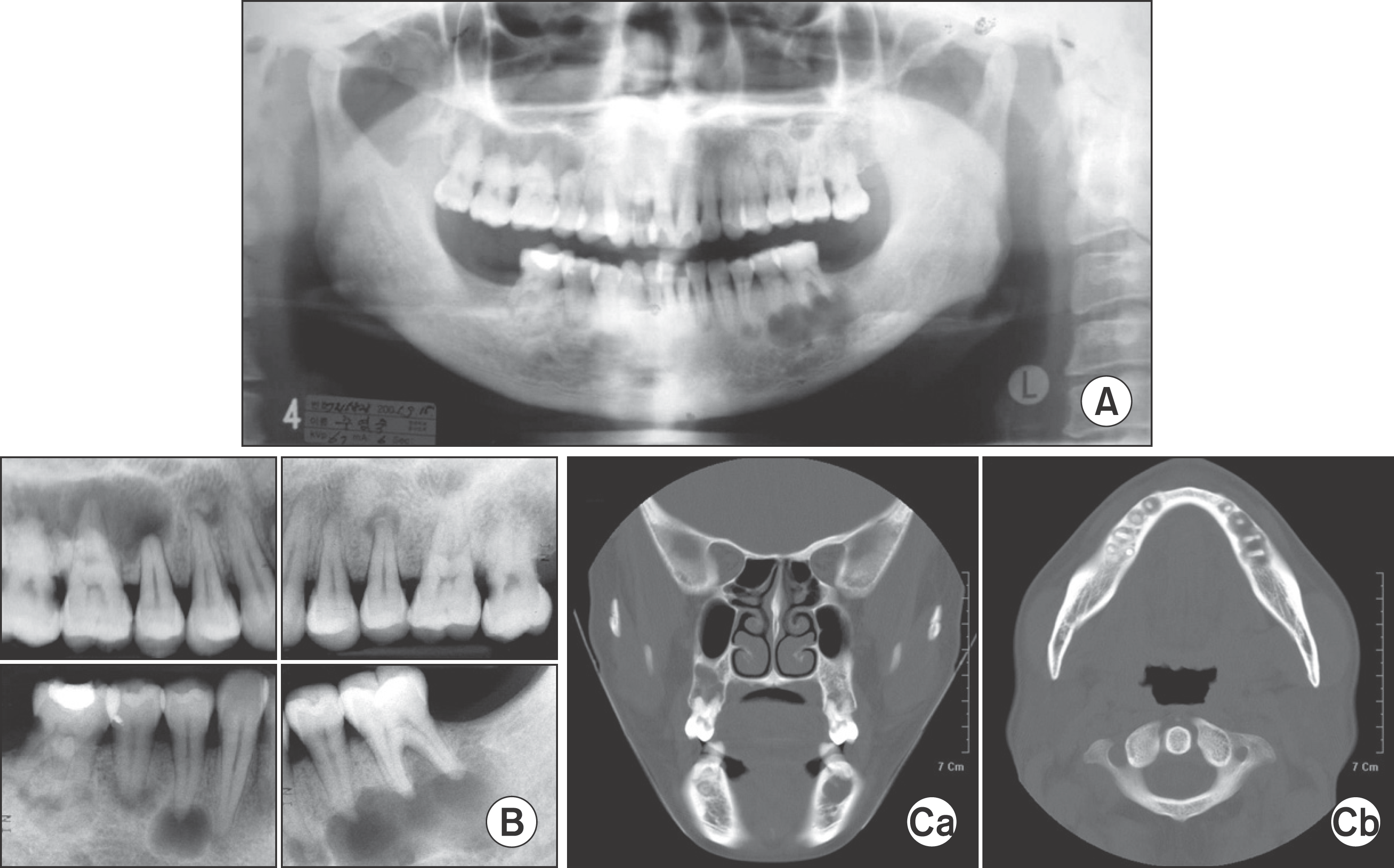 | Fig. 4.Pre-operation. Panoramic (A), periapical view (B), computer tomography findings (Ca, Cb) demonstrated multiple radiopaque/radiolucent admixed lesion on maxilla and mandible. Nam-Kyun Kim et al: Florid cemento-osseous dysplasia: a report of two cases. J Korean Assoc Oral Maxillofac Surg 2011
|


 PDF
PDF ePub
ePub Citation
 Print
Print


 XML Download