

 PDF
PDF ePub
ePub Citation
 Print
Print


Abstract
Introduction
Bone regeneration of cystic defects of the jaws after a cyst treatment requires lengthy healing periods. Generally, the bony changes are observed periodically through a visual radiographic reading as well as by the clinical opinion and radiographic images (panorama, periapical view), but it is difficult to compare the objective bony changes using only the radiographic density. In addition, it is difficult to observe minute bony changes through a visual radiographic reading, which can lead to a subjective judgment. This study exmined the bone density after the enucleation of a jaw cyst by fractal analysis.
Materials and Methods
Eighteen patients with a cystic lesion on the jaw were assessed. Panoramic radiographs were taken preoperatively, immediately postoperatively, and 1, 3, 6 and 12 months after cyst enucleation. The images were analyzed by fractal analysis.
Go to : 
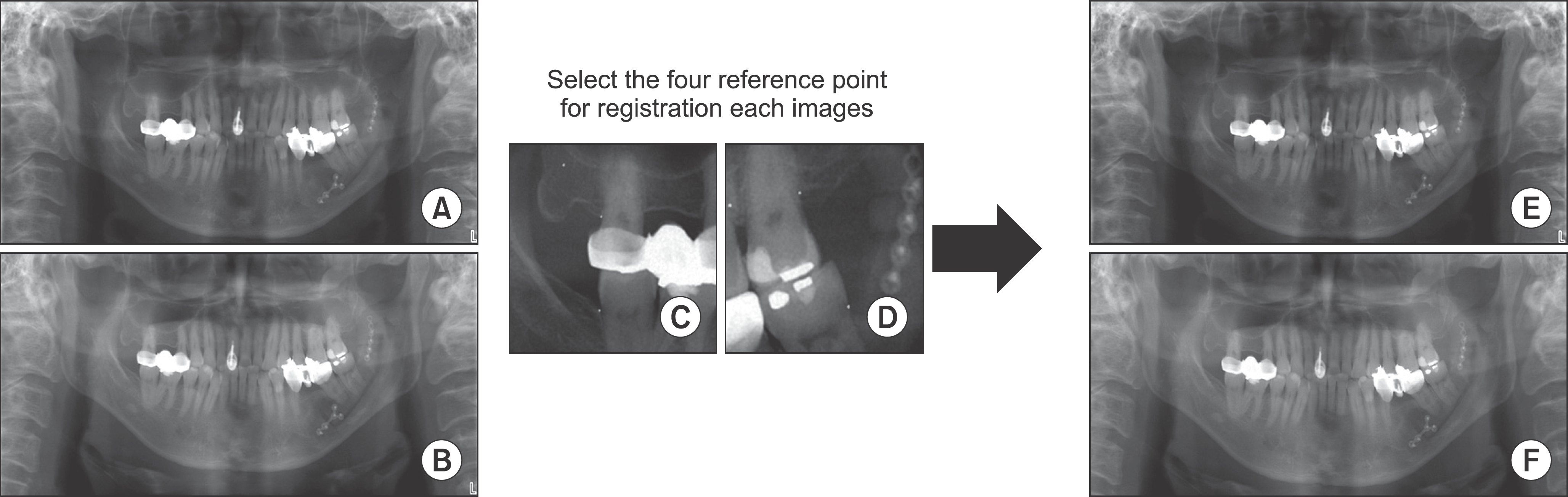 | Fig. 1.Image registration processing by image subtraction tool program (Sunny, Biomedisys Co. Ltd., Seoul, Korea). A, B. Before registration. C, D. Reference point selection. E, F. After registration. Hun-Jun Lim et al: Study on bone healing process following cyst enucleation using fractal analysis. J Korean Assoc Oral Maxillofac Surg 2011
|
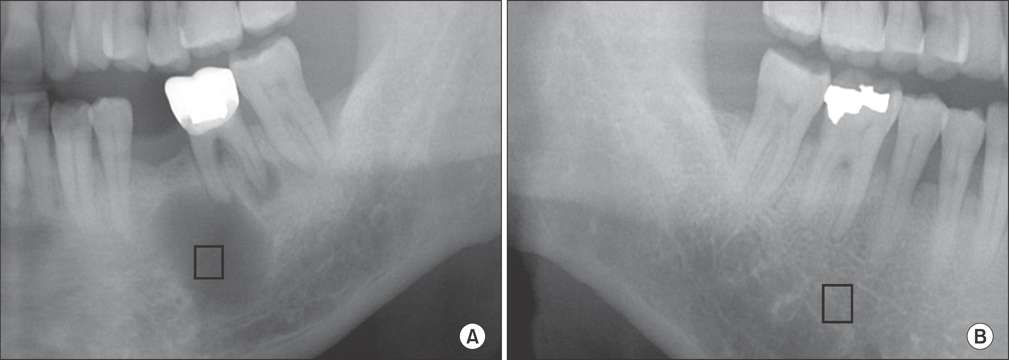 | Fig. 2.Selection of region of interest. A. Selection of lesion region of interest (ROI). B. Selection of control ROI. Hun-Jun Lim et al: Study on bone healing process following cyst enucleation using fractal analysis. J Korean Assoc Oral Maxillofac Surg 2011
|
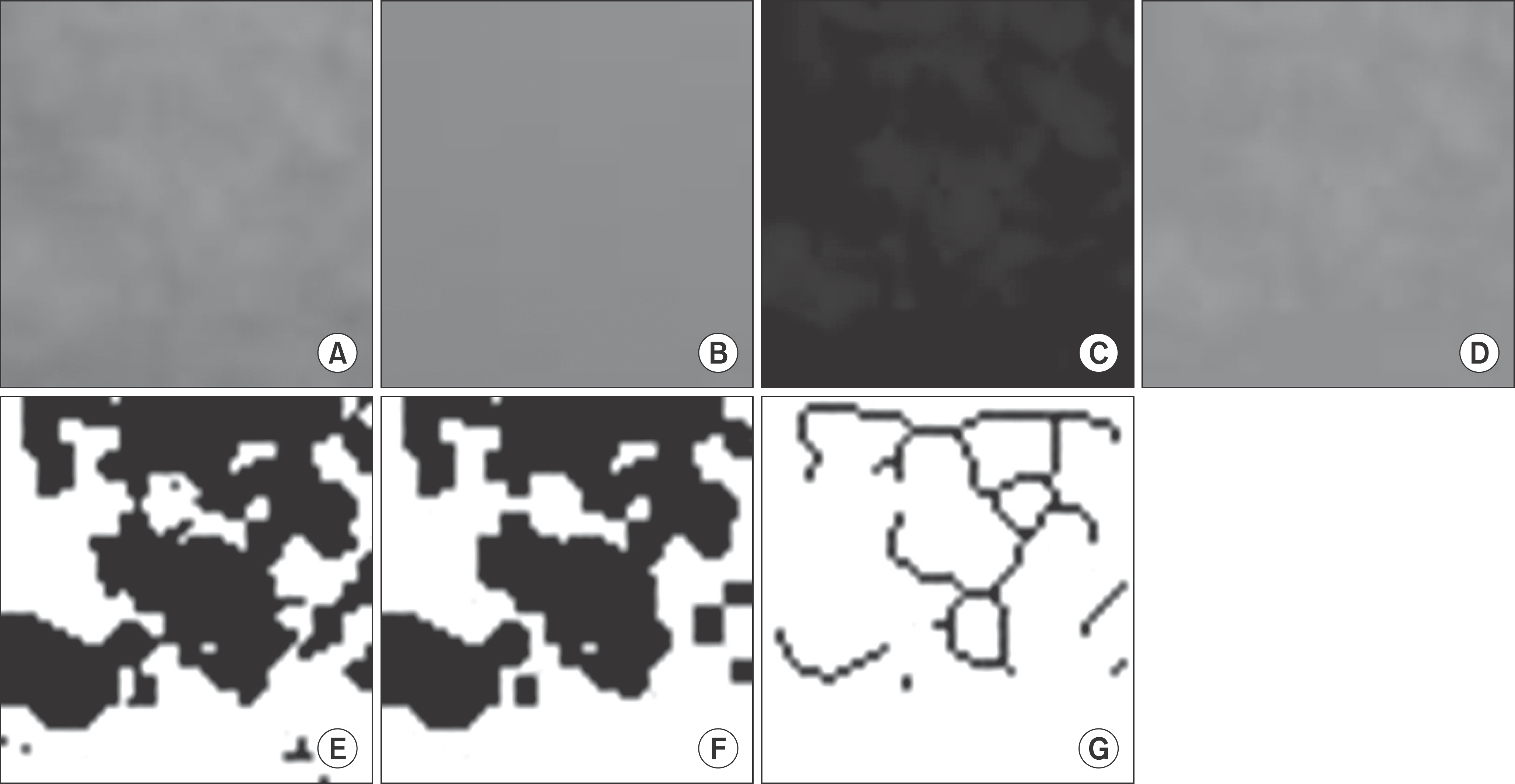 | Fig. 3.Image processing. A. Original region of interest image. B. Gaussian blurring. C. Subtract between (A) and (B). D. Add 128. E. Conversion to binary image. F. Erode and dilation. G. Skeletonize. Hun-Jun Lim et al: Study on bone healing process following cyst enucleation using fractal analysis. J Korean Assoc Oral Maxillofac Surg 2011
|
Table 1.
The change of fractal dimension over time
Table 2.
Comparison with pre-operative fractal dimensions
| Time (I) | Time (J) | Average difference (I-J) | Standard deviation | P-value |
|---|---|---|---|---|
| Pre-operative | Post 1 day | −0.067 | 0.147 | 0.070 |
| Post 1 month | −0.029 | 0.150 | 0.456 | |
| Post 3 months | −0.067* | 0.095 | 0.010 | |
| Post 6 months | −0.112* | 0.108 | 0.001 | |
| Post 12 months | −0.149* | 0.092 | 0.000 |
Table 3.
Comparison with control group fractal dimensions
| Time (I) | Time (J) | Average difference (I-J) | Standard deviation | P-value |
|---|---|---|---|---|
| Control | Pre-operative | 0.151 | 0.106 | 0.000 |
| Post 1day | 0.088 | 0.146 | 0.024 | |
| Post 1 month | 0.133 | 0.139 | 0.002 | |
| Post 3 months | 0.100 | 0.103 | 0.001 | |
| Post 6 months | 0.049* | 0.115 | 0.132 | |
| Post 12 months | −0.006* | 0.074 | 0.754 |
 XML Download