Abstract
Introduction
The current study examined the morphological characteristics of maxillary sinus septum by computed tomography (CT).
Materials and Methods
Two hundred and four patients (408 maxillary sinuses) who visited dental clinic were evaluated. CT were examined. The height of the septum measured from the sinus floor to the apex of the septum more than 3 mm was defined as “sinus septum”.
Results
The prevalence of sinus septa was 21.3% (87/408), and 31.4% (64/204) of patients had more than 1 sinus septum. Females showed higher and thinner sinus septa than males. The anatomic location of the septa were distributed in the 2nd molar region (43.7%), 1st molar region (31.0%), 2nd premolar region (21.8%) and 1st premolar region (3.5%). In 57 patients with chronic disease, there was no significant difference between sinus disease and the presence of sinus septa. The loss of remaining teeth and teeth adjacent to the sinus septum area was not related to the presence of sinus septa. Older subjects showed a reduced height and length of the septum, and a thicker septum.
Go to : 
References
1. Chanavaz M. Maxillary sinus: anatomy, physiology, surgery, and bone grafting related to implantology-eleven years of surgical experience (1979–1990). J Oral Implantol. 1990; 16:199–209.
2. Tatum H Jr. Maxillary and sinus implant reconstructions. Dent Clin North Am. 1986; 30:207–29.
3. Boyne PJ, James RA. Grafting of the maxillary sinus floor with autogenous marrow and bone. J Oral Surg. 1980; 38:613–6.
4. Betts NJ, Miloro M. Modification of the sinus lift procedure for septa in the maxillary antrum. J Oral Maxillofac Surg. 1994; 52:332–3.

5. van den Bergh JP, ten Bruggenkate CM, Disch FJ, Tuinzing DB. Anatomical aspects of sinus floor elevations. Clin Oral Implants Res. 2000; 11:256–65.

6. Andersson JE, Svartz K. CT-scanning in the preoperative planning of osseointegrated implants in the maxilla. Int J Oral Maxillofac Surg. 1988; 17:33–5.

7. Kasabah S, Sleza ′ k R, Simu � nek A, Krug J, Lecaro MC. Evaluation of the accuracy of panoramic radiograph in the definition of maxillary sinus septa. Acta Medica (Hradec Kralove). 2002; 45:173–5.

8. Reddy MS, Mayfield-Donahoo T, Vanderven FJ, Jeffcoat MK. A comparison of the diagnostic advantages of panoramic radiography and computed tomography scanning for placement of root form dental implants. Clin Oral Implants Res. 1994; 5:229–38.

9. Tal H, Moses O. A comparison of panoramic radiography with computed tomography in the planning of implant surgery. Dentomaxillofac Radiol. 1991; 20:40–2.

10. Krennmair G, Ulm CW, Lugmayr H, Solar P. The incidence, location, and height of maxillary sinus septa in the edentulous and dentate maxilla. J Oral Maxillofac Surg. 1999; 57:667–71. discussion 671–2.

11. Underwood AS. An inquiry into the anatomy and pathology of the maxillary sinus. J Anat Physiol. 1910; 44:354–69.
12. Krennmair G, Ulm C, Lugmayr H. Maxillary sinus septa: incidence, morphology and clinical implications. J Craniomaxillofac Surg. 1997; 25:261–5.

13. Vela ′ squez-Plata D, Hovey LR, Peach CC, Alder ME. Maxillary sinus septa: a 3-dimensional computerized tomographic scan analysis. Int J Oral Maxillofac Implants. 2002; 17:854–60.
14. Kim MJ, Jung UW, Kim CS, Kim KD, Choi SH, Kim CK, et al. Maxillary sinus septa: prevalence, height, location, and morphology. A reformatted computed tomography scan analysis. J Periodontol. 2006; 77:903–8.

15. Oh HK, Ryu SY. Clinico-anatomical study of septum in the maxillary sinus. J Korean Assoc Oral Maxillofac Surg. 1998; 24:208–12.
16. Ulm CW, Solar P, Krennmair G, Matejka M, Watzek G. Incidence and suggested surgical management of septa in sinus-lift procedures. Int J Oral Maxillofac Implants. 1995; 10:462–5.
17. So H, Jeong DK, Kwon JH, Ryu SH, Kim HS. Maxillary sinus septum: panoramic radiographic and dental computed tomographic analyses in the planning of implant surgery. J Korean Acad Periodontol. 2006; 36:147–54.

18. Gonza ′ lez-Santana H, Pen arrocha-Diago M, Guarinos-Carbo ′J, Sornl′-Bro¨ker M. A study of the septa in the maxillary sinuses and the subantral alveolar processes in 30 patients. J Oral Implantol. 2007; 33:340–3.
19. Maestre-Ferrl′n L, Gala ′ n-Gil S, Rubio-Serrano M, Penarrocha-Diago M, Penarrocha-Oltra D. Maxillary sinus septa: a systematic review. Med Oral Patol Oral Cir Bucal. 2010; 15:e383–6.
Go to : 
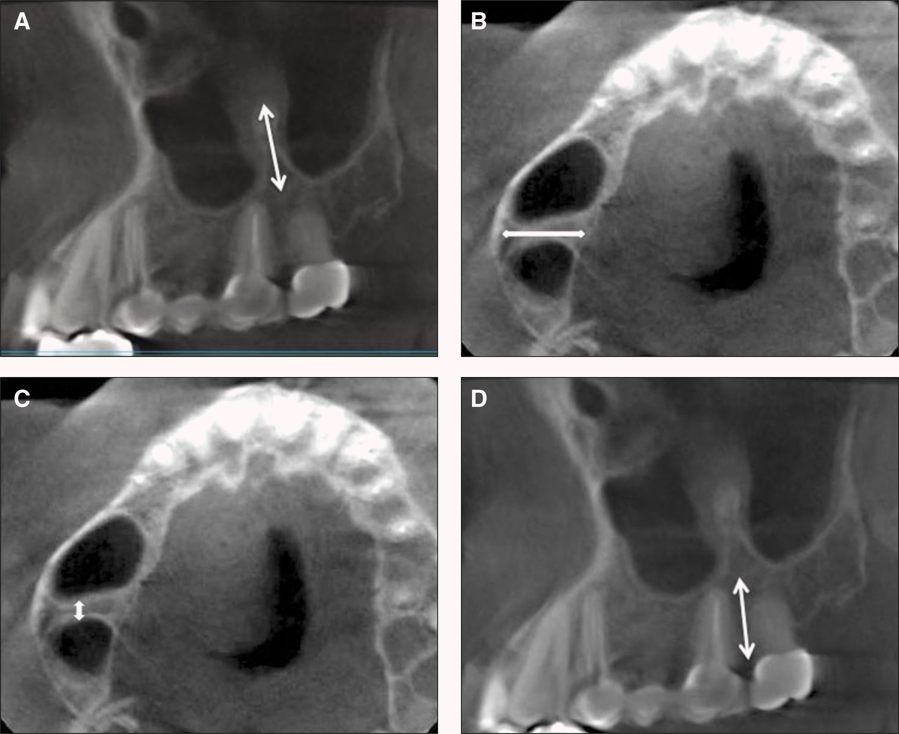
 | Fig. 1.Measured the height from sinus floor to apex of septum, length from medial wall to lateral wall of sinus, thickness at the center point of septum, and vertical height of residual alveolar bone from sinus floor to alveolar ridge where septum. A. Height of maxillary sunus septum. B. Length of maxillary sinus septum. C. Thickness of maxillary sinus septum bone. D. Vertical height of residual alveolar. |
Table 1.
Prevalence of septum in male and female
| Septum | Number of patients | Total | Number of sinus | Total | ||
|---|---|---|---|---|---|---|
| Male | Female | Male | Female | |||
| Presence | 33 (32.00%) | 31 (30.70%) | 64 (31.40%) | 42 (20.40%) | 45∗ (22.30%) | 87 (21.30%) |
| Absence | 70 (68.00%) | 70 (69.30%) | 140 (68.60%) | 164 (79.60%) | 157 (77.70%) | 321 (78.70%) |
| Total (%) | 103 (50.50%) | 101 (49.50%) | 204 (100%) | 206 (50.50%) | 202 (49.50%) | 408 (100%) |
Table 2.
Height, length and thickness of septum and vertical height of alveolar bone
| mm | Male (n=42) | Female (n=45*) | Total (n=87) | P value |
|---|---|---|---|---|
| Height | 4.45±1.31 | 5.92±3.34 | 5.21±2.66 | 0.009∗∗∗ |
| Length | 7.91±2.19 | 8.40±2.59 | 8.13±2.40 | 0.34 |
| Thickness | 1.50±0.87 | 1.18±0.59 | 1.33±0.75 | 0.046∗∗ |
| Vertical height of alveolar bone | 10.18±4.23 | 10.45±3.14 | 10.32±3.69 | 0.737 |
Table 3.
Distribution of sinus septum-categorized by tooth position
| 1st premolar | 2nd premolar | 1st molar | 2nd molar | Total | |
|---|---|---|---|---|---|
| Incidence (%) | 3 (3.5%) | 19 (21.8%) | 27 (31.0%) | 38 (43.7%) | 87 (100%) |
Table 4.
Relationship between age of patients, number of residual teeth and presence of septum
| Septum | Age of patients | Number of residual teeth |
|---|---|---|
| Presence (n=87) | 46.0±16.0 | 4.3±1.3 |
| Absence (n=321 | ) 43.6±14.3 | 4.4±1.2 |
| P value | 0.217 | 0.664 |
Table 5.
Presence of septum and sinus disease
| Septum | Sinus disease | Total | |
|---|---|---|---|
| Presence | Absence | ||
| Presence | 10 (17.5%) | 77 (21.9%) | 87 (21.3%) |
| Absence | 47 (82.5%) | 274 (78.1%) | 321 (78.7%) |
| Total (%) | 57 (14.0%) | 351 (86.0%) | 408 (100%) |
Table 6.
Correlations between age of patient, height, length, thickness of septum, number of residual teeth, and vertical height of residual alveolar bone (n=87)
| Correlation efficient | Age | Height | Length | Thickness | Number of RT | Number of LAT |
|---|---|---|---|---|---|---|
| Height | −0.307∗∗ | |||||
| Length | −0.291∗∗ | 0.603∗∗ | ||||
| Thickness | 0.283∗∗ | −0.217∗ | −0.218∗ | |||
| Number of RT | −0.424∗∗ | 0.209 | 0.085 | −0.029 | ||
| Number of LAT | 0.430∗∗ | −0.192 | −0.079 | 0.002 | −0.887∗∗ | |
| VH | −0.058 | −0.039 | −0.191 | 0.044 | 0.374∗∗ | −0.305∗∗ |


 PDF
PDF ePub
ePub Citation
 Print
Print


 XML Download