Abstract
Introduction
Ameloblastoma is a benign but locally invasive tumor with a high recurrence rate. The aim of this study was to make an easier diagnosis and treatment planning of ameloblastoma.
Materials and Methods
From January 1993 to October 2009, 139 cases from 123 patients, who had been diagnosed with ameloblastoma through radiologic and biopsy in the department of oral and maxillofacial surgery of Kyunpook National University, were selected as the subjects in this study. According to the medical charts, 9 factors (age, gender, location, chief complaints, duration, radiographic findings, size and recurrence) concerned in deciding the treatment method and the relevance between each factor and the treatment methods were examined.(Conservative treatments were marsupialization, enucleation, curettage and lateral decortication. Radical treatments included block excision, resection and hemisection)
Results
In the patients under the age of 20, 77.14% had conservative treatments, whereas 22.86% underwent radical treatments. In the patients over the age of 20, 44.23% were treated conservatively treatments, and 55.77% underwent radical treatments. For unilocular types, 28.57% had conservative treatments, whereas 71.43% had radical treatments. For the multilocular types, 66.67% underwent conservative treatments, and 33.33% had radical treatments. For the primary cases, 58.68% were treated conservatively and 41.32% had radical treatments. For the recurrent cases, 16.67% and 83.33% underwent conservative and radical treatments, respectively.
Go to : 
REFERENCES
1. Small IA, Waldron CA. Ameloblastomas of the Jaws. Oral Surg Oral Med Oral Pathol. 1955; 8:281–97.

2. Dhiravarangkura P. Roentgenographic survey of ameloblastoma in Osaka University Dental School. J Dent Assoc Thailand. 1969; 19:163–78.
3. Regezi JA, Sciubba JJ. Oral Pathology: clinical-pathologic correlations. 1st ed.Philadelphia: WB Saunders;1989. p. 363–374.
4. Hoffman PJ, Baden E, Rankow RM, Potter GD. The fate of the uncontrolled ameloblastoma. Oral Surg Oral Med Oral Pathol. 1968; 26:419–426.

5. Lee FM. Ameloblastoma of the maxilla with probable origin in a residual cyst. Oral Surg Oral Med Oral Pathol. 1970; 29:799–805.

6. Porter J, Miller R, Stratigos GT. Ameloblastoma of the maxilla. Oral Surg Oral Med Oral Pathol. 1977; 44:34–8.

7. Reaume C, Wesley RK, Jung B, Grammer FC. Clinicopathological conference. Case 31, part 2. Ameloblastoma of the maxillary sinus. J Oral Surg. 1980; 38:520–1.
8. Gardner DG. A pathologist's approach to the treatment of ameloblastoma. J Oral Maxilofac Surg. 1984; 42:161–6.

9. Min BI. Clinical study of the recurred ameloblastomas of the oral cavity. J Korean Oral Maxillofac Surg. 1991; 17:18–24.
10. Kim KS. Clinicopathological study of ameloblastomas. J Korean Histo Dent. 1980; 18:1021–29.
11. Ko YH, Kim CS. Clinico-statistical analysis of the odontogenic ectodermal tumor in oral and maxillofacial region. J Korean Oral Maxillofac Surg. 1988; 14:61–76.
12. Leider AS, Eversole LR, Barkin ME. Cystic ameloblastoma. A clinicopathologic analysis. Oral Surg Oral Med Oral Pathol. 1985; 60:624–30.
13. Heffez L, Mafee MF, Vaiana J. The role of magnetic resonance imaging in the diagnosis and management of ameloblastoma. Oral Surg Oral Med Oral Pathol. 1988; 65:2–12.

14. Yokobayashi Y, Yokobayashi T, Nakajima T, Oyama T, Fukushima M, Ishiki T. Marsupialization as a possible diagnostic aid in cystic ameloblastoma. J Maxillofacial Surg. 1983; 11:13741.

15. Gardner AF, Apter MB, Axelrod JH. A study of twenty-one in-stances of ameloblastoma, a tumor of odontogenic origin. J Oral Surg Anesth Hosp Dent Serv. 1963; 21:230–7.
16. Apple NB, Verbin RS. Surgical pathology of the Head and Neck. 1st edition, New York,. 1986. 1331–409.
17. Hager RC, Taylor CG, Allen PM. Ameloblastic fibroma: report of case. J Oral Surg. 1978; 36:66–9.
18. Shafer WG, Hine MK, Levy BM. A Textbook of Oral Pathology. 3rd ed.Philadelphia: Saunders;1985. p. 251–8.
19. Rapidis AD, Angelopoulos AP, Skouteris CA, Papanicolaou S. Mural(intracystic) ameloblastoma. Int J Oral Surg. 1982; 11:166–74.
20. Stanley HR, Diehl DL. Ameloblastoma potential of follicular cysts. Oral Surg Oral Med Oral Pathol. 1965; 20:260–8.

21. Castner DV Jr, McCully AC, Hiatt WR. Intracystic ameloblastoma in the young patient. Oral Surg Oral Med Oral Pathol. 1967; 23:127–34.

22. Hutton CE. Occurence of ameloblastoma within a dentigerous cyst. Report of a case. Oral Surg Oral Med Oral Pathol. 1967; 24:147–50.
23. Lee CK, Yoon JH, Kim J, Lee YH. En bloc excision of the mural ameloblastoma arising from odontogenic keratocyst. J Korean Assoc Maxillofac Plast Reconstr Surg. 1985; 7:113–9.
24. Dresser WJ, Segal E. Ameloblastoma associated with a dentigerous cyst in a 6-year-old child. Report of a case. Oral Surg Oral Med Oral Pathol. 1967; 24:388–91.
25. Gardner DG, Pecak AM. The treatment of ameloblastoma based on pathologic and anatomic principles. Cancer. 1980; 46:2514–9.

26. Kameyama Y, Takehana S, Mizohata M, Nonobe K, Hara M, Kawai T, Fukaya M. A clinicopathological study of ameloblastomas. Int J Oral Maxillofac Surg. 1987; 16:706–12.

27. Adekeye EO. Ameloblastoma of the jaws: a survey of 109 Nigerian patients. J Oral Surg. 1980; 38:36–41.
28. Seldin SD. Ameloblastoma in Young patient: report of two cases. J Oral Surg Anesth Hosp Dent Serv. 1961; 19:508–12.
29. Komisar A. Plexiform ameloblastoma of the maxilla with extension to the skull base. Head Neck Surg 7. 172–5.
30. Potdar GG. Ameloblastoma of the jaw as seen in Bombay, India. Oral Surg Oral Med Oral Pathol. 1969; 28:297–303.

31. Stout RA, Lynch JB, Lewis SR. The conservative surgical approach to ameloblastomas of the mandible. Plast Reconstr Surg. 1963; 31:554–62.

33. Muller H, Slootweg PJ. The ameloblastoma, the controversial approach to therapy. J Maxillofac Surg. 1985; 13:79–84.
34. Petriella VM, Rogow PN, Baden E, Williams AC. Gigantic ameloblastoma of the mandible: report of case. J Oral Surg. 1974; 32:44–9.
35. Möller H, Slootweg P. Clear cell differentiation in an ameloblastoma. J Maxillofac Surg. 1986; 14:158–60.
36. Waldron CA. Ameloblastoma in perspective. J Oral Surg 24. 331–3.
37. Shatkin S, Hoffmeister FS. Ameloblastoma: a rational approach to therapy. Oral Surg Oral Med Oral Pathol. 1965; 20:421–35.

38. Lee EW, Park HS, Cha IH, Kim J. Unicystic Ameloblastoma: case report. J Korean Assoc Maxillofac Plast Reconstr Surg. 1991; 13:160–6.
39. Isacsson G, Andersson L, Forsslund H, Bodin I, Thomsson M. Diagnosis and treatment of the unicystic ameloblastoma. Int J Oral Maxillofac Surg. 1986; 15:759–64.

40. Ronald Marks, Mike Block I. Daniel Sanusi, Bruce Lowe, Bob D. Gross: Unicystic ameloblastoma. International J of Oral Surg. Vol 12:Issue. (3):June 1983; Page. 186–9.
Go to : 
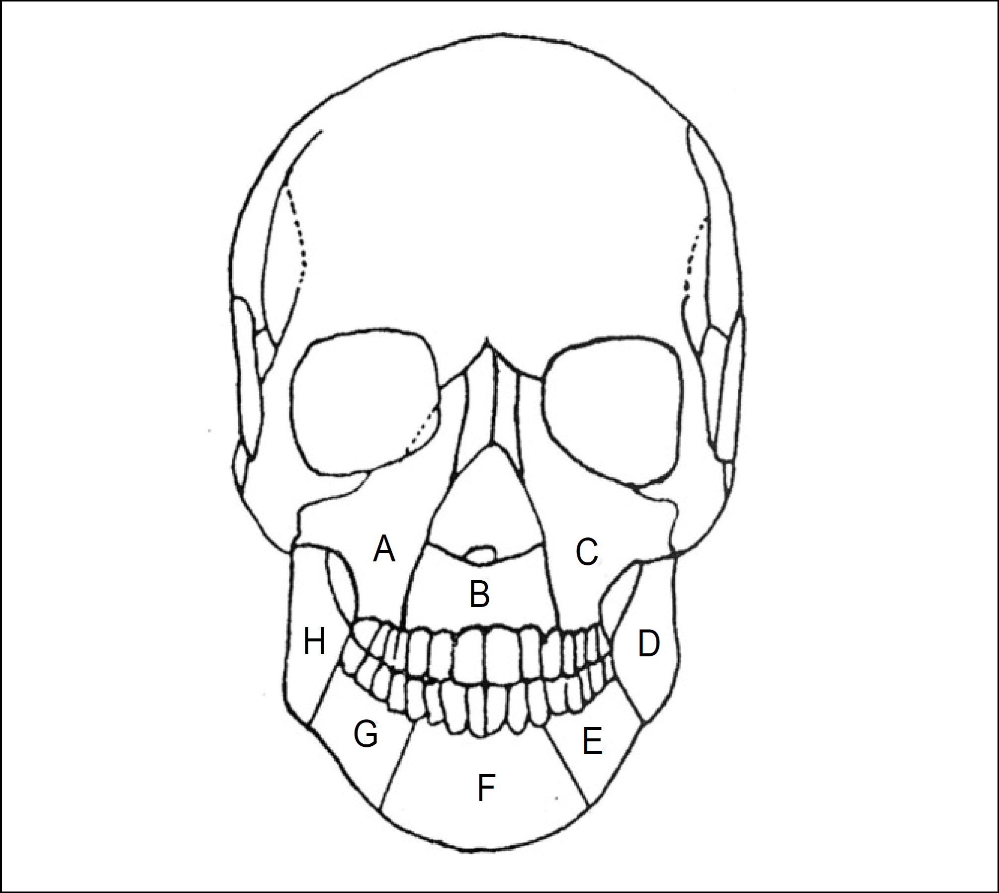
 | Fig. 1.Diagrammatic representation of the various surgical treatment of the Ameloblastoma. A) Marsupialization: Removal of the upper part of the lesion for the making of intraoral opening, B) Enucleation: Removal of a lesion by shelling it out intact, C) Curettage: Surgical scraping of the wall of a cavity within soft tissue or bone for the removal of its contents, D) Lateral decortications with reposition: Surgical curettage after lateral bony window opening following reposition, E) Block excision: Surgical removal of a tumor intact, with a rim of uninvolved bone with maintaining continuity of the inferior or posterior borders of the mandible, F) Resection: Surgical removal of a segment of the mandible or maxilla without maintaining the continuity of the bone, G) Hemisection: Surgical removal of one side of the mandible or maxilla. |
Table 1.
Use of treatment method
Table 2.
Frequency of the treatment in relation to all factors


 PDF
PDF ePub
ePub Citation
 Print
Print


 XML Download