

 PDF
PDF ePub
ePub Citation
 Print
Print


Abstract
This study evaluated the mineral crystalline structure of an autogenous tooth bone graft material. The crystalline structures of the autogenous tooth bone graft material enamel (AutoBT E+), dentin (AutoBT D+), xenograft (BioOss), alloplastic material (MBCP), allograft (ICB) and autogenous mandibular cortical bone were compared using XRD. The XRD pattern of AutoBT dentin and ICB was similar to that of autogenous bone.
Go to : 
References
1. Kim YK, Yeo HH, Ryu CH, Lee HB, Byun UR, Cho JO. An experimental study on the tissue reaction of toothash implanted in mandible body of the mature dog. J Korean Assoc Maxillofac Plast Reconstr Surg. 1993; 15:129–36.
2. Kim YK, Yeo HH, Yang IS, Seo JH, Cho JO. Implantation of toothash combined with plaster of paris: experimental study. J Korean Assoc Maxillofac Plast Reconstr Surg. 1994; 16:122–9.
3. Kim YK, Kim SG, Byeon JH, Lee HJ, Um IU, Lim SC, et al. Development of a novel bone grafting material using autogenous teeth. Oral Surg Oral Med Oral Pathol Oral Radiol Endod. 2010; 109:496–503.

4. Kim YK, Lee HJ, Kim SG, Um IW, Lim SC, Kim SY. Analysis of inorganic component and SEM analysis of autogenous teeth bone graft material and histomorphometric analysis after graft. J Korean Acad Implant Dent. 2009; 28:1–9.
5. Kim YK, Lee JY. The evaluation of postoperative safety of autogenous teeth bone graft. J Korean Acad Implant Dent. 2009; 28:29–35.
6. Klug HP, Alexander LE. X-ray diffraction procedures for polycrystalline and amorphous materials. 2nd ed.New York: Wiley-Interscience;1974.
7. Kim YK, Kim SG, Lee BG. Bone graft and implant: vol 1. Bone biology and bone graft material. Seoul: Narae Pub Co.;2007. p. 171–224.
8. Tadic D, Epple M. A thorough physicochemical characterisation of 14 calcium phosphate-based bone substitution materials in comparison to natural bone. Biomaterials. 2004; 25:987–94.
9. Kirik SD, Solovyov LA, Blokhin AI, Yakimov IS. Structures of. Acta Crystallogr B. 2000; 56:419–25.
10. Balasundaram G, Sato M, Webster TJ. Using hydroxyapatite nanoparticles and decreased crystallinity to promote osteoblast adhesion similar to functionalizing with RGD. Biomaterils. 2006; 27:2798–805.
11. Kim YK, Kim SG, Jin SC, Son JS, Kim SY, Um IW. Analysis of the Inorganic Component of Autogenous Tooth Bone Graft Material. J Nanosci Nanotechnol 2011 accepted.
12. Glimcher MJ. Molecular biology of mineralized tissues with particular reference to bone. Rev Mod Phys. 1959; 31:359–93.
13. Lee SH. Low Crystalline hydroxyl carbonate apatite. J Korean Dental Assoc. 2006; 44:524–33.
Go to :
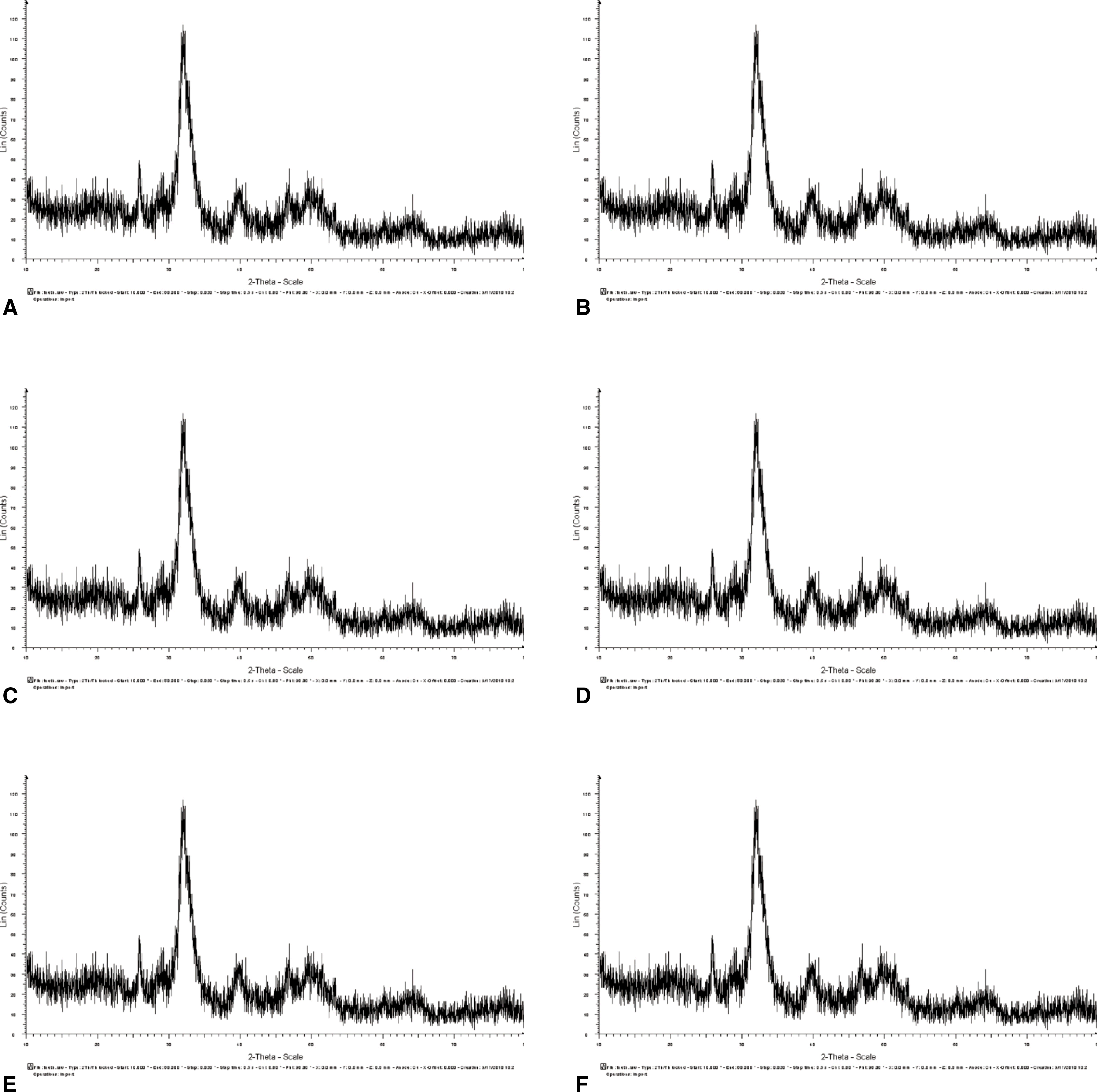 | Fig. 2.XRD patterns of a variety of bone graft materials. A: XRD pattern of autogenous bone, B: XRD pattern of AutoBT dentin, C: ARD pattern of AutoBT enamel, D: XRD pattern of ICB, E: XRD pattern of BioOss, F: XRD pattern of MBCP.(XRD: X-ray diffraction) |
Table 1.
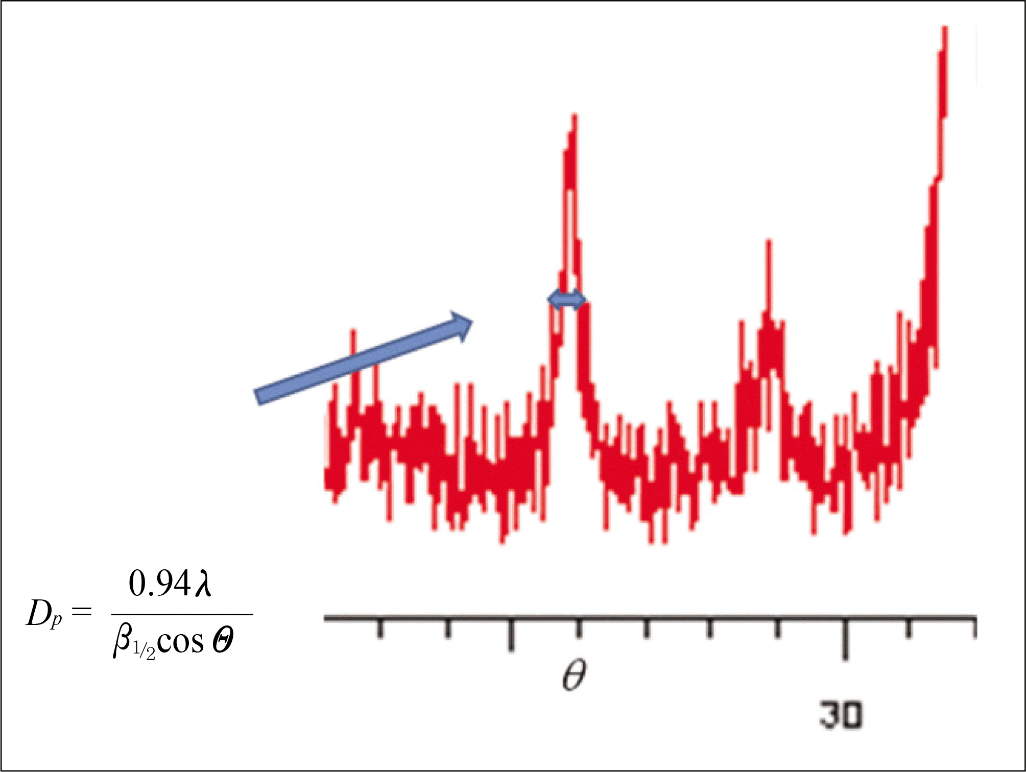
Estimation of domain size form diffraction peak broadening
 XML Download