Abstract
Introduction
The enlargement and deformation of the orbit give rise to a visible enophthalmos. As a consequence, a disturbance of eye motility together with double images is likely to occur. This study examined the degree of enophthalmos according to the extent of orbital wall fracture and volume of herniated orbital tissue in blowout fractures of the medial and inferior orbital wall.
Materials and Methods
This study was performed on patients diagnosed with medial and inferior orbital wall fractures at the Department of Oral and maxillofacial surgery, Chonbuk National University Hospital from 2007 to 2009. The patients' age, gender, etiology of fracture and degree of enophthalmos were investigated. The changes in the degree of enophthalomos, diplopia and ocular motility restriction after operation were examined.
Results
The degree of enophthalomos increased with increasing extent of orbital wall fracture and volume of herniated orbital tissue.
Conclusion
Whether to perform the operation is decided after measuring the extent of the orbital wall fracture and volume of herniated orbital tissue using computed tomography (CT), time for the decision of operation can be shortened. This can cause a decrease in the complications of orbital wall fractures.
Go to : 
References
1. Manolidis S, Weeks BH, Kirby M, Scarlett M, Hollier L. Classification and surgical management of orbital fractures: experience with 111 orbital reconstructions. J Craniofac Surg. 2002; 13:726–37.

2. Girotto JA, MacKenzie E, Fowler C, Redett R, Robertson B, Manson PN. Long-term physical impairment and functional outcomes after complex facial fractures. Plast Reconstr Surg. 2001; 108:312–27.

3. Cunningham LL, Peterson GP, Haug RH. The relationship between enophthalmos, linear displacement, and volume change in experimentally recreated orbital fractures. J Oral Maxillofac Surg. 2005; 63:1169–73.

4. Manson PN, Grivas A, Rosenbaum A, Vannier M, Zinreich J, Iliff N. Studies on enophthalmos: II.The measurement of orbital injuries and their treatment by quantitative computed tomography. Plast Reconstr Surg. 1986; 77:203–14.
5. Hammer B, Prein J. Correction of posttraumatic orbital deformities: operative techniques and review of 26 patients. J Craniomaxillofac Surg. 1995; 23:81–90.

6. Baumann A, Ewers R. Transcaruncular approach for reconstruction of medial orbital wall fracture. Int J Oral Maxillofac Surg. 2000; 29:264–7.

7. Kim DG, Kim HS, Lee SC. Two cases of blowout fracture of the medial orbital wall. Korean J Otolaryngol. 1987; 30:307–12.
8. Greenwald HS Jr, Keeney AH, Shannon GM. A review of 128 patients with orbital fracture. Am J Ophthalmol. 1974; 78:655–64.
9. Rubin PA, Bilyk JP, Shore JW. Management of orbital trauma: fractures, hemorrhage, and traumatic optic neuropathy. Am Acad Ophthalmol Focal Points. 1994; 7:1–8.
10. Dutton JJ. Management of blowout fractures of the orbital floor. Surv Ophthalmol. 1991; 35:279–80.
11. Dulley B, Fells P. Long-term follow-up of orbital blowout fractures with and without surgery. Mod Probl Ophthalmol. 1970; 14:467–70.
12. Osguthorpe JD. Orbital wall fractures: evaluation and management. Otolaryngol Head Neck Surg. 1991; 105:702–7.

13. Hawes MJ, Dortzbach RK. Surgery on orbital floor fracture: influence of time of repair and fracture size. Ophthalmology. 1983; 90:1066–70.
14. Jin HR, Shin SO, Choo MJ, Choi YS. Relationship between the extent of fracture and the degree of enophthalmos in isolated blowout fracture of the medial orbital wall. J Oral Maxillofac Surg. 2000; 58:617–20.
15. Kakibuchi M, Fukazawa K, Fukuda K, Yamada N, Matsuda K, Kawai K, et al. Combination of transconjunctival and endonasal transantral approach in the repair of blowout fractures involving the orbital floor. Br J Plastic Surg. 2004; 57:37–44.
16. Parson G, Mathog RH. Orbital wall and volume relationships. Arch Otolaryngol Head Neck Surg. 1988; 114:743–7.
17. Harris GJ, Garcia GH, Logani SC, Murphy ML, Sheth BP, Seth AK. Orbital blowout fracture: correlation of preoperative computed tomography and postoperative ocular motility. Trans Am Ophthalmol Soc. 1998; 96:329–47.
18. Biesman BS, Hornblass A, Lisman R, Kazlas M. Diplopia after surgical repair of orbital floor fractures. Ophthal Plast Reconstr Surg. 1996; 12:9–16.

19. Putterman AM, Stevens T, Urist MJ. Nonsurgical management of blowout fractures of the orbital floor. Am J Ophthalmol. 1974; 77:232–9.

20. Emory JN, van Noorden GK, Schlernitzauer DA. Orbital floor fractures: longterm follow-up of cases with and without surgical repair. Trans Am Acad Ophthalmol Otolaryngol. 1971; 75:802–12.
Go to : 
 | Fig. 1.Enophthalmos measurement. A: Oculus exophthalmometry, B: Enophthalmos measurement method. |
 | Fig. 2.The area of fracture site is calculated from measurements on coronal (A, C), axial (B), and sagittal (D) CT scan. a: Height of medial wall fracture, b: Length of medial wall fracture, c: Degree of medial displacement of the herniated orbital tissue, d: Width of inferior wall fracture, e: Length of inferior wall fracture, f: Degree of inferior displacement of the herniated orbital tissue. Area of fracture site – medial wall: π×a/2×b/2, inferior wall: π×d/2×e/2 Volume of herniated orbital tissue – medial wall: π×abc/6, inferior wall: π×def/6 |
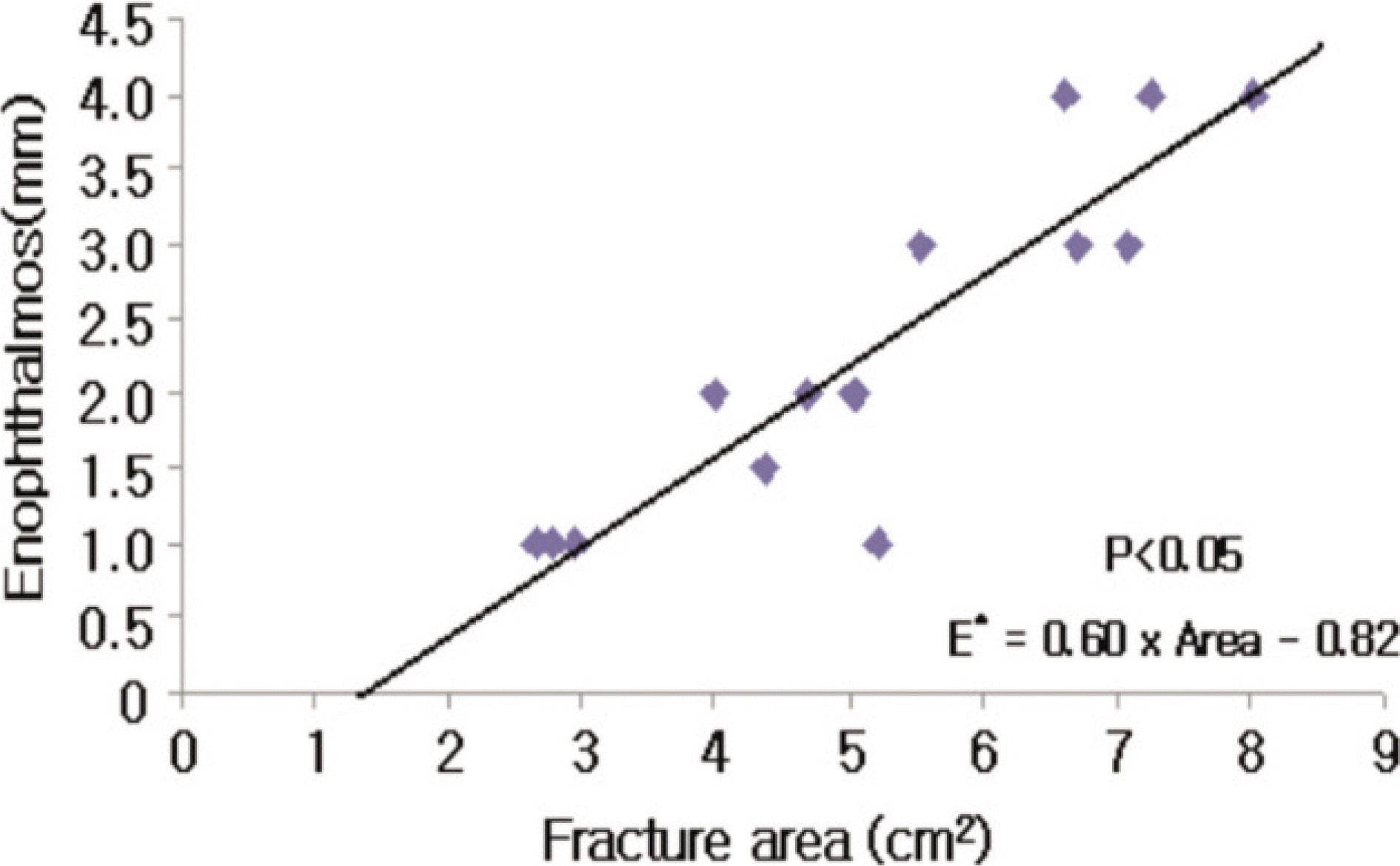
 | Fig. 3.Positive correlation between the area of fracture and the degree of enophthalmos (E*). A: Medial wall fracture, B: Inferior wall fracture. |
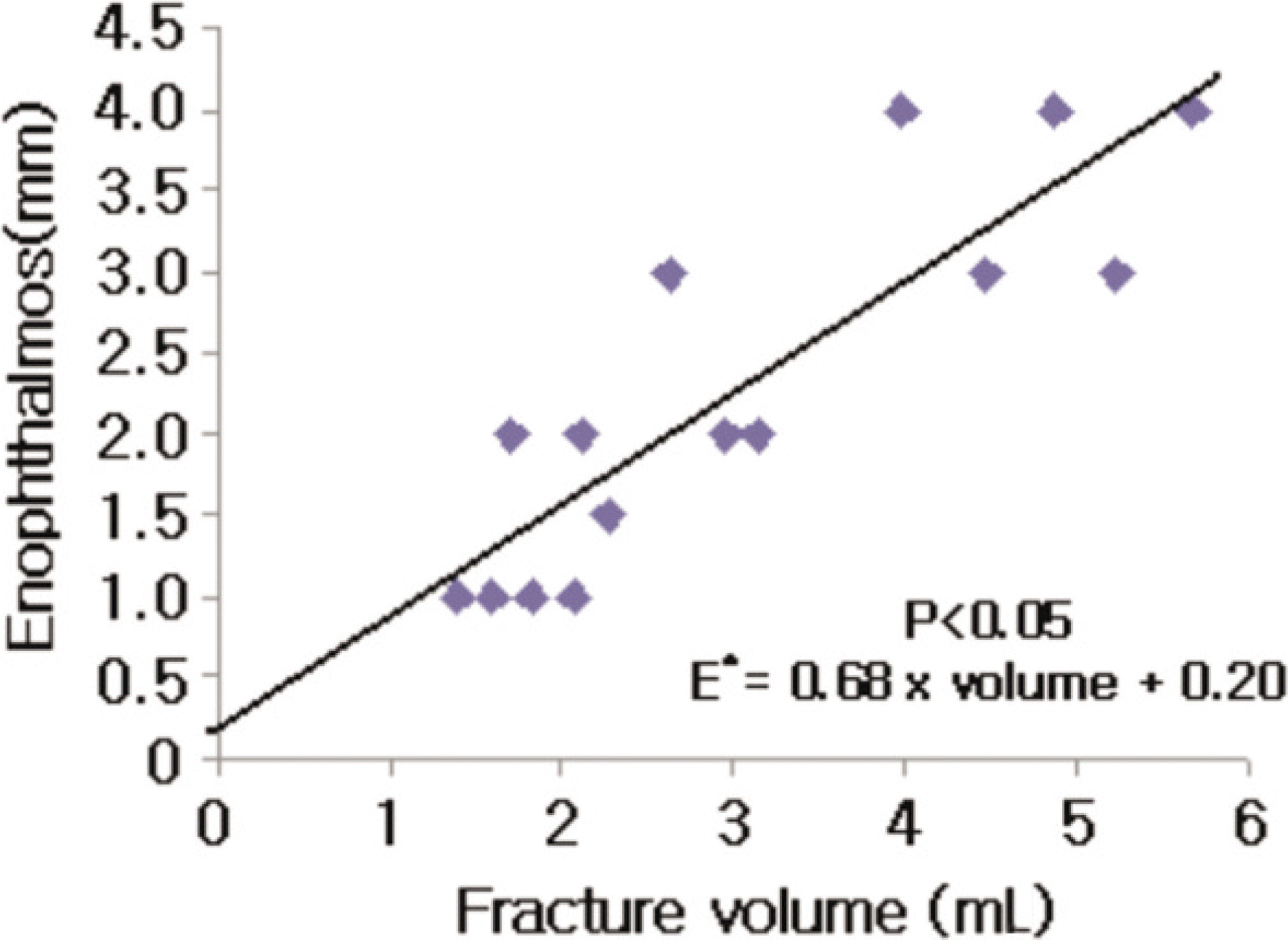
 | Fig. 4.Positive correlation between the volume of herniated orbital tissue and the degree of enophthalmos (E*). A: Medial wall fracture, B: Inferior wall fracture. |
 | Fig. 5.Positive correlation between the area of fracture (medial+inferior) and the degree of enophthalmos (E*). |
 | Fig. 6.Positive correlation between the volume of herniated orbital tissue (medial+inferior) and the degree of enophthalmos (E*). |
Table 1.
Site of fractures of orbital wall
| Fracture site | No. of patients (%) |
|---|---|
| Medial | 31 (35.6) |
| Inferior | 18 (20.7) |
| Medial & Inferior | 38 (43.7) |
| Total | 87 (100) |
Table 2.
Site of operation according to fractures of orbital wall
| Fracture site | No. of patients (%) | |
|---|---|---|
| Medial | 10 (33.4) | |
| Inferior | 3 (10.0) | |
| Medial & Inferior | only medial | 3 (10.0) |
| only inferior | 7 (23.3) | |
| both | 7 (23.3) | |
| Total | 30 (100) |
Table 3.
Preoperative and postoperative values of enophthalmos measured by Oculus exophthalmometry
Table 4.
EOM limitation
| EOM limitation | No. of patients (%) | |
|---|---|---|
| Preoperative | Postoperative* | |
| Positive | 5 (16.7) | 1 (3.3) |
| Negative | 25 (83.3) | 29 (96.7) |
| Total | 30 (100) | 30 (100) |
Table 5.
Diplopia
| Diplopia | No. of patients (%) | |
|---|---|---|
| Preoperative | Postoperative* | |
| Positive | 9 (30.0) | 4 (13.3) |
| Negative | 21 (70.0) | 26 (86.7) |
| Total | 30 (100) | 30 (100) |
Table 6.
Preoperative and postoperative values of enophthalmos measured by Oculus exophthalmometry
| Degree of enophthalmos (mm) | No. of patients (%) | |
|---|---|---|
| Preoperative | Postoperative | |
| E=0 | 0 (0) | 4 (66.6) |
| 0<E<1 | 0 (0) | 0 (0) |
| 1≤E<2 | 0 (0) | 1 (16.7) |
| 2≤E<3 | 4 (66.7) | 1 (16.7) |
| 3≤E | 2 (33.3) | 0 (0) |
| Total | 6 (100) | 6 (100) |
Table 7.
EOM limitation
| EOM limitation | No. of patients (%) | |
|---|---|---|
| Preoperative | Postoperative* | |
| Positive | 2 (33.3) | 0 (0) |
| Negative | 4 (66.7) | 6 (100) |
| Total | 6 (100) | 6 (100) |
Table 8.
Diplopia
| Diplopia | No. of patients (%) | |
|---|---|---|
| Preoperative | Postoperative* | |
| Positive | 1 (16.7) | 0 (0) |
| Negative | 5 (83.3) | 6 (100) |
| Total | 6 (100) | 6 (100) |


 PDF
PDF ePub
ePub Citation
 Print
Print


 XML Download