Abstract
Introduction
Very high aggressiveness and recurrence are important clinical characteristics of ameloblastoma compared to the other benign tumors. Therefore, an accurate diagnosis and treatment plan is important. This study examined the association of the clinical findings and recurrence based on the radiological findings of ameloblastoma. In recurrent cases, these results are expected to help in the diagnosis and treatment of ameloblastoma to examine the relevance with the clinical characteristics and radiological features.
Materials and Methods
For a clinical (gender, age) and radiological (location, internal pattern, size, perforation, border pattern, impacted tooth, root resorption) evaluation, this study examined 156 cases of 147 patients diagnosed with ameloblastoma, who had been treated and in most cases regularly checked at the department of oral and maxillofacial surgery, Kyungpook National University Hospital, between January 1993 and December 2009. For a recurrent rate evaluation, a more than 3 years follow-up period is needed. Accordingly, 116 patients diagnosed with ameloblastoma between January 1994 and December 2007 were investigated.
Results
The recurrence rate in all cases was 6.1% but was 7.8% in cases with follow-up periods more than 3 years. The male-to-female ratio was 3:2, showing a slight male predilection. Ameloblastoma had a peak occurrence in the second decade of life followed by the fourth decade of life. The mandibular angle area is the most frequent site of ameloblastoma (50.8%) in the jaws. Six cases of unilocular (7.8%) and 3 cases of multilocular (7.7%) ameloblastomas recurred. Seven cases of smooth (10%) and 2 cases of irregular (4.3%) ameloblastomas recurred. No cases of ameloblastomas without perforation of the cortical bone (0%) and 9 cases with a perforation of cortical bone (11.1%) recurred. Four cases of the ameloblastomas with impacted teeth (11.4%) and 5 cases of ameloblastomas without impacted tooth (6.2%) recurred. Seven cases of ameloblastomas with root resorption (10.9%) and 2 cases of ameloblastomas without root resorption (3.8%) recurred.
Go to : 
References
1. Park NB, Shin SW, Kim CS. Clinico-statical study on the radiographic findings by the clinical findings of 115 ameloblastomas. J Korean Assoc Maxillofac Plast Reconstr Surg. 1995; 17:415–28.
2. Choi HB, You DS. A study of ameloblastoma on the relationship between histopathologic patterns and radiographic characteristics. Korean J Oral Maxillofac Radiol. 1992; 22:339–50.
3. Churchill HR. Histological differentiation between certain dentigerous cysts and ameloblastoma. Dent Cosmos. 1934; 76:1173–8.
4. Mansson JK, McDonald JR, Figi FA. Adamantinoma of the jaws; a clinicopathologic study of 100 histologically proved cases. Plast Reconstr Surg Transplant Bull. 1959; 23:510–25.
5. Gorlin RJ, Chaudhry AP, Pindborg JJ. Odontogenic tumors. Classification, histopathologiy and clinical behavior in man and domasticated animals. Cancer. 1961; 14:73–101.
6. Shafer WG, Hine MK, Levy BM. A textbook of oral pathology. 4th ed.Philadelphia: WB Saunders;1983. p. 276–85.
7. Struthers P, Shear M. Root resorption by ameloblastomas and cysts of the jaws. Int J Oral Surg. 1976; 5:128–32.

8. Park TW. The incidence and classification of ameloblastoma. Korean J Oral Maxillofac Radiol. 1985; 15:21–6.
10. Eversole LR, Leider AS, Strub D. Radiographic characteristics of cystogenic ameloblastoma. Oral Surg Oral Med Oral Pathol. 1984; 57:572–7.

11. Lucas RB. Pathology of tumours of the oral tissues. 4th ed.New York: Churchill Livingstone;1984. p. 31–60.
12. Robinson HBG. Ameloblastoma; a survey of 379 cases from the literature. Arch Pathol. 1937; 23:831–45.
13. Small IA, Waldron CA. Ameloblastoma of the jaws. Oral Surg Oral Med Oral Pathol. 1955; 8:281–97.
14. Gardner DG. Plexiform unicystic ameloblastoma; a diagnostic problem in dentigerous cyst. Cancer. 1981; 47:1358–63.
15. Gardner DG, Pecak AM. The treatment of ameloblastoma based on pathologic and anatomic principles. Cancer. 1980; 46:2514–19.

16. Gardner DG. A pathologist's approach to the treatment of ameloblastoma. J Oral Maxillofac Surg. 1984; 42:161–6.

17. Gardner DG, Corio RL. The relationship of plexiform unicystic ameloblastoma to conventional ameloblastoma. Oral Surg Oral Med Oral Pathol. 1983; 56:54–60.

18. Gardner DG, Corio RL. Plexiform unicystic ameloblastoma; a variant of ameloblastoma with a low recurrence after enucleation. Cancer. 1984; 53:1730–5.
19. Hendler BH, Abaza NA, Moon AC, Herrod NW. Case 33, part 2. Ameloblastoma of the mandible. J Oral Surg. 1981; 39:208–13.
20. Mehlisch DR, Dahlin DC, Masson JK. Ameloblastoma: a clinicopathologic report. J Oral Surg. 1972; 30:9–22.
21. Tsaknis PJ, Nelson JF. The maxillary ameloblastoma: an analysis of 24 cases. J Oral Surg. 1980; 38:336–42.
22. Smith JF. Ameloblastoma: Report of thirty cases. Oral Surg. 1960; 13:1253–7.
23. Shteyer A, Lustman J, Lewin-Epstein J. The mural ameloblastoma: a review of the literature. J Oral Surg. 1978; 36:866–72.
24. Robinston L, Martinez MG. Unicystic ameloblastoma: a prognostically distinct entity. Cancer. 1977; 40:2278–85.
25. Adekeye EO. Ameloblastoma of the jaws: a survey of 109 Nigerian patients. J Oral Surg. 1980; 38:36–41.
26. Sirichitra V, Dhiravarangkura P. Intrabony ameloblastoma of the jaws: an analysis of 147 Thai patients. Int J Oral Surg. 1984; 13:187–93.
27. Park H, Jeong HG, Kim KD, Park CS. A radiologic study of ameloblastoma using computed tomography. Korean J Oral Maxillofac Radiol. 2005; 35:77–82.
28. Stafne EC. Value of roentgenograms in diagnosis of tumors of the jaws. Oral Surg Oral Med Oral Pathol. 1953; 6:82–92.

29. Sehdev MK, Huvos AG, Strong EW, Gerold FP, Willis GW. Proceedings: Ameloblastoma of maxilla and mandible. Cancer. 1974; 33:324–33.
30. Dolan EA, Angellilo JC, Georgiade NG. Recurrent ameloblastoma in autogenous rib graft: report of a case. Oral Surg Oral Med Oral Pathol. 1981; 51:357–60.
31. Mu ¨ller H, Slootweg PJ. The ameloblastoma, the controversial approach to therapy. J Maxillofac Surg. 1985; 13:79–84.
32. Hayward JR. Recurrent ameloblastoma 30 years after surgical treatment. J Oral Surg. 1973; 31:368–70.
33. Gardner DG. Peripheral ameloblastoma: a study of 21 cases, including 5 reported as basal cell carcinoma of the gingiva. Cancer. 1977; 39:1625–33.
Go to : 
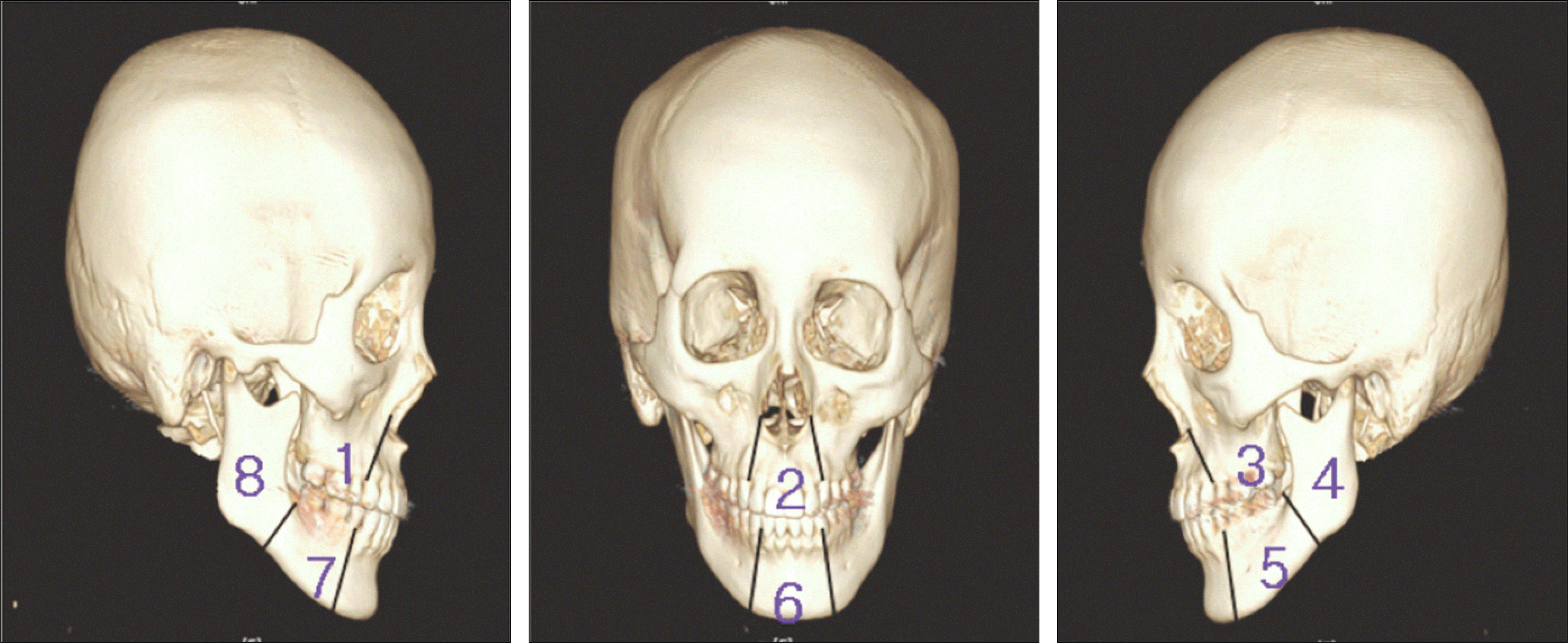
 | Fig. 1.Classification of anatomical location of lesions. Maxilla (1: right posterior, 2: anterior, 3: left posterior). Mandible (4: left angle & ramus, 5: left body, 6: anterior, 7: right body, 8: right angle & ramus). |
Table 1.
Distribution of age and gender
Table 2.
Distribution of anatomic location
Table 3.
Internal pattern of lesion
| Internal pattern | Male | Female | ||
|---|---|---|---|---|
| No. of cases | % | No. of cases | % | |
| Unilocular | 52 | 60.0 | 45 | 74.0 |
| Multilocular | 34 | 40.0 | 16 | 26.0 |
| Total | 86 | 100 | 61 | 100 |
Table 4.
Size of lesion on the panoramic view
Table 5.
Perforation of cortical bone
| Perforation | Male | Female | ||
|---|---|---|---|---|
| No. of cases | % | No. of cases | % | |
| Yes | 27 | 31.4 | 16 | 26.2 |
| No | 59 | 68.6 | 45 | 73.8 |
| Total | 86 | 100 | 61 | 100 |
Table 6.
Bord er pattern o f lesion
| Border pattern | Male | Female | ||
|---|---|---|---|---|
| No. of cases | % | No. of cases | % | |
| Smooth | 51 | 59.3 | 37 | 60.7 |
| Irregular | 35 | 40.7 | 24 | 39.3 |
| Total | 86 | 100 | 61 | 100 |
Table 7.
The impacted tooth
| Impacted tooth | Male | Female | ||
|---|---|---|---|---|
| No. of patients | % | No. of patients | % | |
| Yes | 22 | 25.6 | 20 | 32.8 |
| No | 64 | 74.4 | 41 | 67.2 |
| Total | 86 | 100 | 61 | 100 |
Table 8.
The root resorption
| Root resorption | Male | Female | ||
|---|---|---|---|---|
| No. of patients | % | No. of patients | % | |
| Yes | 50 | 58.1 | 26 | 42.6 |
| No | 36 | 41.9 | 35 | 57.4 |
| Total | 86 | 100 | 61 | 100 |
Table 9.
Distribution of anatomic location and recurrence
Table 10.
Internal pattern of lesion and recurrence
| Internal pattern | Primary | Recurrent | ||
|---|---|---|---|---|
| No. of cases | % | No. of cases | % | |
| Unilocular | 77 | 66.4 | 6 | 7.8 |
| Multilocular | 39 | 33.6 | 3 | 7.7 |
| Total | 116 | 100 | 9 | 7.8 |
Table 11.
Size of lesion and recurrence
Table 12.
Perforation of cortical bone and recurrence
| Perforation | Primary | Recurrent | ||
|---|---|---|---|---|
| No. of cases | % | No. of cases | % | |
| Yes | 35 | 30.2 | 0 | 0 |
| No | 81 | 69.8 | 9 | 11.1 |
| Total | 116 | 100 | 9 | 7.8 |
Table 13.
Border pattern of lesion and recurrence
| Border pattern | Primary | Recurrent | ||
|---|---|---|---|---|
| No. of cases | % | No. of cases | % | |
| Smooth | 70 | 60.3 | 7 | 10 |
| rregular | 46 | 39.7 | 2 | 4.3 |
| Total | 116 | 100 | 9 | 7.8 |


 PDF
PDF ePub
ePub Citation
 Print
Print


 XML Download