

 PDF
PDF ePub
ePub Citation
 Print
Print


Abstract
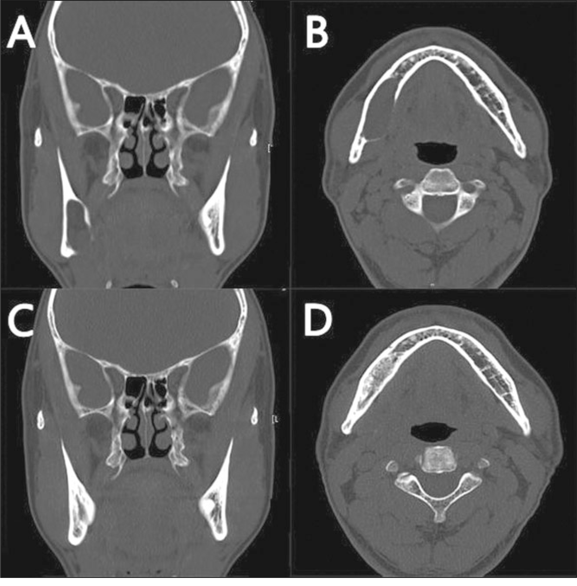
Cystic lesions on the jaws with expansion can invade the adjacent anatomical structure, infiltrate and expand the jaws, cause facial deformity, etc. There is great potential for pathologic fractures after cyst enucleation, and damage to the major structures like the nerve, artery. For these reasons, marsupialization and decompression are commonly used to reduce the cystic size. In 1947, Thomas first mentioned decompression that reduces the osmotic pressure in a cyst by making a hole in the cyst and insert a drain. In our cases, a large sized cystic lesion was treated with a specific device made from an orthodontic band and spinal needle. This device is easy and effective for applications and self irrigation.
References
1. The Korean Association of Oral and Maxillofacial Surgeons. Textbook of oral and maxillofacial surgery. 2nd ed.Seoul: Dental and Medical Publishing Co.;2005.
2. Partsch C. Uber kietercysten. Dtsch Monatsschr Zahnheilkd. 1892; 10:271–3.
3. Thomas EH. Cysts of the jaws: saving involved vital teeth by tube drainage. J Oral Surg (Chic). 1947; 5:1–9.
4. Enislidis G, Fock N, Sulzbacher I, Ewers R. Conservative treatment of large cystic lesions of the mandible: a prospective study of the effect of decompression. Br J Oral Maxillofac Surg. 2004; 42:546–50.

5. van Doorn ME. Enucleation and primary closure of jaw cysts. Int J Oral Surg. 1972; 1:17–25.
6. Dammer R, Niederdellmann H, Dammer P, Nuebler-Moritz M. Conservative or radical treatment of keratocysts: a retrospective review. Br J Oral Maxillofac Surg. 1997; 35:46–8.
7. Eyre J, Zakrzewska JM. The conservative management of large odontogenic keratocysts. Br J Oral Maxillofac Surg. 1985; 23:195–203.
8. Takase T, Wada M, Nagahama F, Yamazaki M. Treatment of large radicular cysts by modified marsupialization. J Nihon Univ Sch Dent. 1996; 38:161–8.
9. Ziccardi VB, Eggleston TI, Schneider RE. Using fenestration technique to treat a large dentigerous cyst. J Am Dent Assoc. 1997; 128:201–5.
10. Nakamura N, Mitsuyasu T, Mitsuyasu Y, Taketomi T, Higuchi Y, Ohishi M. Marsupialization for odontogenic keratocysts: longterm follow-up analysis of the effects and changes in growth characteristics. Oral Surg Oral Med Oral Pathol Oral Radiol Endod. 2002; 94:543–53.
11. Marker P, Br�ndum N, Clausen PP, Bastian HL. Treatment of large odontogenic keratocysts by decompression and later cystectomy: a longterm follow-up and a histologic study of 23 cases. Oral Surg Oral Med Oral Pathol Oral Radiol Endod. 1996; 82:122–31.
12. Br�ndum N, Jensen VJ. Recurrence of keratocysts and decompression treatment. A longterm follow-up of forty-four cases. Oral Surg Oral Med Oral Pathol. 1991; 72:265–9.
13. Takahashi K, Miyauchi K, Sato K. Treatment of ameloblastoma in children. Br J Oral Maxillofac Surg. 1998; 36:453–6.
14. Nakamura N, Higuchi Y, Tashiro H, Ohishi M. Marsupialization of cystic ameloblastoma: a clinical and histopathologic study of the growth characteristics before and after marsupialization. J Oral Maxillofac Surg. 1995; 53:748–54. discussion 755–6.
Fig. 1.
A: Die model with orthodontic band, B: Sixteen gauge spinal needle, C: Side view of decompression appliance, D: Over view of decompression appliance.
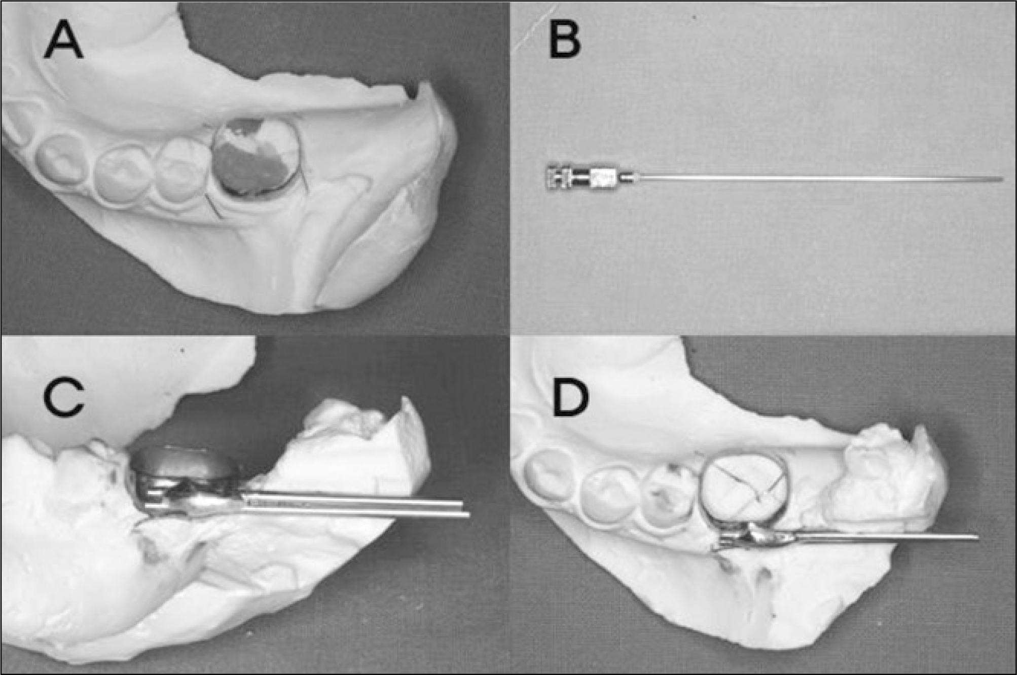
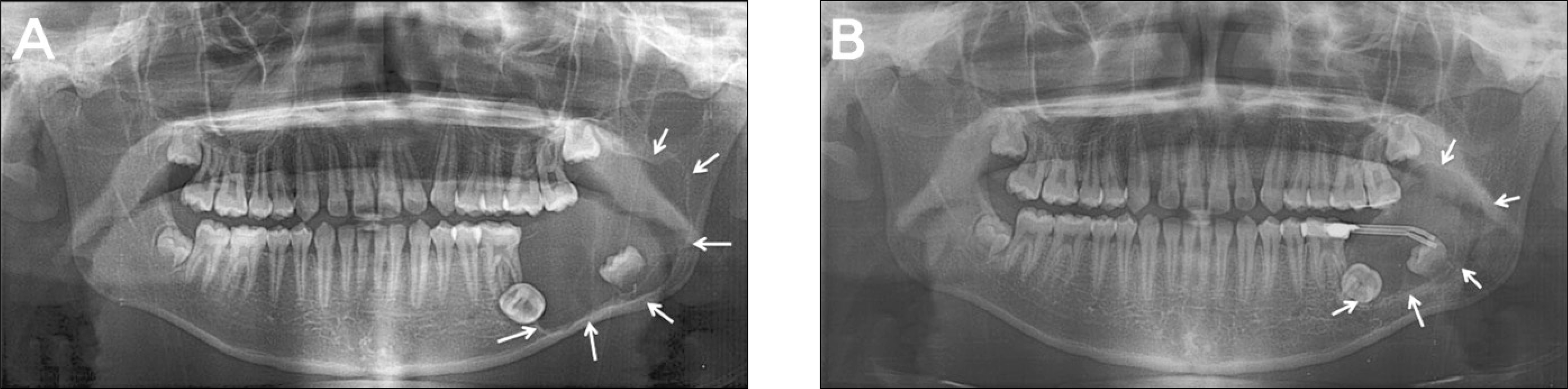
Fig. 2.
Panoramic X-rays of case 1. A: Preoperative panoramic X-ray. Huge radiolucent cystic lesion was observed on right body of mandible, B: Three months follow up panoramic X-ray. The cavity size of the cystic lesion was decreased about 50%, C: Six months follow up panoramic X-ray. The cavity size of the cystic lesion was decreased about 75%.

 XML Download