

 PDF
PDF ePub
ePub Citation
 Print
Print


Abstract
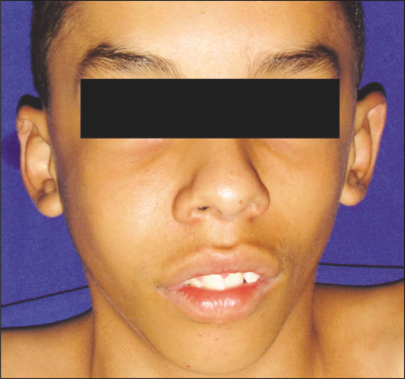
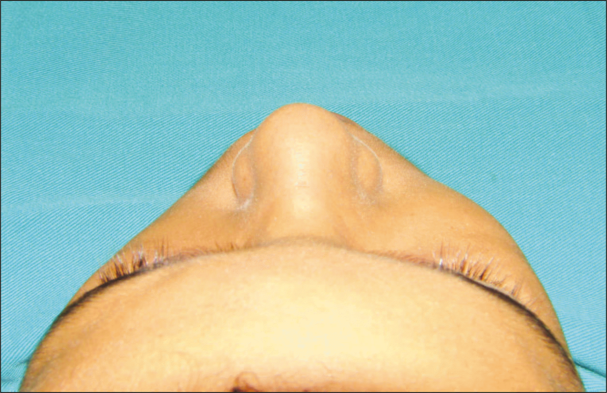
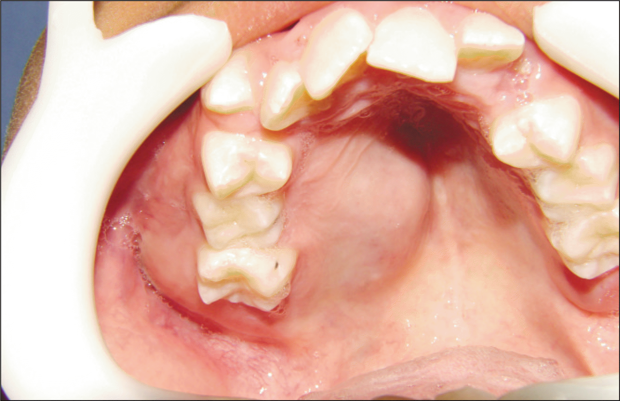
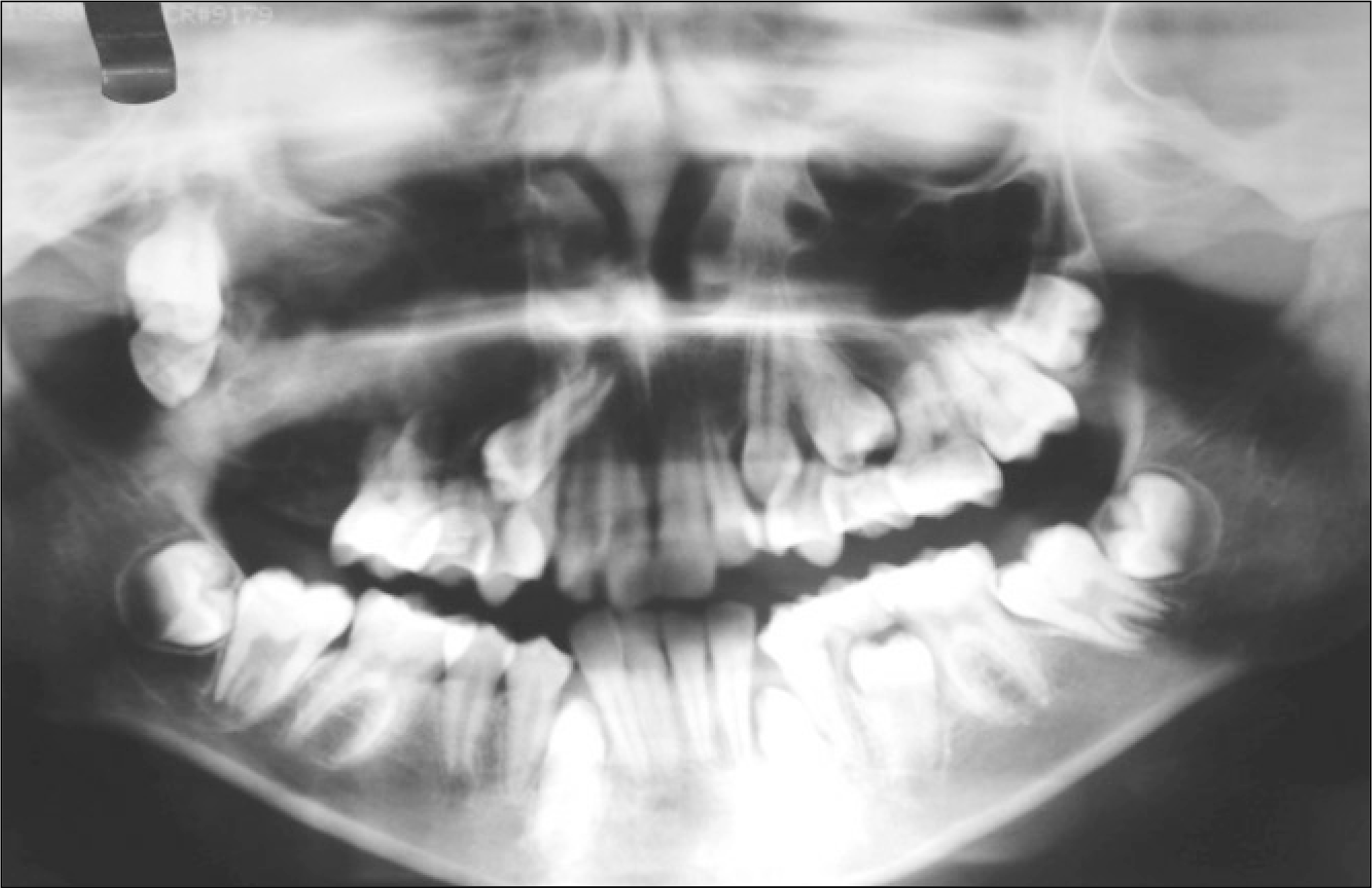
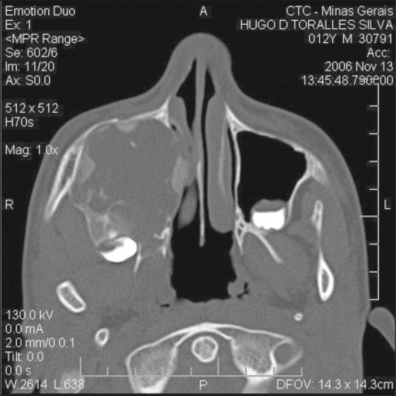
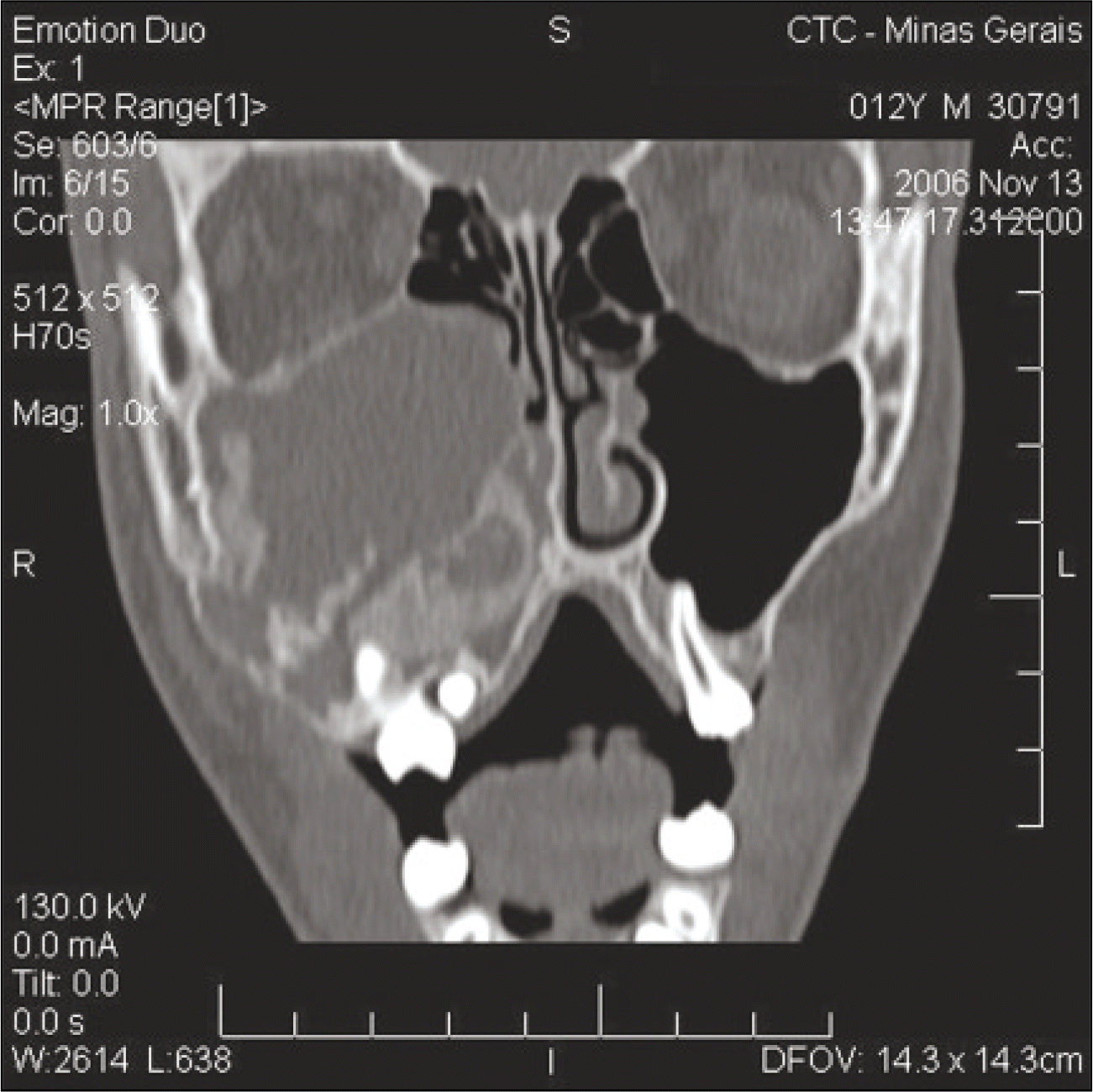
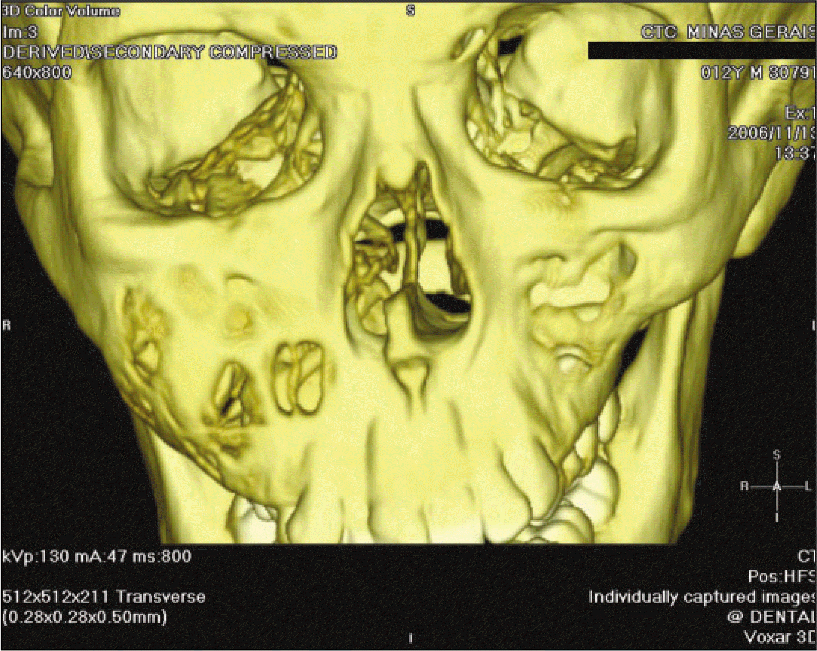
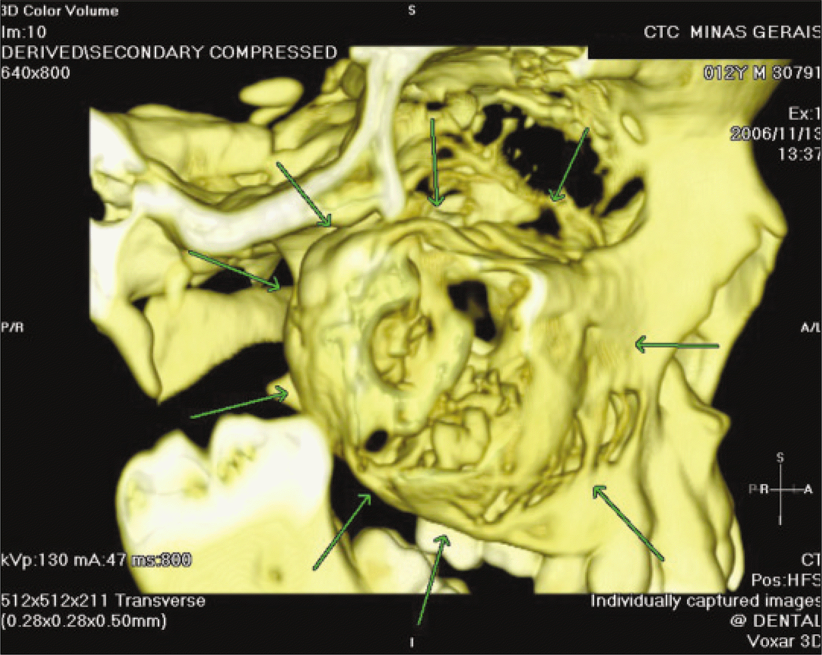
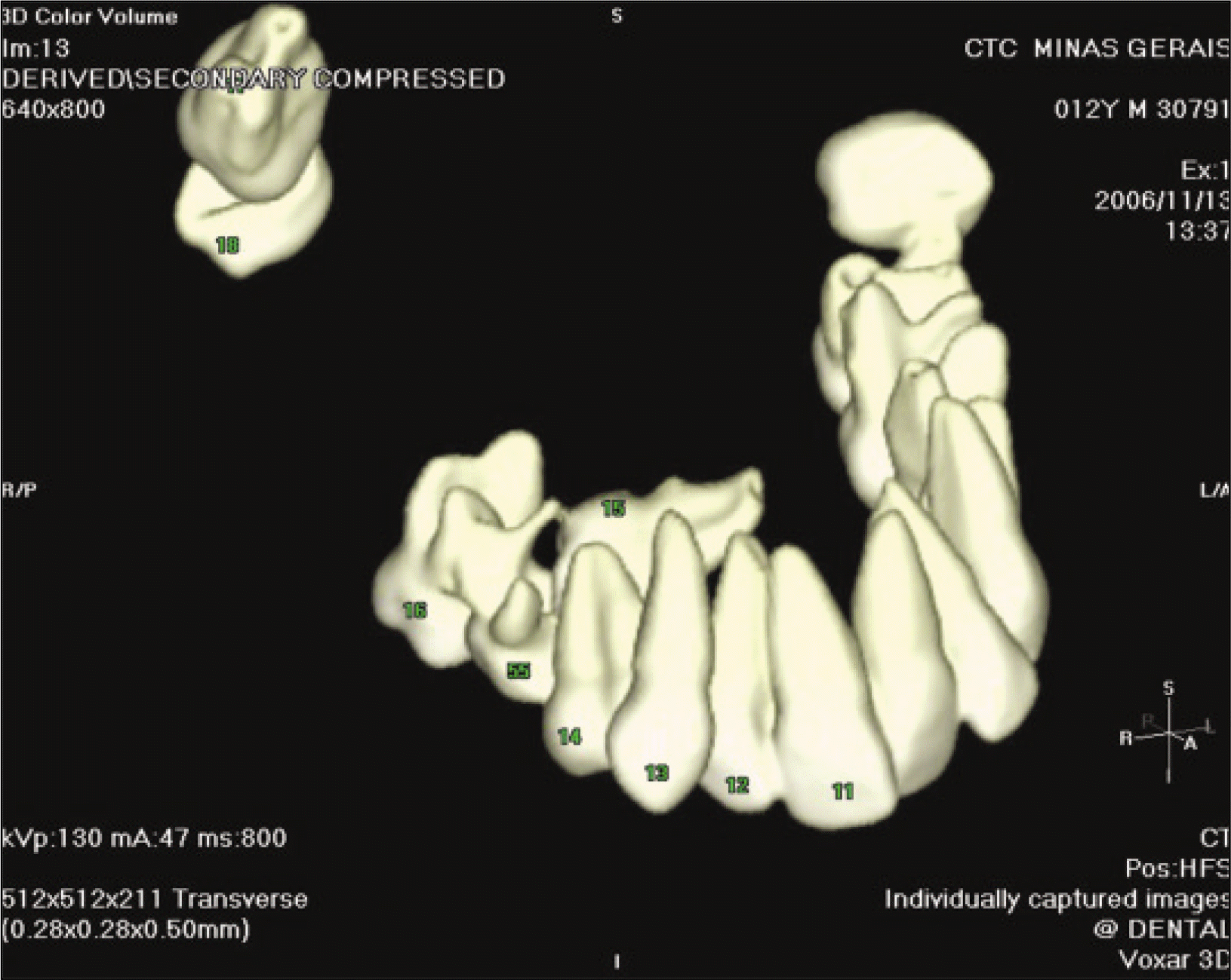
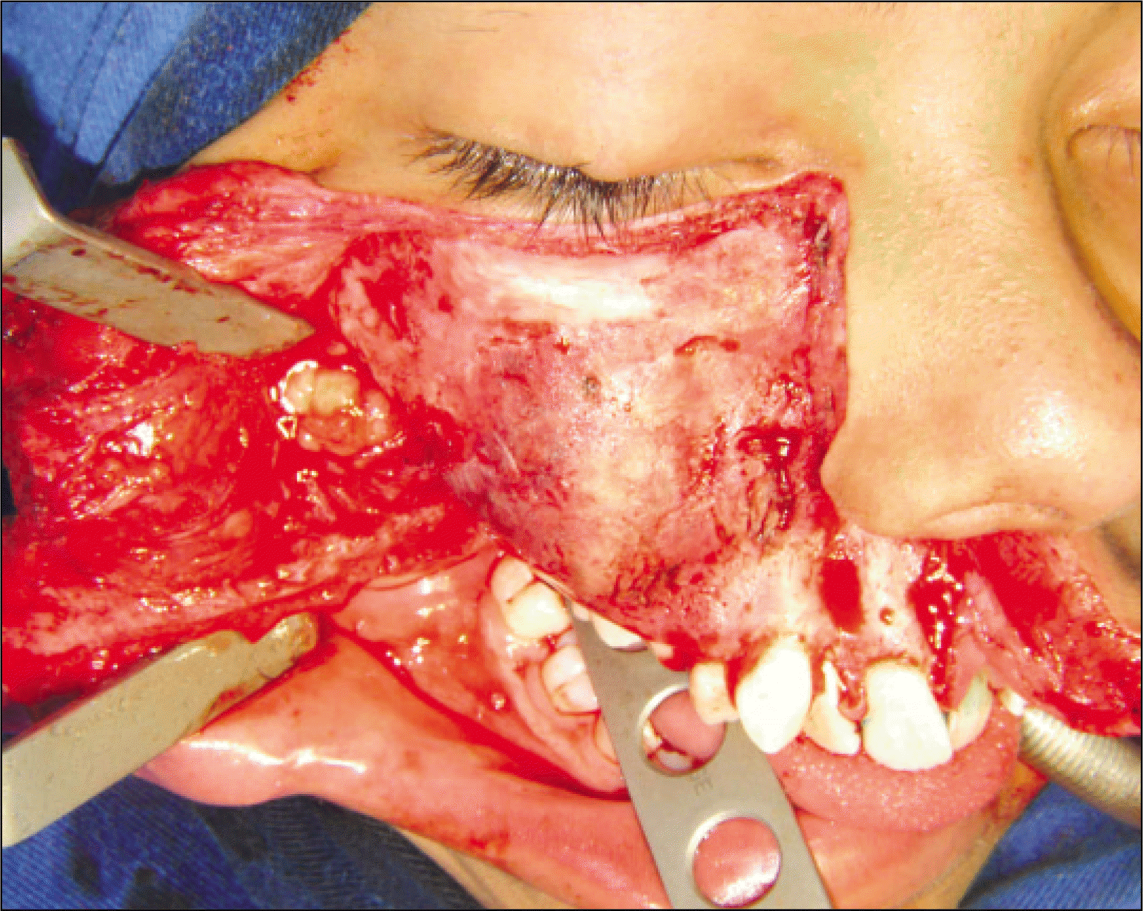
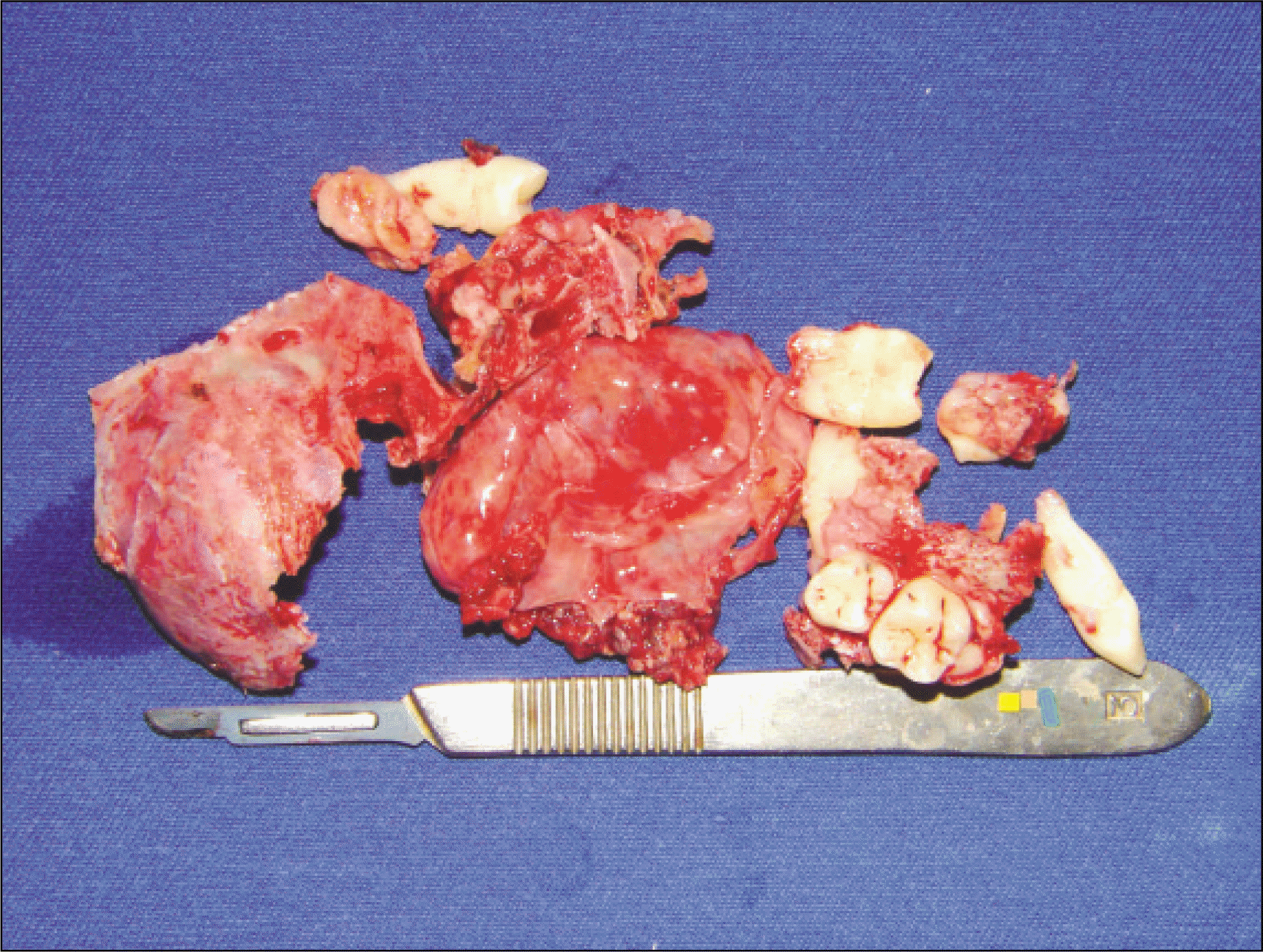
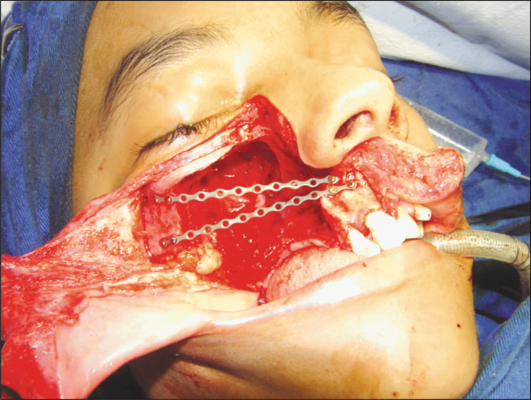
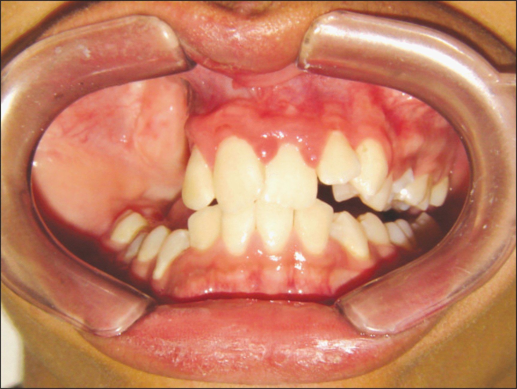
Juvenile ossifying fibroma is an expansive intraosseous lesion of the bones. In most patients, the tumors are located in the facial bones. The main characteristics of juvenile ossifying fibroma are the early age of onset, localization of the tumor, radiological pattern and a tendency for recurrence. This article describes a case of expanded juvenile ossifying fibroma in the right maxilla in a 12-year old boy. The lesion was removed totally by surgery under general anesthesia. The patient showed no radiological signals of recurrence approximately two years after surgery.
Go to : 
References
1. Scholl RJ, Kellett HM, Neumann DP, Lurie AG. Cysts and cystic lesions of the mandible: clinical and radiologic-histopathologic review. Radiographics. 1999; 19:1107–24.

2. Knox GW, Roth M, Saleh H, Stiles W. A unique temporal bone lesion resembling juvenile active ossifying myxoma. Am J Otol. 1996; 17:297–300.
3. Johnson LC, Yousefi M, Vinh TN, Heffner DK, Hyams VJ, Hartman KS. Juvenile active ossifying fibroma. Its nature, dynamics and origin. Acta Otolaryngol Suppl. 1991; 488:1–40.
4. Bendet E, Bakon M, Talmi YP, Tadmor R, Kronenberg J. Juvenile cementoossifying fibroma of the maxilla. Ann Otol Rhinol Laryngol. 1997; 106:75–8.
5. Khoury NJ, Naffaa LN, Shabb NS, Haddad MC. Juvenile ossifying fibroma: CT and MR findings. Eur Radiol. 2002; 12(Suppl 3):S109–13.
6. Lawton MT, Heiserman JE, Coons SW, Ragsdale BD, Spetzler RF. Juvenile active ossifying fibroma. Report of four cases. J Neurosurg. 1997; 86:279–85.
7. Slootweg PJ, Mu ¨ller H. Juvenile ossifying fibroma. Report of four cases. J Craniomaxillofac Surg. 1990; 18:125–9.
8. Kramer IRH, Pindborg JJ, Shear M. Histological typing of odontogenic tumours. 2nd ed.Berlin: Springer-Verlag;1992.
9. Leimola-Virtanen R, Va ¨ha ¨talo K, Syrja ¨nen S. Juvenile active ossifying fibroma of the mandible: a report of 2 cases. J Oral Maxillofac Surg. 2001; 59:439–44.
10. Ferris NJ, Tien RD. Ethmoid mucocele in an infant with a benign fibroosseous lesion. AJNR Am J Neuroradiol. 1995; 16:473–5.
11. Slootweg PJ, Panders AK, Koopmans R, Nikkels PG. Juvenile ossifying fibroma. An analysis of 33 cases with emphasis on histopathological aspects. J Oral Pathol Med. 1994; 23:385–8.
12. Brannon RB, Fowler CB. Benign fibroosseous lesions: a review of current concepts. Adv Anat Pathol. 2001; 8:126–43.
13. Bertrand B, Eloy P, Cornelis JP, Gosseye S, Clotuche J, Gilliard C. Juvenile aggressive cementoossifying fibroma: case report and review of the literature. Laryngoscope. 1993; 103:1385–90.
14. Cawson RA, Binnie WH, Eveson JW. Color atlas of oral disease: clinical and pathologic correlations. 2nd ed.London: Wolfe Publishing;1994.
15. Kuta AJ, Worley CM, Kaugars GE. Central cementoossifying fibroma of the maxillary sinus: a review of six cases. AJNR Am J Neuroradiol. 1995; 16:1282–6.
16. Regezi JA, Sciubba JJ. Oral pathology: clinical pathologic correlations. 2nd ed.Philadelphia: Saunders;1993.
17. Fakadej A, Boynton JR. Juvenile ossifying fibroma of the orbit. Ophthal Plast Reconstr Surg. 1996; 12:174–7.
18. Zama M, Gallo S, Santecchia L, Bertozzi E, de Stefano C. Juvenile active ossifying fibroma with massive involvement of the mandible. Plast Reconstr Surg. 2004; 113:970–4.
Go to :
 | Fig. 5.Histological sections. A: Numerous osseous spicules. B: Giant cells adjacent to the bony spicules, which are lined by lightly eosinophilic material, suggestive of osteoid. |
 XML Download