

 PDF
PDF ePub
ePub Citation
 Print
Print


Abstract
Introduction
Materials and Methods
Results
References
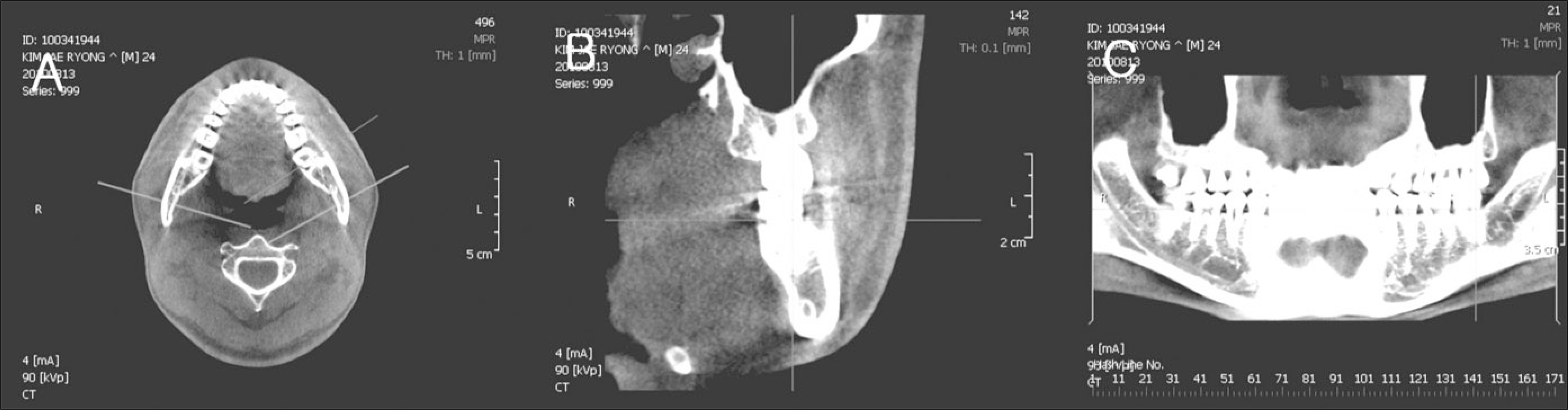 | Fig. 1.Converted images by using Ondemand program. A: Axial image, B: Cross-sectional image, C: Panoramic image. |
 | Fig. 2.Measuring points and measurements in the cross sectional images. (A: distance from the buccal aspect of the mandibular canal to outer aspect of the buccal cortex, B: distance from the buccal aspect of the mandibular canal to inner aspect of the buccal cortex, C: distance from the inferior aspect of the mandibular canal to inferior border of the mandible, D: thickness of the mandible, E: cross-sectional surface area of the mandible) |
Table 1.
| Group | Male/Female | Mean age | Chin deviation |
|---|---|---|---|
| Non-asymmetry (n=17) | 9/8 | 23.7±4.28 | |
| Asymmetry (n=20) | 7/13 | 22.6±3.55 | 6.2±2.9 (Rt./Lt.=8/12) |
Table 2.
| Deviated side | Opposite side | P value | |||||||
|---|---|---|---|---|---|---|---|---|---|
| 1st molar | 2nd molar | Ramus | 1st molar | 2nd molar | Ramus | 1st molar | 2nd molar | Ramus | |
| A | 4.68±1.87 | 6.36±1.47 | 4.88±1.36 | 5.09±1.76 | 6.24±1.56 | 4.68±1.30 | 0.155 | 0.687 | 0.497 |
| B | 2.33±1.48 | 3.87±1.55 | 2.56±1.29 | 2.54±1.27 | 3.55±1.57 | 2.40±1.19 | 0.448 | 0.264 | 0.58 |
| C | 8.81±3.19 | 8.18±1.94 | 10.30±3.36 | 8.48±2.97 | 8.17±1.85 | 11.51±4.00 | 0.503 | 0.822 | 0.033* |
| D | 10.78±2.76 | 12.87±1.87 | 11.38±2.17 | 11.12±1.56 | 12.70±1.80 | 11.10±1.78 | 0.513 | 0.477 | 0.361 |
| E | 331.18±48.62 | 364.70±78.99 | 374.27±90.36 | 332.94±57.72 | 353.74±84.80 | 368.67±79.05 | 0.762 | 0.335 | 0.49 |
A: distance from the buccal aspect of the mandibular canal to outer aspect of the buccal cortex, B: distance from the buccal aspect of the mandibular canal to inner aspect of the buccal cortex, C: distance from the inferior aspect of the mandibular canal to inferior border of the mandible, D: thickness of the mandible, E: cross-sectional surface area of the mandible)
Table 3.
(A: distance from the buccal aspect of the mandibular canal to outer aspect of the buccal cortex, B: distance from the buccal aspect of the mandibular canal to inner aspect of the buccal cortex, C: distance from the inferior aspect of the mandibular canal to inferior border of the mandible, D: thickness of the mandible, E: cross-sectional surface area of the mandible)
Table 4.
| 1st molar | 2nd molar | Ramus | |
|---|---|---|---|
| A | 0.171 | 0.054 | 0.151 |
| B | 0.046* | 0.013* | 0.089 |
| C | 0.724 | 0.569 | |
| D | 0.009** | 0.019* | 0.031* |
| E | 0.013* | 0.019* | 0.010* |
A: distance from the buccal aspect of the mandibular canal to outer aspect of the buccal cortex, B: distance from the buccal aspect of the mandibular canal to inner aspect of the buccal cortex, C: distance from the inferior aspect of the mandibular canal to inferior border of the mandible, D: thickness of the mandible, E: cross-sectional surface area of the mandible)
 XML Download