

 PDF
PDF ePub
ePub Citation
 Print
Print


Abstract
The purpose of this study was to examine the soft tissue changes in skeletal class II patients after mandibular advancement by bilateral sagittal split ramus osteotomy (BSSRO). In Asian population, the incidence of skeletal class II malocclusion is lower than that of skeletal class III malocclusion unlike the caucasians. This study was conducted to figure out the ratio at which hard tissue and soft tissue changes after mandibular advancement by analyzing cephalograms of 13 patients that have undergone the mandibular advancement surgery. As a result, change ratios of Li, B′, Pog′ according to the movement of li, B, Pog were found to be 0.59, 1.06, 0.82. Also, vertical height of vermilion zone (Si-Vb) and lower lip and chin (Si-Me′) were measured to evaluate vertical changes. Vermilion zone showed tendency to decrease by 1.02 mm on the average postoperatively, whereas vertical length of lower lip and chin showed tendency to increase by 3.57 mm on the average.
Go to : 
References
1. Ewing M, Ross BR. Soft tissue response to mandibular advancement and genioplasty. Am J Orthod Dentofacial Orthop. 1992; 101:550–5.

2. Thu ¨er U, Ingervall B. Vuillemin T Stability and effect on the soft tissue profile of mandibular advancement with sagittal split osteotomy and rigid internal fixation. Int J Adult Orthodon Orthognath Surg. 1994; 9(3):175–85.
3. Blomqvist JE, Ahlborg G, Isaksson S, Svartz K. A comparison of skeletal stability after mandibular advancement and use of two rigid internal fixation techniques. J Oral Maxillofac Surg. 1997; 55:568–74.
4. Quast DC, Biggerstaff RH, Haley JV. The short-term and longterm soft-tissue profile changes accompanying mandibular advancement surgery. Am J Orthod. 1983; 84:29–36.
5. van Sickels JE, Larsen AJ, Thrash WJ. A retrospective study of relapse in rigidly fixated sagittal split osteotomies: contributing factors. Am J Orthod Dentofacial Orthop. 1988; 93:413–8.
6. Mommaerts MY, Marxer H. A cephalometric analysis of the longterm, soft tissue profile changes which accompany the advancement of the mandible by sagittal split ramus osteotomies. J Craniomaxillofac Surg. 1987; 15:127–31.
7. Joss CU, Thu ¨er UW. Stability of the hard and soft tissue profile after mandibular advancement in sagittal split osteotomies: a longitudinal and longterm follow-up study. Eur J Orthod. 2008; 30:16–23.
8. Mobarak KA, Espeland L, Krogstad O, Lyberg T. Soft tissue profile changes following mandibular advancement surgery: predictability and longterm outcome. Am J Orthod Dentofacial Orthop. 2001; 119:353–67.
9. Veltkamp T, Buschang PH, English JD, Bates J, Schow SR. Predicting lower lip and chin response to mandibular advancement and genioplasty. Am J Orthod Dentofacial Orthop. 2002; 122:627–34.
10. Talbott JP. Soft tissue response to mandibular advancement surgery [dissertation]. Lexington: University of Kentucky;1975.
11. Dermaut LR, de Smit AA. Effects of sagittal split advancement osteotomy on facial profiles. Eur J Orthod. 1989; 11:366–74.
12. McDonnel JP, McNeill RW, West RA. Advancement genioplasty: a retrospective cephalometric analysis of osseous and soft tissue changes. J Oral Surg. 1977; 35:640–7.
13. Joss CU, Vassalli IM, Thu ¨er UW. Stability of soft tissue profile after mandibular setback in sagittal split osteotomies: a longitudinal and longterm follow-up study. J Oral Maxillofac Surg. 2008; 66:1610–6.
Go to :
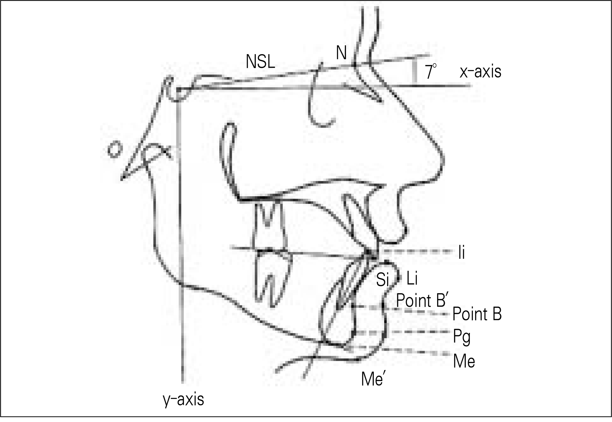
Table 1.
Cephalometric landmarks and measurement definitions
Table 2.
Postoperative change of the soft and hard tissue
Table 3.
The amount of anterior movement in Group 1 and Group 2 (T2-T1)
Table 4.
Ratio of the soft tissue changes in relation to the hard tissue changes
 XML Download