

 PDF
PDF ePub
ePub Citation
 Print
Print


Abstract
In the presence of acute pericoronitis of mandilbular third molar, antibiotic therapy and early incision and drainage are the method of choice, followed by definitive surgical extraction of the tooth as soon as it becomes subacute.
If excision of the overlying tissues is decided on, it should be done adequately. All overlying tissues must be throughly excised, and the crown portion of the unerupted tooth should be completely exposed. After excision has been completed, the wound should be managed with a surgical dressing. This should be allowed to remain approximately 7 days. And then, surgical extraction of the impacted mandibular third molar can be done usually. In this operation, there are many complications, such as, postoperative bleeding, infection, trismus, dysphasia and paresthesia. The surgeon are discredited and medicolegal problem may be occurred in the presence of many distressed complications. Therefore, the relatively nonsurgical treatment is the method of choice. So, authors selected the conservative treatment methods of incision and drainage, primary endodontic drainage, operculectomy without surgical extraction of the mandibular third molars. The results were more favorable without the postoperative complication in Wonju old offender prison.
References
1. Robinson RA. Clinical aspects of diseases associated with impacted mandibular third molars. Arch Clin Oral Pathol. 1940; 4:348–54.
2. Falace DA. Emergency dental care. 1st ed.Baltimore: Williams & Wilkins;1995. p. 228–53.
3. Yoon JH, Lee CK. Color atlas of teeth extraction in dental practice. 1st ed.Seoul: Jee Sung Publishing;1995. p. 109–58.
4. Bruce RA, Frederickson GC, Small GS. Age of patients and morbidity associated with mandibular third molar surgery. J Am Dent Assoc. 1980; 101:240–5.
5. Glickman I. Clinical periodontology. 4th ed.Philadelphia: W. B. Saunders;1972. p. 143–5.
6. Blair VP. The gingival operculum and the erupting lower third molar. Arch Clin Oral Pathol. 1940; 4:283–4.
7. Laskin DM. Surgical aids to the eruption of teeth. Laskin DM, editor. Oral and maxillofacial surgery. Ⅱ. St. Louis: Mosby;1985. p. 99–117.
8. Kruger GO. Textbook of oral and maxillofacial surgery. 6th ed.St. Louis: Mosby;1984. p. 65–105.
9. Pogrel MA. Complications of third molar surgery. Oral Maxillofac Surg Clin North Am. 1990; 2:441–8.
10. Goldberg MH, Nemarich AN, Marco WP. 2nd. Complications after mandibular third surgery: a statistical analysis of 500 consecutive procedures in private practice. J Am Dent Assoc. 1985; 111:277–9.
11. Lapeyrolerie F. Management of dentoalveolar hemorrhage. Dent Clin North Am. 1973; 17:523–32.
12. Topazian RG, Goldberg MH. Management of infections of the oral and maxillofacial regions. 1st ed.Philadelphia: W. B. Saunders;1981. p. 329–50.
13. Whang JS, Kim GY, Kim DY, Kim MH, Kim YJ, Kim WH, et al. Comprehension of stress science. 2nd ed.Seoul: Shin Kwang Publication;2007. p. 245–321.
14. Shafer WG, Hine MK, Levy BM, Tomich CE. A textbook of oral pathology. 4th ed.Philadelphia: W. B. Saunders;1983. p. 719–59.
15. Osborn TP, Frederickson G, Small IA. Torgerson TS. A prospective study of complications related to mandibular third molar surgery. J Oral Maxillofac Surg. 1985; 43:767–9.
16. Kim SN, Yum KW, Lee MS, Lee SW, Lee SJ. Emergency care in dental office. 3rd ed.Seoul: Jee Sung Publishing;2000. p. 198–201.
17. Grossman LI. Endodontic practice. 8th ed.Philadelphia: Lea & Febiger;1974. p. 151–68.
18. Lim SS. Clinical endodontics. 1st ed.Seoul: Medical and Dental publication;1994. p. 1–15.
19. Conley JJ. Blood vessel complications. Conley JJ, editor. Complications of head and neck surgery. Philadelphia: W. B. Saunders;1979. p. 66–80.
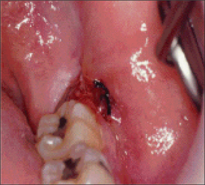
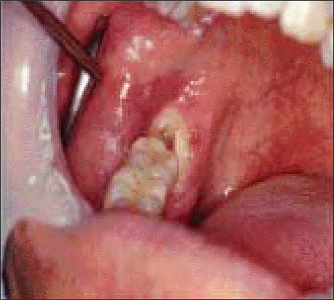
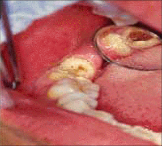
Table 1.
Factors in wound infection.
 XML Download