

 PDF
PDF ePub
ePub Citation
 Print
Print


Abstract
The submandibular gland is the second largest major salivary gland, which secretes 40% of the total daily saliva. Owing to its anatomic characteristics as well as the high viscosity and basicity of the saliva, sialolithiasis is found most commonly in the submandibular gland. Sialolithiasis that cannot be treated by conservative treatment is conventionally removed by an excision of the submandibular gland. Generally, an excision of the submandibular gland is performed via an extra-oral approach but the disadvantages of this treatment include a risk of injuring the facial nerve and scar formation. Case reports have revealed an even less invasive intraoral surgical technique for the removal of sialolith that does not affect the submandibular gland function. The functional recovery of the gland, complications and recurrence rates after surgery with this conservative intraoral procedure were all successful.
We report 5 patients from the department of Oral and Maxillofacial Surgery at Dental Hospital, Yonsei University, who had undergone a resection of the sialolith though the intraoral approach with successful results.
Go to : 
REFERENCES
1. Neville BW, Damm DD, Allen CW, Bouquot JE. Oral and maxillofacial pathology. 3rd ed.St. Louis, Mo: Saunders;2009.
2. Peterson LJ, Indresano AT, Marciani RD, Roser SM. Principles of oral and maxillofacial surgery. Philadelphia: JB Lippincott;1992.
3. McGurk M, Makdissi J, Brown JE. Intraoral removal of stones from the hilum of the submandibular gland: report of technique and morbidity. Int J Oral Maxillofac Surg. 2004; 33:683–6.

4. McGurk M. Surgical release of a stone from the hilum of the submandibular gland: a technique note. Int J Oral Maxillofac Surg. 2005; 34:208–10.
5. Combes J, Karavidas K, McGurk M. Intraoral removal of proximal submandibular stones–an alternative to sialadenectomy? Int J Oral Maxillofac Surg. 2009; 38:813–6.
6. Marchal F, Dulguerov P, Becker M, Barki G, Disant F, Lehmann W. Specificity of parotid sialendoscopy. Laryngoscope. 2001; 111:264–71.
7. Chung IK, Kim JR, Kim UK, Shin SH, Kim YD, Byun JH, et al. A clinical study of submandibular gland excision. J Korean Assoc Oral Maxillofac Surg. 2004; 30:545–50.
8. Beahm DD, Peleaz L, Nuss DW, Schaitkin B, Sedlmayr JC, Rivera-Serrano CM, et al. Surgical approaches to the submandibular gland: a review of literature. Int J Surg. 2009; 7:503–9.
9. Capaccio P, Torretta S, Pignataro L. The role of adenectomy for salivary gland obstructions in the era of sialendoscopy and lithotripsy. Otolaryngol Clin North Am. 2009; 42:1161–71.
10. Downton D, Qvist G. Intraoral excision of the submandibular gland. Proc R Soc Med. 1960; 53:543–4.
11. Hong KH, Kim YK. Intraoral removal of the submandibular gland: a new surgical approach. Otolaryngol Head Neck Surg. 2000; 122:798–802.
12. van den Akker HP, Busemann-Sokole E. Submandibular gland function following transoral sialolithectomy. Oral Surg Oral Med Oral Pathol. 1983; 56:351–6.
13. Nishi M, Mimura T, Marutani K, Noikura T. Evaluation of submandibular gland function by sialo-scintigraphy following sialolithectomy. J Oral Maxillofac Surg. 1987; 45:567–71.
14. Makdissi J, Escudier MP, Brown JE, Osailan S, Drage N, McGurk M. Glandular function after intraoral removal of salivary calculi from the hilum of the submandibular gland. Br J Oral Maxillofac Surg. 2004; 42:538–41.
15. Zenk J, Constantinidis J, Al-Kadah B, Iro H. Transoral removal of submandibular stones. Arch Otolaryngol Head Neck Surg. 2001; 127:432–6.
16. Park JS, Sohn JH, Kim JK. Factors influencing intraoral removal of submandibular calculi. Otolaryngol Head Neck Surg. 2006; 135:704–9.
17. Koch M, Zenk J, Iro H. Algorithms for treatment of salivary gland obstructions. Otolaryngol Clin North Am. 2009; 42:1173–92.
18. Escudier MP, Brown JE, Putcha V, Capaccio P, McGurk M. Factors influencing the outcome of extracorporeal shock wave lithotripsy in the management of salivary calculi. Laryngoscope. 2010; 120:1545–9.
Go to :
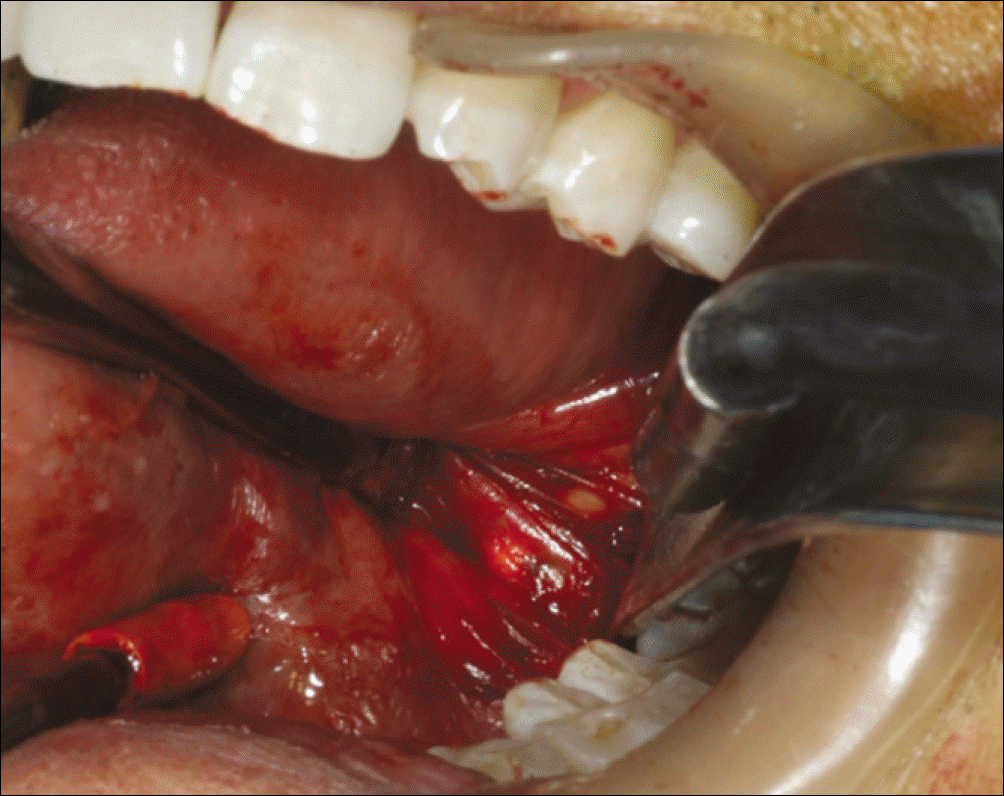
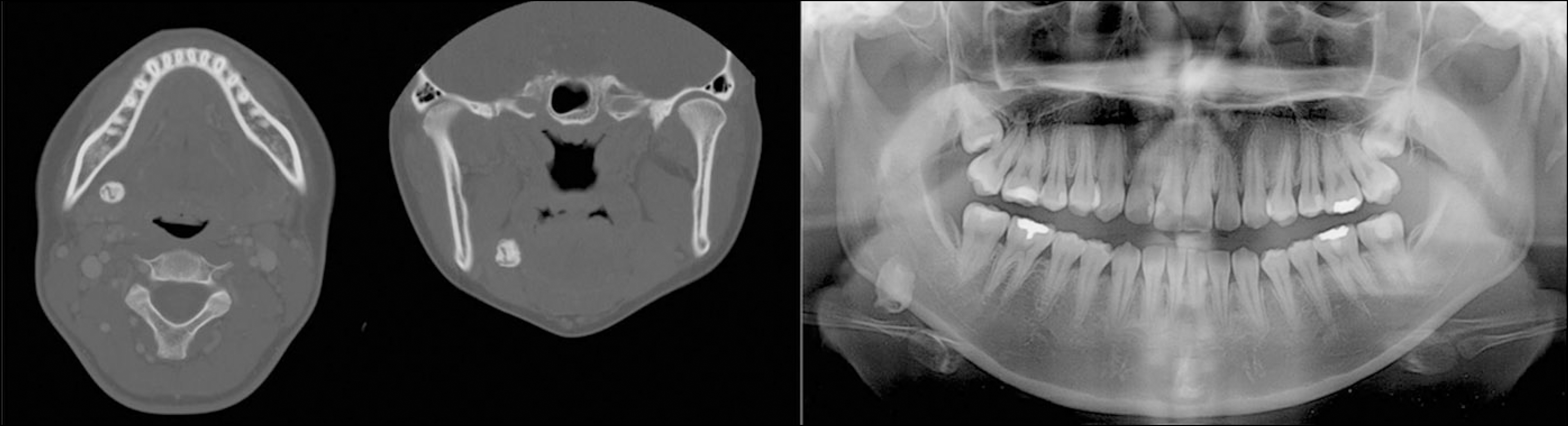
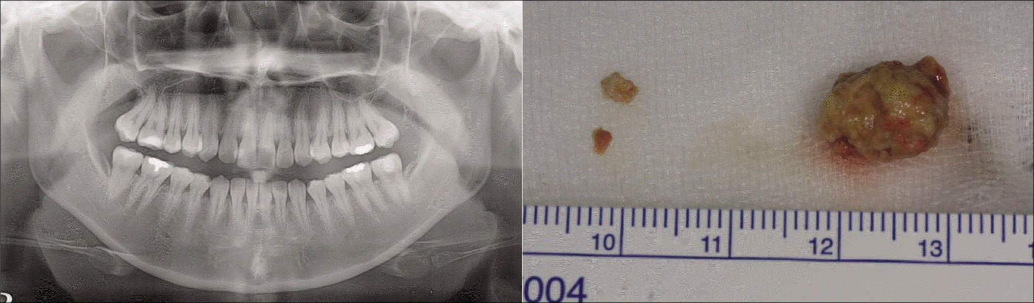
Table 1.
Patients and clinical information
 XML Download