

 PDF
PDF ePub
ePub Citation
 Print
Print


Abstract
Carcinoma ex pleomorphic adenoma is transformed at the incidence of 1-20% in pleomorphic adenoma and frequently recurred. It accounts for 10% of all malignant salivary tumors and its average age of occurrence is 60s. It will present in a large, long-standing pleomorphic adenoma or in one that was previously treated but has recurred. According to cell composition in malignant cell carcinoma, and clear cell adenocarcinoma. Most (75%) occur in parotid gland, while about 20% occur in the minor gland of the oral mucosa. The metastasis rate to regional lymph node is about 25%, and to distant organs about 33% and the 5-year survival rates are 40%. Though the treatment of the carcinoma ex pleomorphic adenoma is not established, it is treated ideally with and extensive resection, neck dissection, postoperative radiotherapy, and chemotherapy. When occurred in parotid gland, facial paralysis is reported. With a review of literatures, we report a case of carcinoma ex pleomorphic adenoma which operated with total parotidectomy and supraomohyoid neck dissection.
References
1. Seifert G. Histopathology of malignant salivary gland tumours. Eur J Cancer B Oral Oncol. 1992; 28B:49–56.

2. Genelhu MC, Gobbi H, Soares FA, Campos AH, Ribeiro CA, Cassali GD. Immunohistochemical expression of p63 in pleomorphic adenomas and carcinomas ex-pleomorphic adenomas of salivary glands. Oral oncol. 2006; 42:154–60.
3. Henriksson G, Westrin KM, Carlsoo B, Silfversward C. Recurrent primary pleomorphic adenomas of salivary gland origin: intrasurgical rupture, histopathologic features, and pseudopodia. Cancer. 1998; 82:617–20.
4. Johns ME, Goldsmith MM. Incidence, diagnosis and classification of salivary gland tumors. Part 1. Oncology (Williston Park). 1989; 3:47–56. discussion 56, 58, 62.
5. Zbaren P, Nuyens M, Loosli H, Stauffer E. Diagnostic accuracy of fine-needle aspiration cytology and frozen section in primary parotid carcinoma. Cancer. 2004; 100:1876–83.
6. Zbaren S, Caversaccio MD, Stauffer E. Carcinoma ex pleomorphic adenoma: diagnostic difficulty and outcome. Otolaryngol Head Neck Surg. 2008; 138:601–5.
7. Lim YC, Lee SY, Kim K, Lee JS, Koo BS, Shin HA, et al. Conservative parotidectomy for the treatment of parotid cancers. Oral oncol. 2005; 41:1021–7.
8. Spiro RH. Salivary neoplasms: overview of a 35-year experience with 2,807 patients. Head Neck Surg. 1986; 8:177–84.
9. Konno A, Numata T, Nagata H, Terada N, Hanazawa T. Effects of TNM extension and histopathological type of the tumor on long-term survival rates of parotid and submandibular gland cancer. Gan To Kagaku Ryoho. 1997; 24:1347–53.
10. Beckhardt RN, Weber RS, Zane R, Garden AS, Wolf P, Carrillo R, et al. Minor salivary gland tumors of the palate: clinical and pathologic correlates of outcome. Laryngoscope. 1995; 105:1155–60.
11. Furukawa M, Suzuki H, Matsuura K, Takahashi E, Suzuki H, Tezuka F. Carcinoma ex pleomorphic adenoma of the palatal minor salivary gland with extension into the nasopharynx. Auris Nasus Larynx. 2001; 28:279–81.
12. Akan H, Yildiz L, Unal R. Carcinoma ex pleomorphic adenoma of the minor salivary gland with pulmonary metastasis. Diagn Interv Radiol. 2008; 14:3–5.
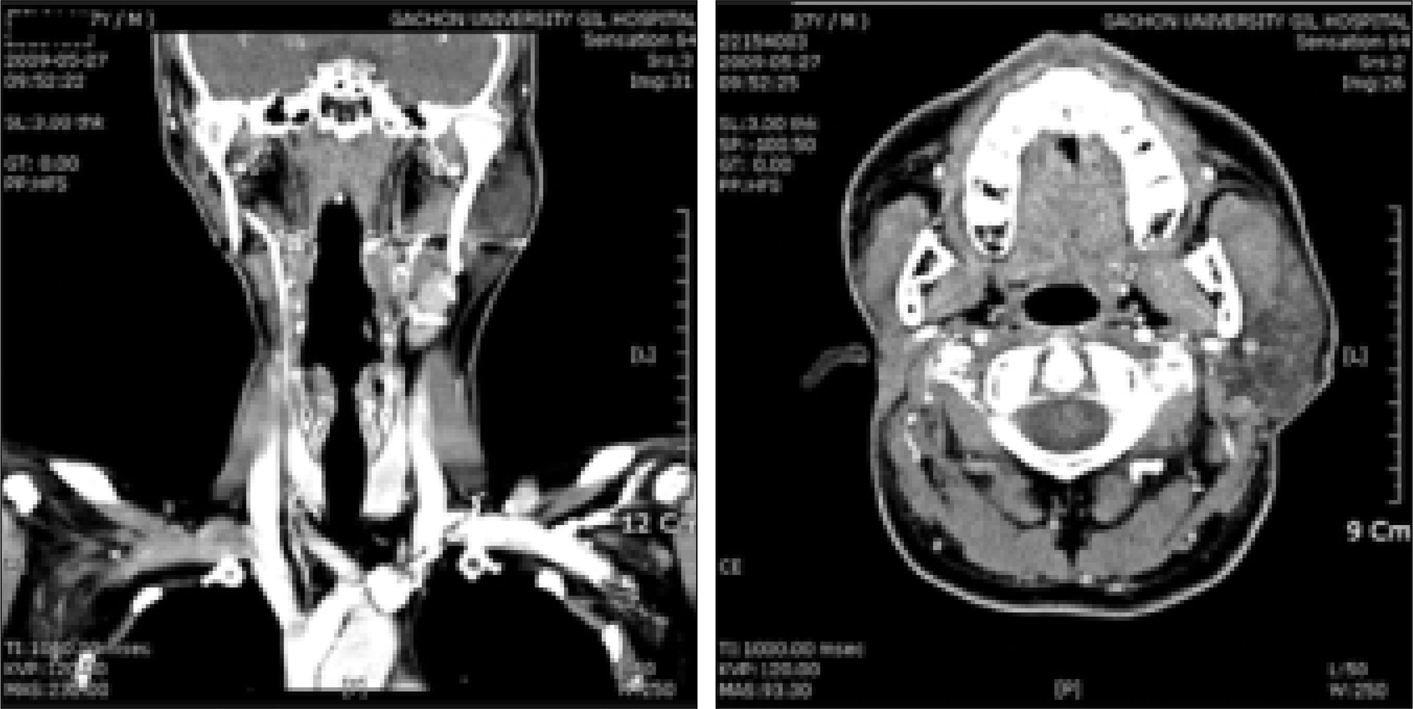
Fig. 1.
About 4 cm sized lobulating well-defined mass with hyperintense on T2WI enhancement on right parotid.
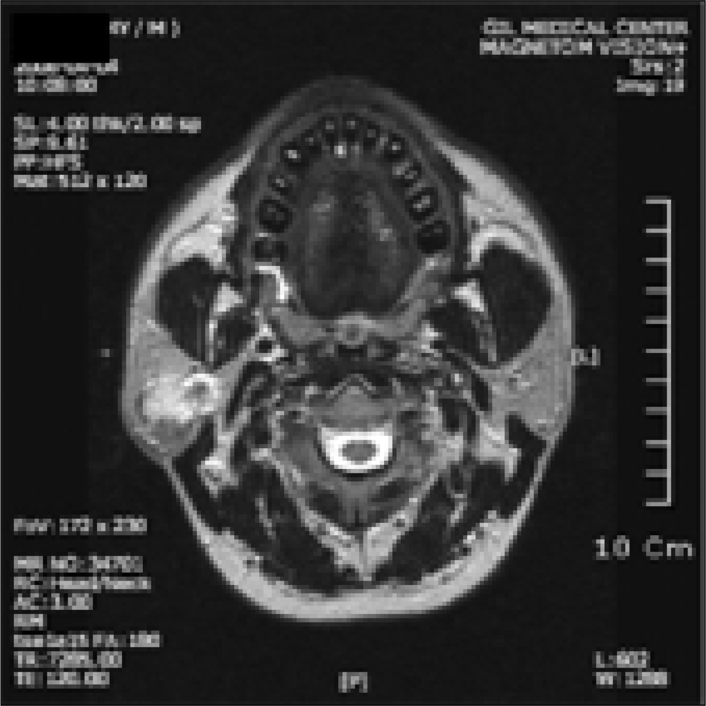
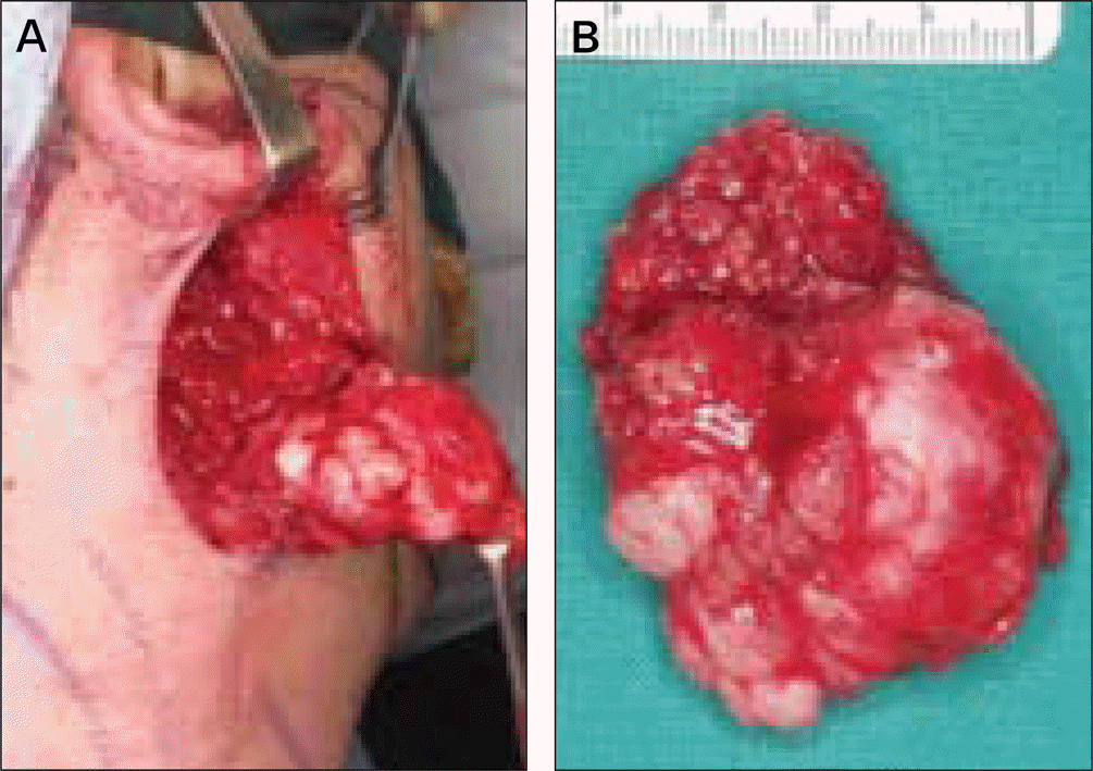
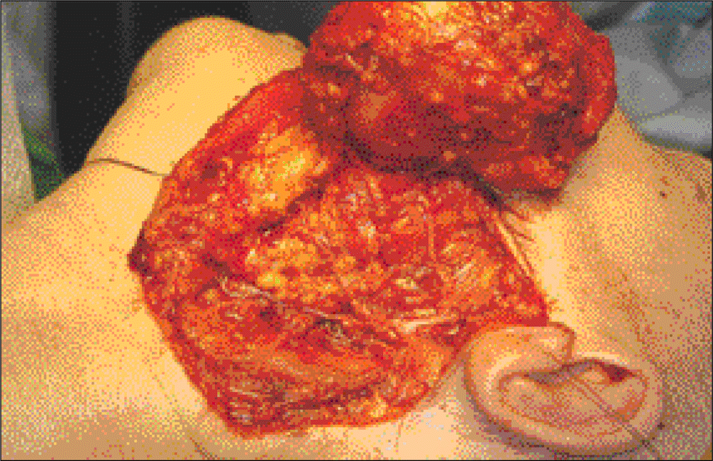
Fig. 3.
Photographs showing confinement within tumor capsule, no lymphatic and vascular tumor emboli, and clear resection margin. By immunohistochemistry, the carcinoma component is positive for p53 and shows high Ki-67 index (up to 30%).
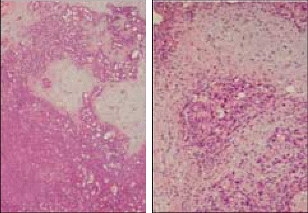
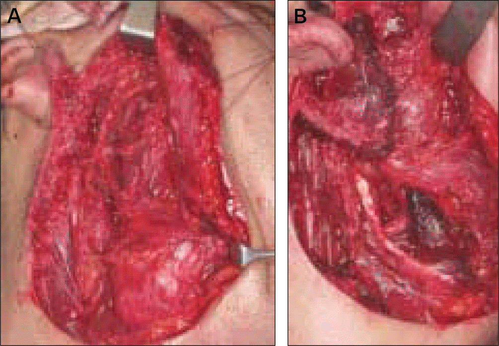
Fig. 4.
Intraoperative photographs. A. Supraomohyoid neck dissection was performed and facial nerve was preserved. B. Facial nerve was covered with rotating sternocleidmas-toid muscle flap.
 XML Download