

 PDF
PDF ePub
ePub Citation
 Print
Print


Abstract
Purpose
To research about maxillofacial traumatic injuries of children in aspects of gender difference, various incidence rates between age, trauma type, cause, monthly and daily incidence rate, type of tooth damage, gingival damage, soft tissue damage, and type of facial bone fracture.
Materials and methods
Study group consisted of children under 15 years of age who visited Dental Hospital, School of Dentistry, Kyung Hee University from 2004/7/1 to 2007/6/30 with chief complaint of oral and maxillofacial traumatic injuries. 1,559 cases of traumatic injuries were studied from 1,556 (1,004 male, 552 female) children.
Conclusion
1. There were slightly more boys than girls, giving a male-to-female ratio of 1.82:1.0. The 1-3 year old boys and girls had the highest number of traumatic injuries.
2. Of the 1,556 patients, 68.63% had soft tissue injuries, 50.22% had periodontal injuries, 29.89% had teeth injuries, and 3.85% had maxillofacial bone fractures.
3. Falling down was the most common cause of injury in both sexes.
4. The months with the highest incidence rates were in order May (12.12%), June (11.74%), and October (11.13%). Most of the injuries occurred on weekends.
5. The most common tooth injury was uncomplicated crown fracture, and the most common periodontal injury was subluxation. The majority of traumatizes teeth were the upper central incisors.
6. The most common soft tissue injury was intraoral lacerations.
7. Mandibular fractures were most frequent in facial bone fractures; symphysis, condylar head, and angle fractures were most frequent in mandibular fractures; maxillary and nasal bone fractures were most frequent in midfacial bone fractures.
References
1. Gassner R, Tuli T, Ha ¨chl O, Rudisch A, Ulmer H. Cranio-maxillofacial trauma: a 10 year review of 9,543 cases with 21,067 injuries. J Craniomaxillofac Surg. 2003; 31:51–61.

2. da Silva AC, Passeri LA, Mazzonetto R, de Moraes M, Moreira RW. Incidence of dental trauma associated with facial trauma in Brazil: a 1-year evaluation. Dent Traumatol. 2004; 20:6–11.
3. Zerfowski M, Bremerich A. Facial trauma in children and adolescents. Clin Oral Investig. 1998; 2:120–4.
4. Rahman RA, Ramli R, Rahman NA, Hussaini HM, Idrus SM, Hamid AL. Maxillofacial trauma of pediatric patients in Malaysia: a retrospective study from 1999 to 2001 in three hospitals. Int J Pediatr Otorhinolaryngol. 2007; 71:929–36.
5. Ferreira PC, Amarante JM, Silva AC, Pereira JM, Cardoso MA, Rodrigues JM. Etiology and patterns of pediatric mandibular fractures in Portugal: a retrospective study of 10 years. J Craniofac Surg. 2004; 15:384–91.
6. Min SK, Choi MG, Oh SH, Lee DK. A clinical study of the pediatric mandibular fractures. J Korean Assoc Maxillofac Plast Reconstr Surg. 2000; 22:555–62.
7. Kim HE, Han KH, Kim TY, Ko SJ, Jeon IS, Yoon KO. Clinical study of mandibular fracture of children. J Korean Assoc Maxillofac Plast Reconstr Surg. 2003; 25:426–31.
8. Choi SC, Prak JH, Lee KH. A study of the traumatic injuries in the primary dentition. J Korean Acad Pediatr Dent. 2003; 30:618–25.
9. Flores MT, Andersson L, Andreasen JO, Bakland LK, Malmgren B, Barnett F, et al. Guidelines for the management of traumatic dental injuries. II. Avulsion of permanent teeth. Dent Traumatol. 2007; 23:130–6.
10. Flores MT, Andersson L, Andreasen JO, Bakland LK, Malmgren B, Barnett F, et al. Guidelines for the management of traumatic dental injuries. I. Fractures and luxations of permanent teeth. Dent Traumatol. 2007; 23:66–71.
11. Flores MT, Malmgren B, Andersson L, Andreasen JO, Bakland LK, Barnett F, et al. Guidelines for the management of traumatic dental injuries. III. Primary teeth. Dent Traumatol. 2007; 23:196–202.
12. Hogg NJ, Horswell BB. Hard tissue pediatric facial trauma: a review. J Can Dent Assoc. 2006; 72:555–8.
13. Hogg NJ, Horswell BB. Soft tissue pediatric facial trauma: a review. J Can Dent Assoc. 2006; 72:549–52.
14. Oh SH. Long-term evaluations of pediatric condyle fracture patients. J Korean Assoc Maxillofac Plast Reconstr Surg. 2002; 24:482–8.
15. Andreasen JO, Andreasen FM, Meja �re I, Cvek M. Healing of 400 intra-alveolar root fractures. 2. Effect of treatment factors such as treatment delay, repositioning, splinting type and period and antibiotics. Dent Traumatol. 2004; 20:203–11.
16. Andreasen JO, Andreasen FM, Meja �re I, Cvek M. Healing of 400 intra-alveolar root fractures. 1. Effect of pre-injury and injury factors such as sex, age, stage of root development, fracture type, location of fracture and severity of dislocation. Dent Traumatol. 2004; 20:192–202.
17. Andreasen JO, Bakland LK, Andreasen FM. Traumatic intrusion of permanent teeth. Part 3. A clinical study of the effect of treatment variables such as treatment delay, method of repositioning, type of splint, length of splinting and antibiotics on 140 teeth. Dent Traumatol. 2006; 22:99–111.
18. Andreasen JO, Bakland LK, Andreasen FM. Traumatic intrusion of permanent teeth. Part 2. A clinical study of the effect of preinjury and injury factors, such as sex, age, stage of root development, tooth location, and extent of injury including number of intruded teeth on 140 intruded permanent teeth. Dent Traumatol. 2006; 22:90–8.
19. Andreasen JO, Bakland LK, Matras RC, Andreasen FM. Traumatic intrusion of permanent teeth. Part 1. An epidemiological study of 216 intruded permanent teeth. Dent Traumatol. 2006; 22:83–9.
20. Sandalli N, Cildir S, Guler N. Clinical investigation of traumatic injuries in Yeditepe University, Turkey during the last 3 years. Dent Traumatol. 2005; 21:188–94.
21. Gassner R, Tuli T, Ha ¨chl O, Moreira R, Ulmer H. Cranioma-xillofacial trauma in children: a review of 3,385 cases with 6,060 injuries in 10 years. J Oral Maxillofac Surg. 2004; 62:399–407.
22. Kargul B, Cag ̆lar E, Tanboga I. Dental trauma in Turkish children, Istanbul. Dent Traumatol. 2003; 19:72–5.
23. Harrington MS, Eberhart AB, Knapp JF. Dentofacial trauma in children. ASDC J Dent Child. 1988; 55:334–8.
24. Caldas AF, Burgos ME. A retrospective study of traumatic dental injuries in a Brazilian dental trauma clinic. Dent Traumatol. 2001; 17:250–3.
25. Sarog ̆lu I, So ¨nmez H. The prevalence of traumatic injuries treated in the pedodontic clinic of Ankara University, Turkey, during 18 months. Dent Traumatol. 2002; 18:299–303.
26. Ferreira PC, Amarante JM, Silva PN, Rodrigues JM, Choupina MP, Silva AC, et al. Retrospective study of 1251 maxillofacial fractures in children and adolescents. Plast Reconstr Surg. 2005; 115:1500–8.
27. Wright G, Bell A, McGlashan G, Vincent C, Welbury RR. Dentoalveolar trauma in Glasgow: an audit of mechanism and injury. Dent Traumatol. 2007; 23:226–31.
28. Skaare AB, Jacobsen I. Etiological factors related to dental injuries in Norwegians aged 7-18 years. Dent Traumatol. 2003; 19:304–8.
29. Skaare AB, Jacobsen I. Primary tooth injuries in Norwegian children (1-8 years). Dent Traumatol. 2005; 21:315–9.
30. Rajab LD. Traumatic dental injuries in children presenting for treatment at the Department of Pediatric Dentistry, Faculty of Dentistry, University of Jordan, 1997-2000. Dent Traumatol. 2003; 19:6–11.
31. Granville-Garcia AF, de Menezes VA, de Lira PI. Dental trauma and associated factors in Brazilian preschoolers. Dent Traumatol. 2006; 22:318–22.
32. Skaare AB, Jacobsen I. Dental injuries in Norwegians aged 7-18 years. Dent Traumatol. 2003; 19:67–71.
33. Castro JC, Poi WR, Manfrin TM, Zina LG. Analysis of the crown fractures and crown-root fractures due to dental trauma assisted by the Integrated Clinic from 1992 to 2002. Dent Traumatol. 2005; 21:121–126.
34. Motamedi MH. An assessment of maxillofacial fractures: a 5-year study of 237 patients. J Oral Maxillofac Surg. 2003; 61:61–4.
35. Iida S, Matsuya T. Paediatric maxillofacial fractures: their aetio-logical characters and fracture patterns. J Craniomaxillofac Surg. 2002; 30:237–41.
36. Qudah MA, Al-Khateeb T, Bataineh AB, Rawashdeh MA. Mandibular fractures in Jordanians: a comparative study between young and adult patients. J Craniomaxillofac Surg. 2005; 33:103–6.
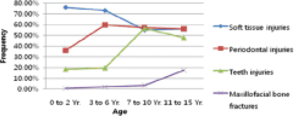
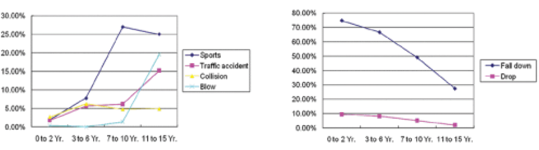
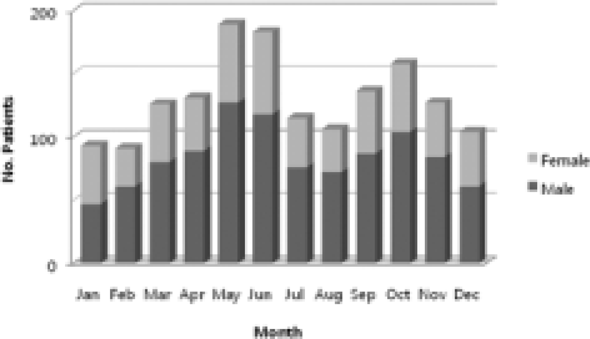
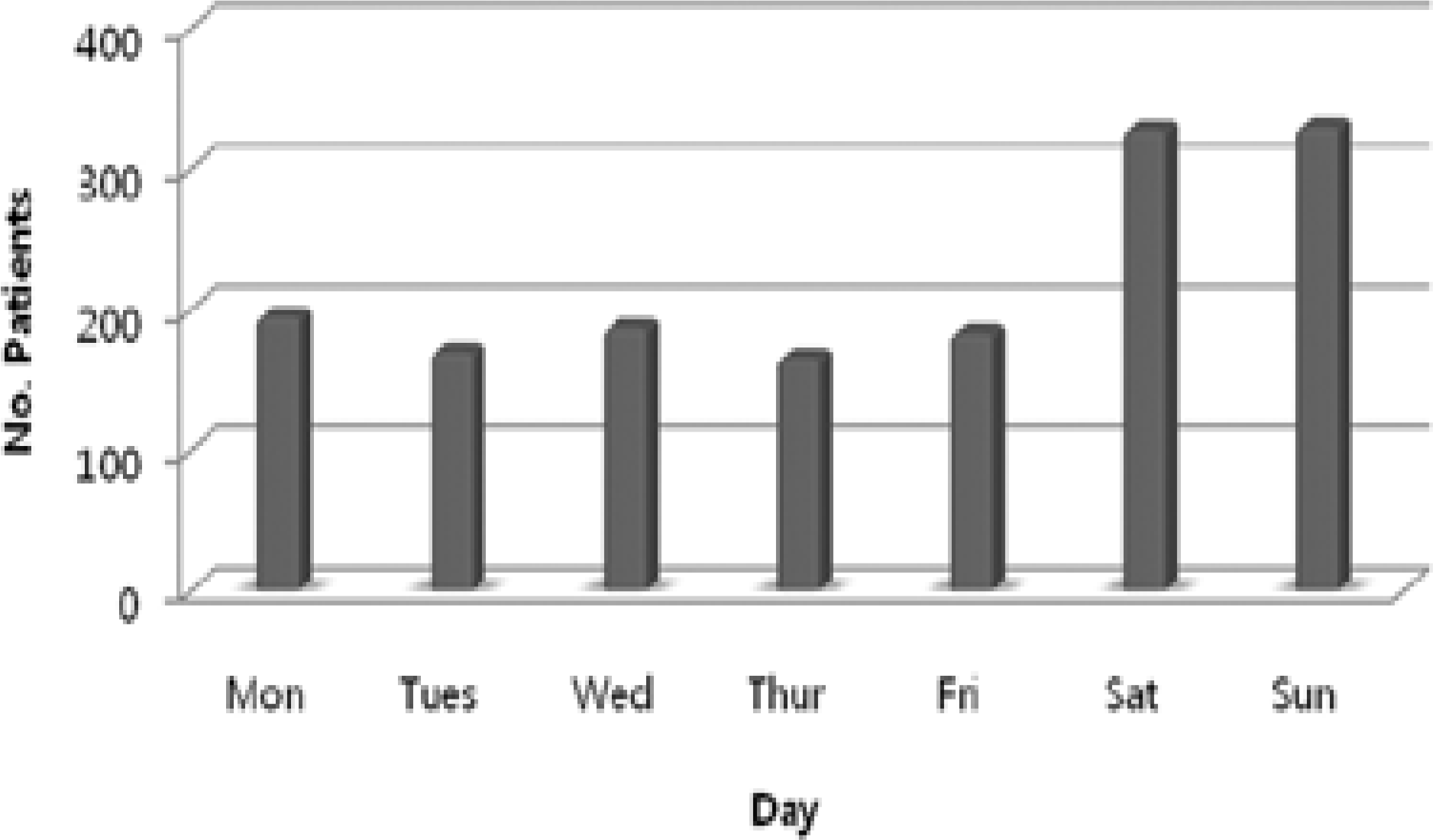
Table 1.
Classification for teeth injuries.
Table 2.
Classification for periodontal injury.
Table 3.
Age and sex distribution of children with trauma.
Table 4.
Category of injury.
Table 5.
Causes of injuries according to sex.
Table 6.
Distribution of injuries according to place of occurrence and sex.
Table 7.
Diagnosis of dental injuries in primary and permanent dentition.
Table 8.
Distribution of dental injuries in primary and permanent dentition.
Table 9.
Diagnosis of periodontal injuries in primary and permanent dentition.
Table 10.
Distribution of periodontal injuries in primary and permanent dentition.
Table 11.
Diagnosis of soft tissue injuries by age.
Table 12.
Treatment methods in maxillofacial fracture.
Table 13.
Causes of maxillofacial fracture according to sex.
 XML Download