Abstract
Introduction
The planning of implant surgery is an important factor for the implant prosthesis. Stereolithographic (SLA) surgical stents based on a computer simulation are quite helpful for clinicians to perform the surgery as planned. Although many clinical and technical trials have been performed for computed tomography (CT)-guided implant stents to improve the surgical procedures and prosthetic treatment, there are still many problems to solve. We developed a system of a surgical guide based on 3 dimensional (3D) CT for implant therapy and achieved satisfactory results in the terms of planning and operation.
Materials and Methods
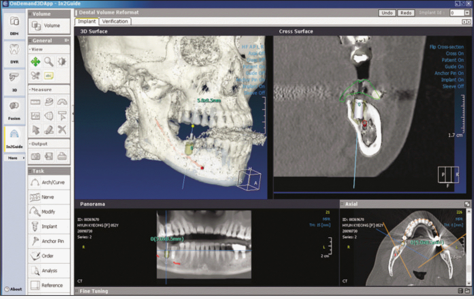
Fifteen patients were selected and 30 implant fixtures were installed. The preoperative CT data for surgical planning were prepared after obtaining informed consent. Surgical planning was performed using the simulation program, Ondemend3D In2Guide. The stents were fabricated based on the simulation data containing information of the residual bone, the location of the nerve, and the expected design of the prostheses. After surgery with these customized stents, the accuracy and reproducibility of implant surgery were evaluated based on the computer simulation. The data of postoperative CT were used to confirm this system using the image fusion technique and compare the implant fixtures between the planned and implanted.
Results
The mean error was 1.18 (±0.73) mm at the occlusal center, 1.23 (±0.67) mm at the apical center, and the axis error between the two fixtures was 3.25� (±3.00). These stents showed superior accuracy in maxilla cases. The lateral side error at the apical center was significantly different from the error at the occlusal center but there were no significant differences between the premolars, 1st molars and 2nd molars.
Go to : 
References
1. Jacobs R, Adriansens A, Naert I, Quirynen M, Hermans R, van Steenberghe D. Predictability of reformatted computed tomography for preoperative planning of endosseous implants. Dentomaxillofac Radiol. 1999; 28:37–41.

2. Ozan O, Turkyilmaz I, Ersoy AE, McGlumphy EA, Rosenstiel SF. Clinical accuracy of 3 different types of computed tomography-derived stereolithographic surgical guides in implant placement. J Oral Maxillofac Surg. 2009; 67:394–401.

3. Jacobs R, Adriansens A, Verstreken K, Suetens P, van Steenberghe D. Predictability of a three-dimensional planning system for oral implant surgery. Dentomaxillofac Radiol. 1999; 28:105–11.

4. Di Giacomo GA, Cury PR, de Araujo NS, Sendyk WR, Sendyk CL. Clinical application of stereolithographic surgical guides for implant placement: preliminary results. J Periodontol. 2005; 76:503–7.
5. Vrielinck L, Politis C, Schepers S, Pauwels M, Naert I. Image-based planning and clinical validation of zygoma and pterygoid implant placement in patients with severe bone atrophy using customized drill guides. Preliminary results from a prospective clinical follow-up study. Int J Oral Maxillofac Surg. 2003; 32:7–14.

6. Verstreken K, van Cleynenbreugel J, Marchal G, Naert I, Suetens P, van Steenberghe D. Computer-assisted planning of oral implant surgery: a three-dimensional approach. Int J Oral Maxillofac Implants. 1996; 11:806–10.
7. Bianchi SD, Lojacono A, Nevins M, Ramieri G, Corrente G, Martuscelli G, et al. The use of replicate resin models in the treatment of maxillary sinus augmentation patients. Int J Periodontics Restorative Dent. 1997; 17:348–57.
8. Heissler E, Fischer FS, Bolouri S, Lehmann T, Mathar W, Gebhardt A, et al. Custom-made cast titanium implants produced with CAD/CAM for the reconstruction of cranium defects. Int J Oral Maxillofac Surg. 1998; 27:334–8.

9. Runte C, Dirksen D, Delere ′H, Thomas C, Runte B, Meyer U, et al. Optical data acquisition for computer-assisted design of facial prostheses. Int J Prosthodont. 2002; 15:129–32.
10. Stoler A. Helical CT scanning for CAD/CAM subperiosteal implant construction. J Oral Implantol. 1996; 22:247–57.
11. Sarment DP, Al-Shammari K, Kazor CE. Stereolithographic surgical templates for placement of dental implants in complex cases. Int J Periodontics Restorative Dent. 2003; 23:287–95.
12. Sarment DP, Sukovic P, Clinthorne N. Accuracy of implant placement with a stereolithographic surgical guide. Int J Oral Maxillofac Implants. 2003; 18:571–7.
13. Voitik AJ. CT data and its CAD and CAM utility in implant planning: part I. J Oral Implantol. 2002; 28:302–3.

14. Valente F, Schiroli G, Sbrenna A. Accuracy of computer-aided oral implant surgery: a clinical and radiographic study. Int J Oral Maxillofac Implants. 2009; 24:234–42.
15. Kramer FJ, Baethge C, Swennen G, Rosahl S. Navigated vs. conventional implant insertion for maxillary single tooth replacement. Clin Oral Implants Res. 2005; 16:60–8.

16. Schneider D, Marquardt P, Zwahlen M, Jung RE. A systematic review on the accuracy and the clinical outcome of computer-guided template-based implant dentistry. Clin Oral Implants Res. 2009; 20(Suppl 4):73–86.

17. D' haese J, van de Velde T, Elaut L, de Bruyn H. A prospective study on the accuracy of mucosally supported stereolithographic surgical guides in fully edentulous maxillae. Clin Implant Dent Relat Res [in press 2009 Nov 10].
Go to : 
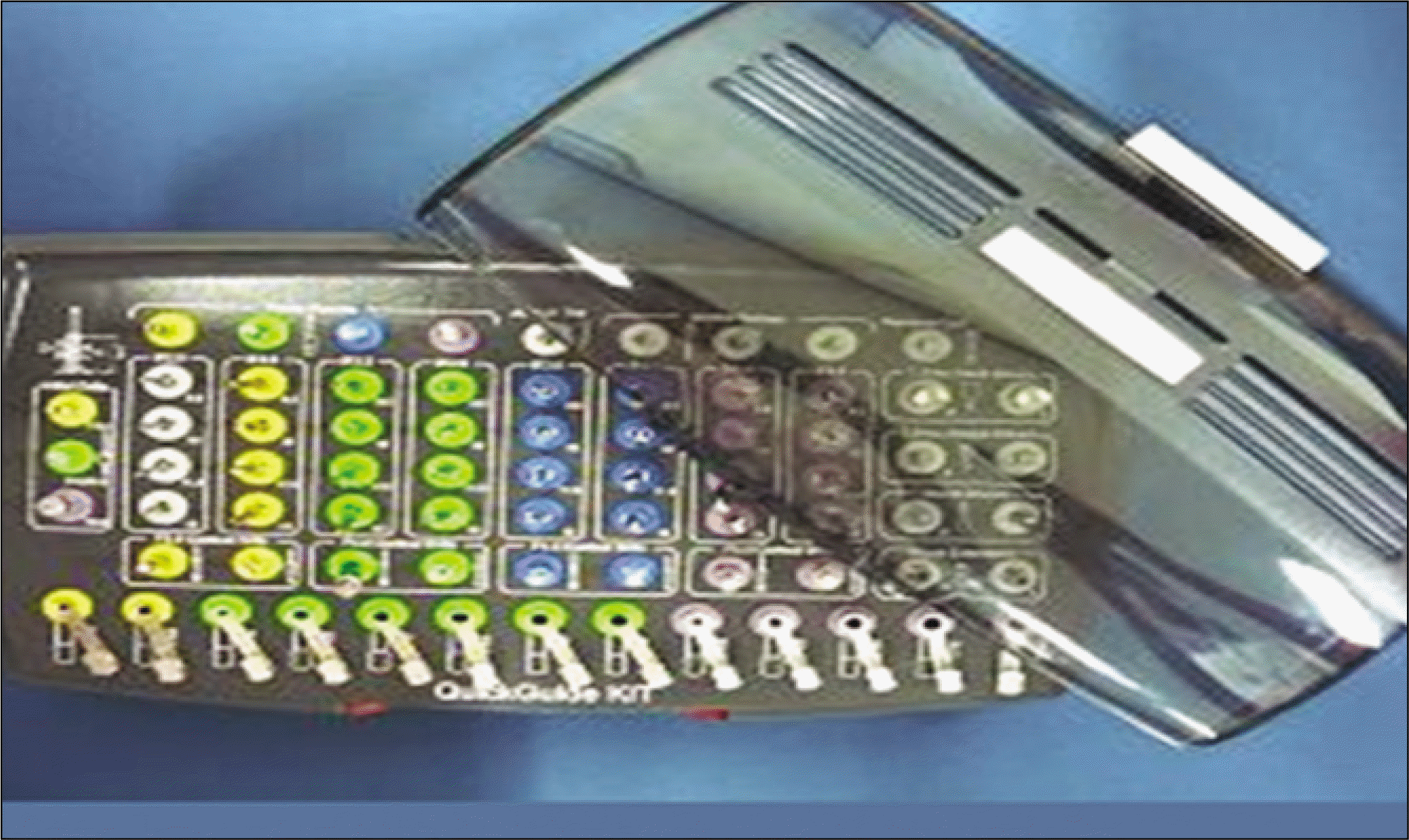
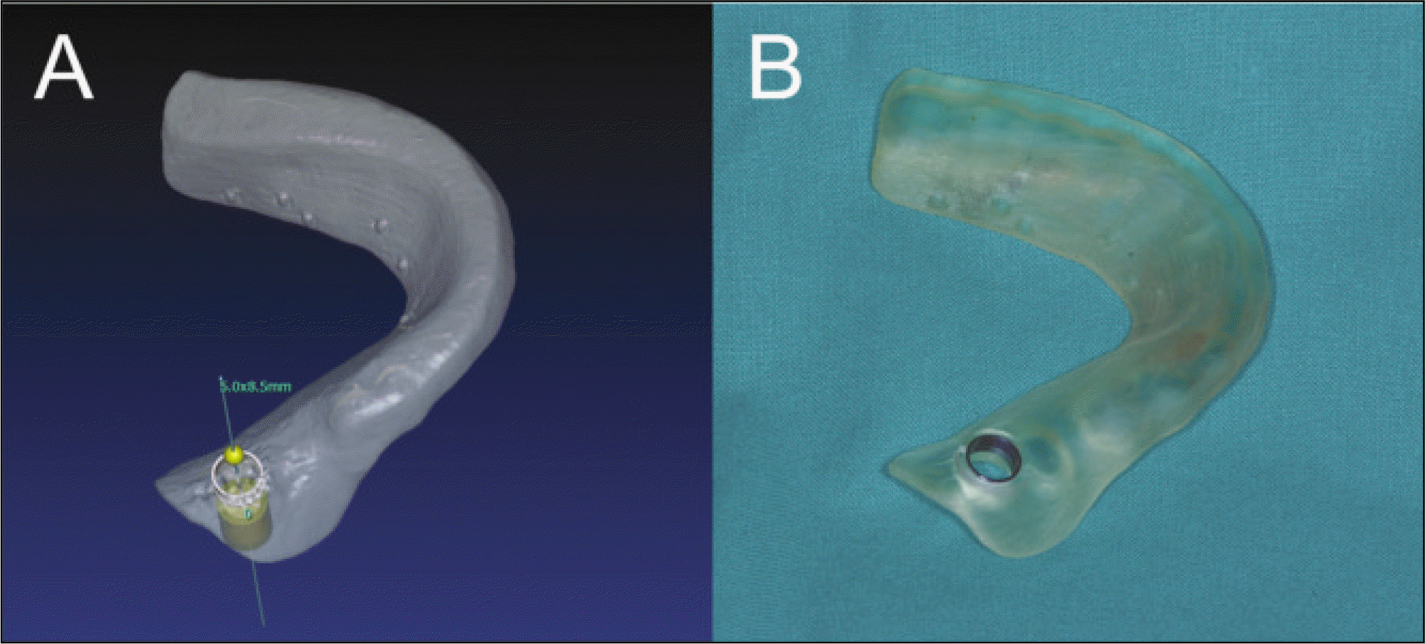 | Fig. 2.Fabrication of the stereolithographic (SLA) surgical guide based on the computer simulation. A. Virtual SLA surgical stent on the computer program. B. Real SLA surgical stent made by rapid prototyping technique with metal sleeve for drill guide and implant fixture. |
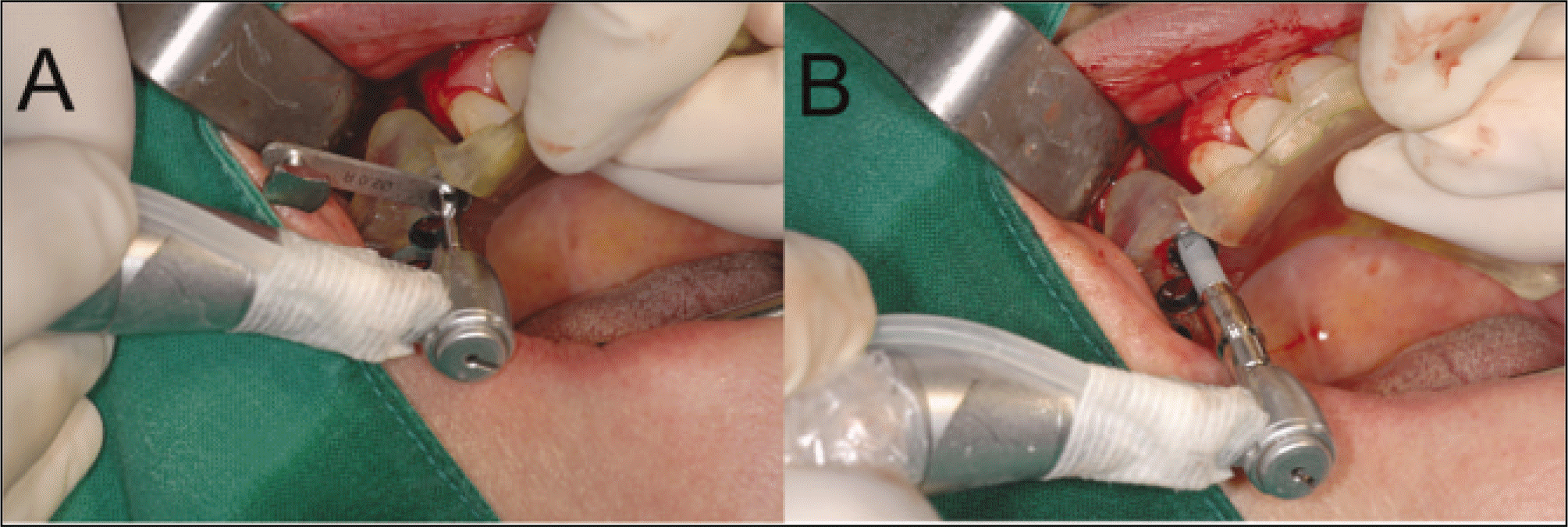 | Fig. 4.Implant site preparation by the guidance of stereolithographic (SLA) surgical stent. A. Sequential drilling through the metal sleeve. B. Guiding of metal sleeve for the fixture insertion. |
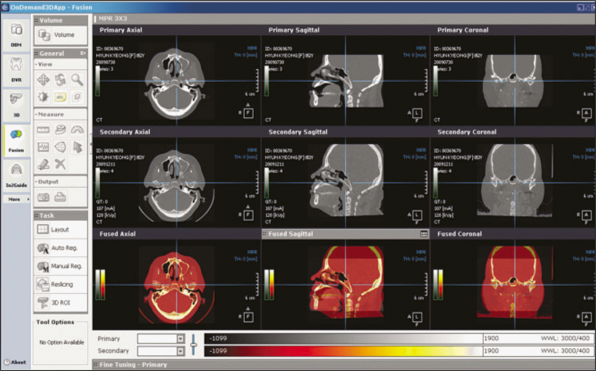 | Fig. 5.Image fusion processing for the comparison of implant positions between the planned and the placed. |
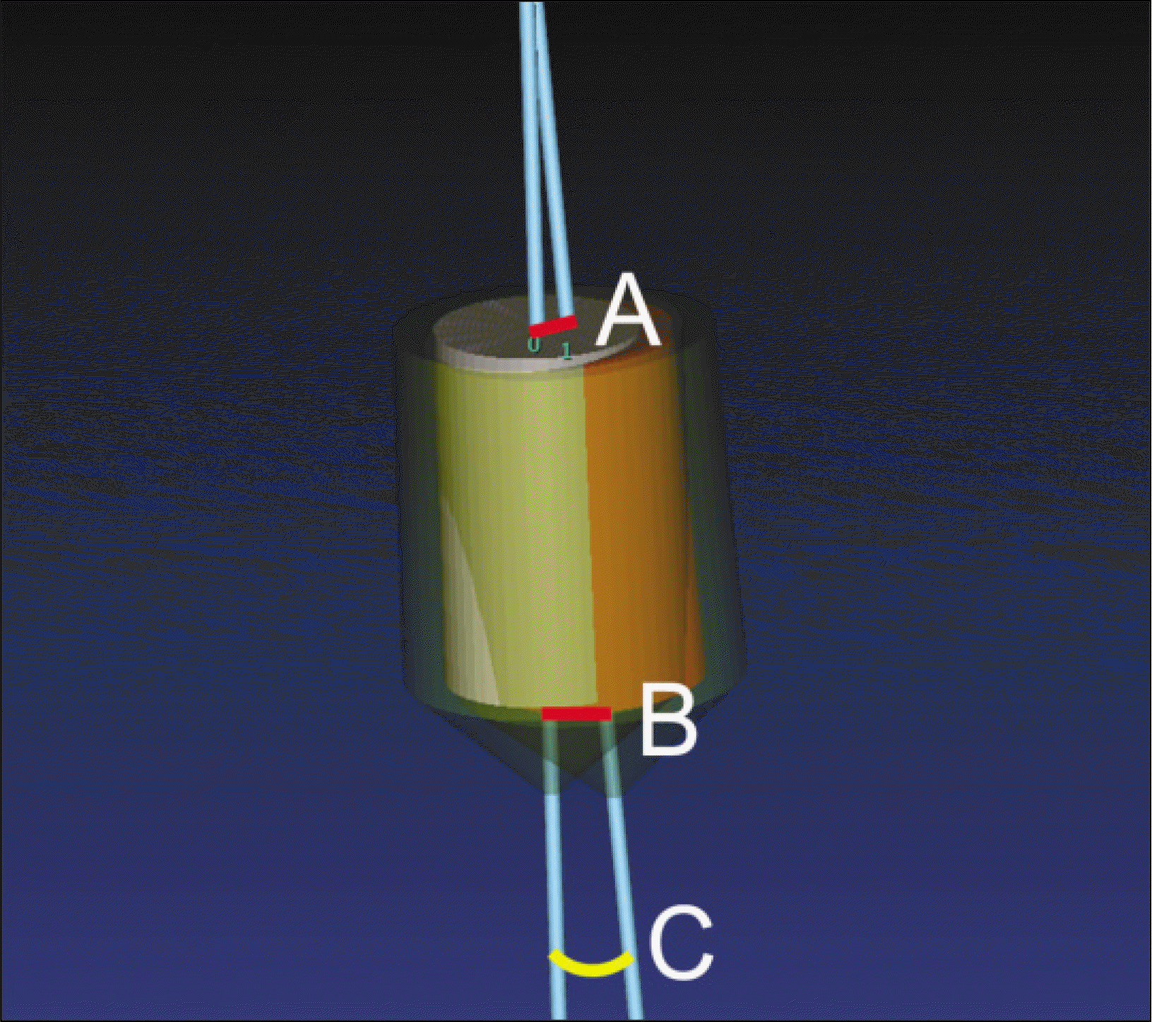 | Fig. 6.The result of image fusion of two implants. A. Occlusal difference of fixture center (mm). B. Apical difference of fixture center (mm). C. Axis difference of implant fixtures (�). |
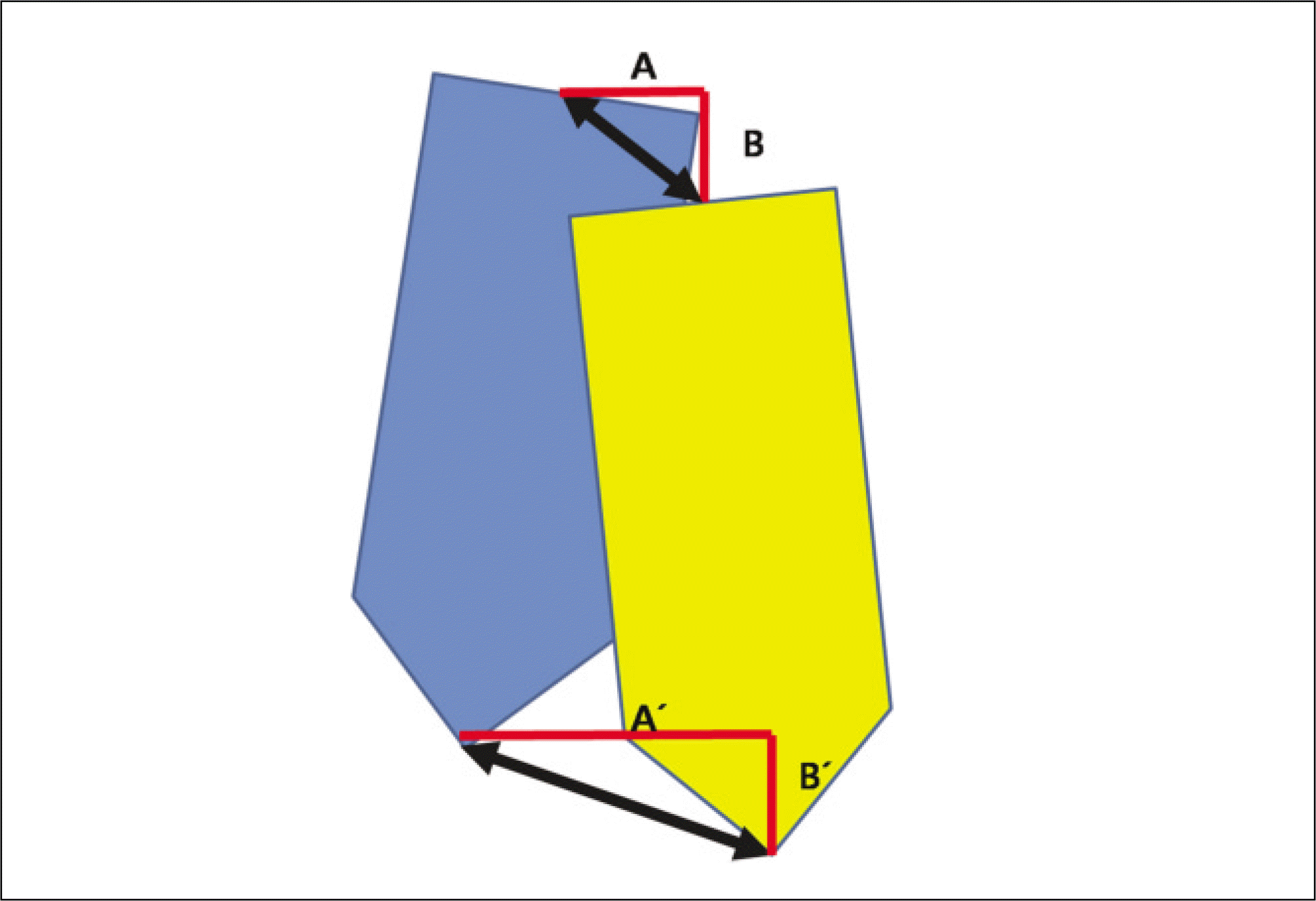 | Fig. 7.The disassembling of errors into the two directions between two implant fixtures. A(A´). Lateral error of the occlusal (apical) center. B(B´). Vertical error of the occlusal (apical) center. |
Table 1.
Demographic data of the patients for implant surgery
Table 2.
The differences of errors between the maxilla and mandible for the angle and the center of occlusal and apical points
Table 3.
The differences of errors according to the positions of implants for the angle and the center of occlusal and apical points


 PDF
PDF ePub
ePub Citation
 Print
Print


 XML Download