

 PDF
PDF ePub
ePub Citation
 Print
Print


Abstract
Introduction
In the management of dentofacial deformities, variable movement of the maxilla can be made possible by a Le Fort I osteotomy. Posterior impaction of the maxilla necessary for rotation of the maxillomandibular complex enhances the functions and esthetic results. In cases of posterior impaction of the maxilla, an increase in the figure of the occlusal plane angle and incisor inclination can occur. This study reports the relationship between the amount of posterior impaction and the change in the occlusal plane angle and incisor inclination in a Le Fort I osteotomy by preoperative and postoperative lateral cephalograms.
Materials and Methods
Twenty patients who had undergone orthognathic surgery in Dong-A University Medical Center participated in this study. Lateral cephalometrics, within 3 weeks prior to surgery and 3 days after surgery, were used for analysis. Pre and postoperative measurements of the occlusal plane angle and incisal inclination based on the Frankfort horizontal (FH) plane were performed. X and Y were defined as the amount of vertical change in the upper incisor tip and the amount of vertical change in the upper first molar mesial cup tip through the operation. The amount of final posterior maxillary impaction was determined by subtracting Y from X, which is the difference in vertical height. According to the amount of posterior maxillary impaction, the change in the occlusal plane angle and incisal inclination was measured.
Results
The average posterior maxillary impaction was 2.91 mm and the average change in the occlusal plane angle and incisal inclination was 6.54�after surgery. As a result, each mm of posterior maxillary impaction changed the occlusal plane angle and incisal inclination by 2.25�. Statistically, there was high significance. Two cases were observed: one with the same amount of posterior maxillary impaction performed on both the right and left showing 2.20�, and the other with a different amount of posterior maxillary impaction performed showing 2.35�. In this case, there was no significance difference between the two cases.
Conclusion
Each mm of posterior maxillary impaction changes the occlusal plane angle and incisal inclination by an average of 2.25�. In posterior maxillary impaction, there was no significant difference in the amount of change in the occlusal plane angle and incisal inclination regardless of whether there was an equal amount of posterior maxillary impaction on both sides. This study is expected to help in the presurgical orthodontic preparation and presurgical treatment planning.
Go to : 
References
1. Blair VP. Operations on the jaw bone and face: a study of etiology and pathological anatomy of developmental malrelations of the maxilla and mandible to each other and to facial outline and of their operative treatment when beyond the scope of the orthodontist. Surg Gynecol Obstet. 1907; 4:67–78.
2. Epker BN, Fish LC. The surgical orthodontic correction of class III skeletal open-bite. Am J orthod. 1978; 73:601–18.
3. Choi SW, Park HS, Cha IH. A study on accuracy of the maxillary repositioning in orthognathic surgery by the external measuring technique. J Korean Assoc Oral Maxillofac Surg. 1996; 22:537–43.
4. Rotter BE, Zeitler DL. Stability of the Le Fort Ⅰ maxillary osteotomy after rigid internal fixation. J Oral Maxillofac Surg. 1999; 57:1080–8. discussion 1089.
5. Moser K, Freihofer HP. Long-term experience with simultaneous movement of the upper and lower jaw. J Maxillofac Surg. 1980; 8:271–7.

6. Choi BH, Yoon JH. Soft tissue changes associated with Le Fort I maxillary advancement. J Korean Assoc Oral Maxillofac Surg. 1984; 10:175–82.
7. Epker BN, Turvey T, Fish LC. Indication for simultaneous mobilization of the maxilla and mandible for the correction of dentofacial deformities. Oral Surg Oral Med Oral Pathol. 1982; 54:369–81.
8. Turvey TA. Simultaneous mobilization of the maxilla and mandible: surgical technique and results. J Oral Maxillofac Surg. 1982; 40:96–9.
9. Sarver DM, Weissman SM, Johnston MW. Diagnosis and treatment planning of hypodivergent skeletal pattern with clockwise occlusal plane rotation. Int J Adult Orthodon Orthognath Surg. 1993; 8:113–21.
10. Naini FB, Hunt NP, Moles DR. The relationship between maxillary length, differential maxillary impaction, and the change in maxillary incisor inclination. Am J Orthod Dentofacial Orthop. 2003; 124:526–9.
11. Turvey T, Hall DJ, Fish LC, Epker BN. Surgical-orthodontic treatment planning for simultaneous mobilization of the maxilla and mandible in the correction of dentofacial deformities. Oral Surg Oral Med Oral Pathol. 1982; 54:491–8.
12. Wolford LM, Chemello PD, Hilliard FW. Occlusal plane alteration in orthognathic surgery. J Oral Maxillofac Surg. 1993; 51:730–40.
13. Lee JH, Lee HJ. Stability of occlusal plane after Le Fort I maxillary osteotomy in patients with skeletal class III maloccusion. J Korean Assoc Oral Maxillofac Surg. 1996; 22:429–36.
14. Reyneke JP, Evans WG. Surgical manipulation of the occlusal plane. Int J Adult Orthodon Orthognath Surg. 1990; 5:99–110.
15. Reitzik M. Skeletal and dental changes after surgical correction of mandibular prognathism. J Oral surg. 1980; 38:109–16.
16. Swinnen K, Politis C, Willems G, de Bruyne I, Fieuws S, Heidbuchel K, et al. Skeletal and dentoalveolar stability after surgical orthodontic treatment of anterior open bite: a retrospective study. Eur J Orthod. 2001; 23:547–57.
Go to :
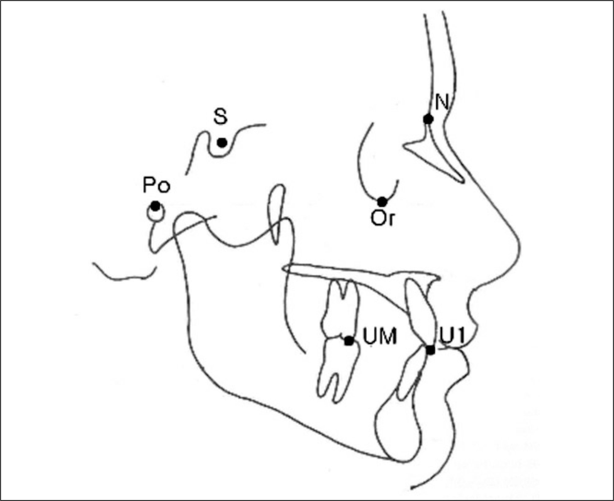 | Fig. 1.Anatomic landmark used in this study. (S: sella, N: nasion, Po: porion, Or: orbitale, UM: upper fist molar mesial cusp tip, U1: upper incisor cusp tip) |
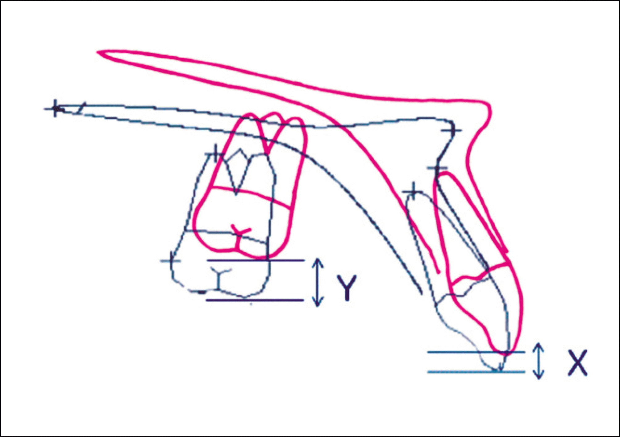 | Fig. 2.Measuring parameters. (X: vertical change of upper incisor, Y: vertical change of upper first molar, Y-X: amount of posterior Mx. impaction) |
Table 1.
Average occlusal plane angle and incisal inclination in presurgical state
| Mean | Range | |
|---|---|---|
| occlusal plane angle | 7.2 | −1.75–12.24 |
| incisal inclination | 122.46 | 114.31–132.7 |
Table 2.
Average amount of posterior impaction and average amount of change of occlusal plane angle, incisal inclination after surgery
| Amount | Mean | Range | SD |
|---|---|---|---|
| PI | 2.91 | 0.26–7.75 | 1.78 |
| OPA, II change | 6.54 | 0.5–16 | 3.77 |
| OPA, II change due to PI | 2.25* | 1.5–3.1 | 0.39 |
Table 3.
Average amount of change of occlusal plane angle, incisal inclination in same posterior impaction and differential posterior impaction
| Mean | SD | N | P value | |
|---|---|---|---|---|
| Same posterior impaction | 2.2 | 0.27 | 10 | 0.393∗ |
| Differential posterior impaction | 2.35 | 0.38 | 10 |
 XML Download