Abstract
The management of teeth in the line of a mandibular fracture is controversial despite the general agreement that most of these teeth can be preserved. Teeth should be retained if bony attachments are adequate for survival, the tooth is sound and important in maintaining fixation of the fractured segment of bone. Teeth should be removed if they are loose and interfere with the reduction of fragments, are devitalized and potentially a source of wound infection, are damaged beyond their usefulness or may become devital and interfere with healing by becoming infected. However, tooth removal will increase the level of trauma, extend the severity of the wound and require expensive prosthetic treatment. Therefore, it is very important to conserve infected teeth in the line of a mandibular fracture through early primary endodontic treatment (pulp extirpation, canal enlargement and canal opening drainage) and splinting. The basic principles underlying the treatment of pulpless teeth are those underlying general surgery. Therefore, debridement of the infected wound (pulp extirpation and canal enlargement), drainage (canal opening) and gentle treatment of the tissues (occlusal reduction and teeth splinting) are the principles of surgery. This is a representative case report of conservative care by the early endodontic drainage of infected teeth in the line of a mandibular fracture.
Go to : 
References
1. Kahnberg KE, Ridell A. Prognosis of teeth involved in the line of mandibular fractures. Int J Oral Surg. 1979; 8:163–72.
2. Kamboozia AH, Punnia-Moorthy A. The fate of teeth in mandibular fracture lines. A clinical and radiographic follow-up study. Int J Oral Maxillofac Surg. 1993; 22:97–101.
3. de Amaratunga NA. The effect of teeth on the line of mandibular fractures on healing. J Oral Maxillofac Surg. 1987; 45:312–4.
4. Neal DC, Wagner WF, Alpert B. Morbidity associated with teeth in the line of mandibular fractures. J Oral Surg. 1978; 36:859–62.
6. Schneider SS, Stern M. Teeth in the line of mandibular fractures. J Oral Surg. 1971; 29:107–9.
7. Lieblich SE, Topazian RG. Infection in the patient with maxillofacial trauma. Fonesca RJ, Walker RV, editors. Oral and Maxillofacial Trauma. Philadelphia: Saunders;1991. p. 1150–71.

8. Dingman RO, Izenberg PH. Complication of facial trauma. Conley JJ, editor. Complications of head and neck surgery. 1st ed.Philadelphia: WB Saunders;1979. p. 358–70.
9. Grossman LI. Endodontic practice. 8th ed.Philadelphia: Lea & Febiger;1974.
10. Lim SS. Clinical endodontics. 1st ed.Seoul: Dental and Medical Publishing;1994. p. 1–15.
11. Kim KW, Kim SK, Kim YG, Kim JR, Kim HG, Min SK, et al. Oral and maxillofacial infections. 1st ed.Seoul: Jee Seung Publishing;2007. p. 248–63.
12. Prein J, Beyer M. Management of infection and nonunion in mandibular fractures. Oral Maxillofac Clin North Am. 1990; 2:187–94.

13. Munster AM. Immunologic response of trauma and burns. An overview. Am J Med. 1984; 76:142–5.
14. Goldberg MH. Prevention and control of infection in the surgical patient. Topazian RG, Goldberg MH, editors. Management of infections of the oral and maxillofacial regions. 1st ed.Philadelphia: WB Saunders;1981. p. 329–42.
15. Koury ME. Complications in the treatment of mandibular fractures. Kaban LB, Pogrel MA, Perrott DH, editors. Complications in oral and maxillofacial surgery. 1st ed.Philadelphia: Saunders;1997. p. 121–63.
16. Shetty V, Freymiller E. Teeth in the line of fracture: a review. J Oral Maxillofac Surg. 1989; 47:1303–6.

Go to : 
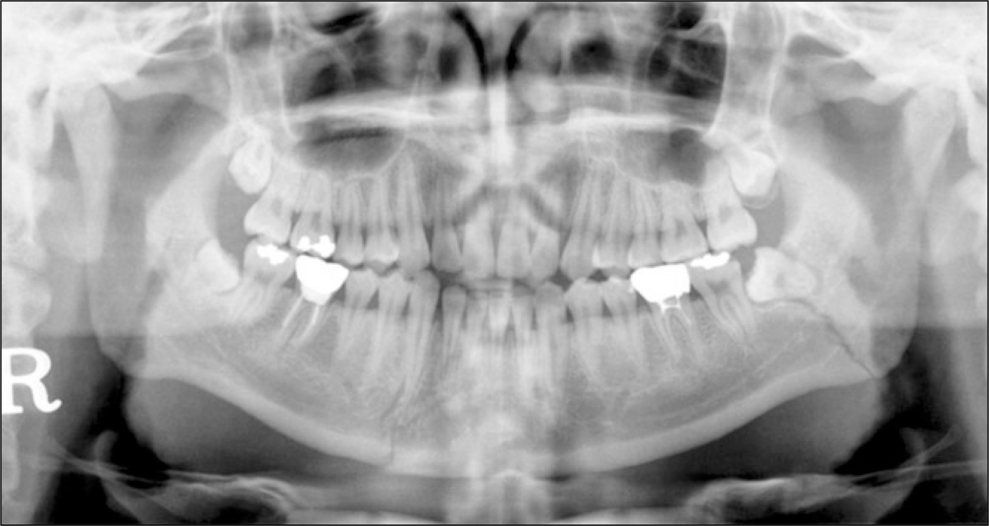
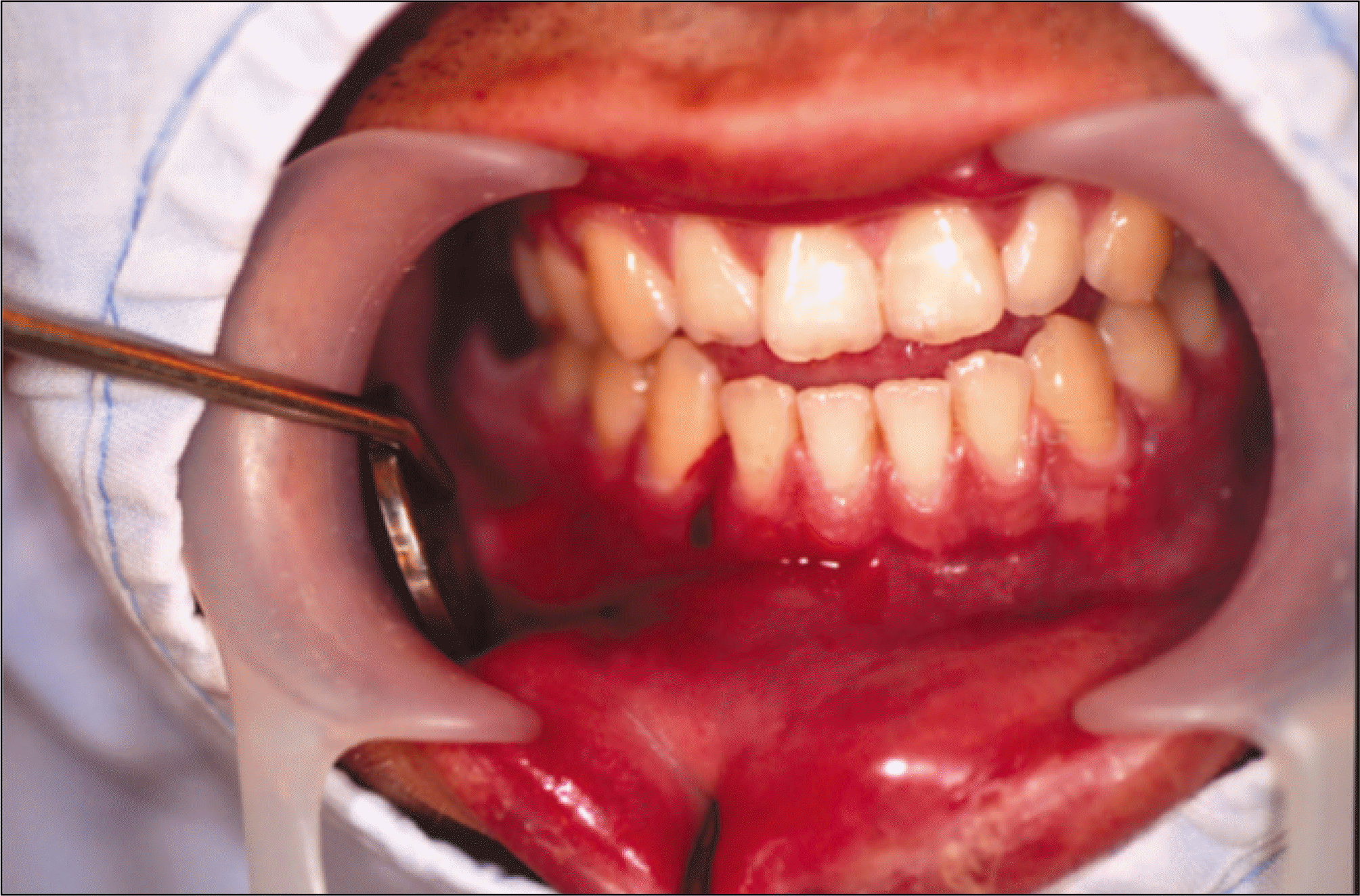 | Fig. 1.Initial view of the infected tooth #42 in the line of a mandibular symphysis fracture. |
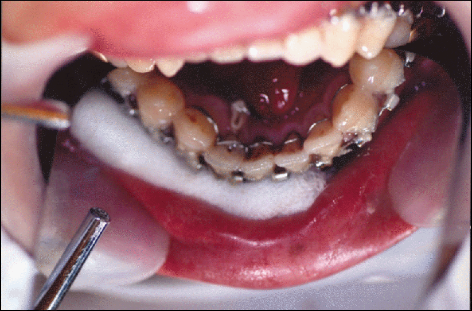
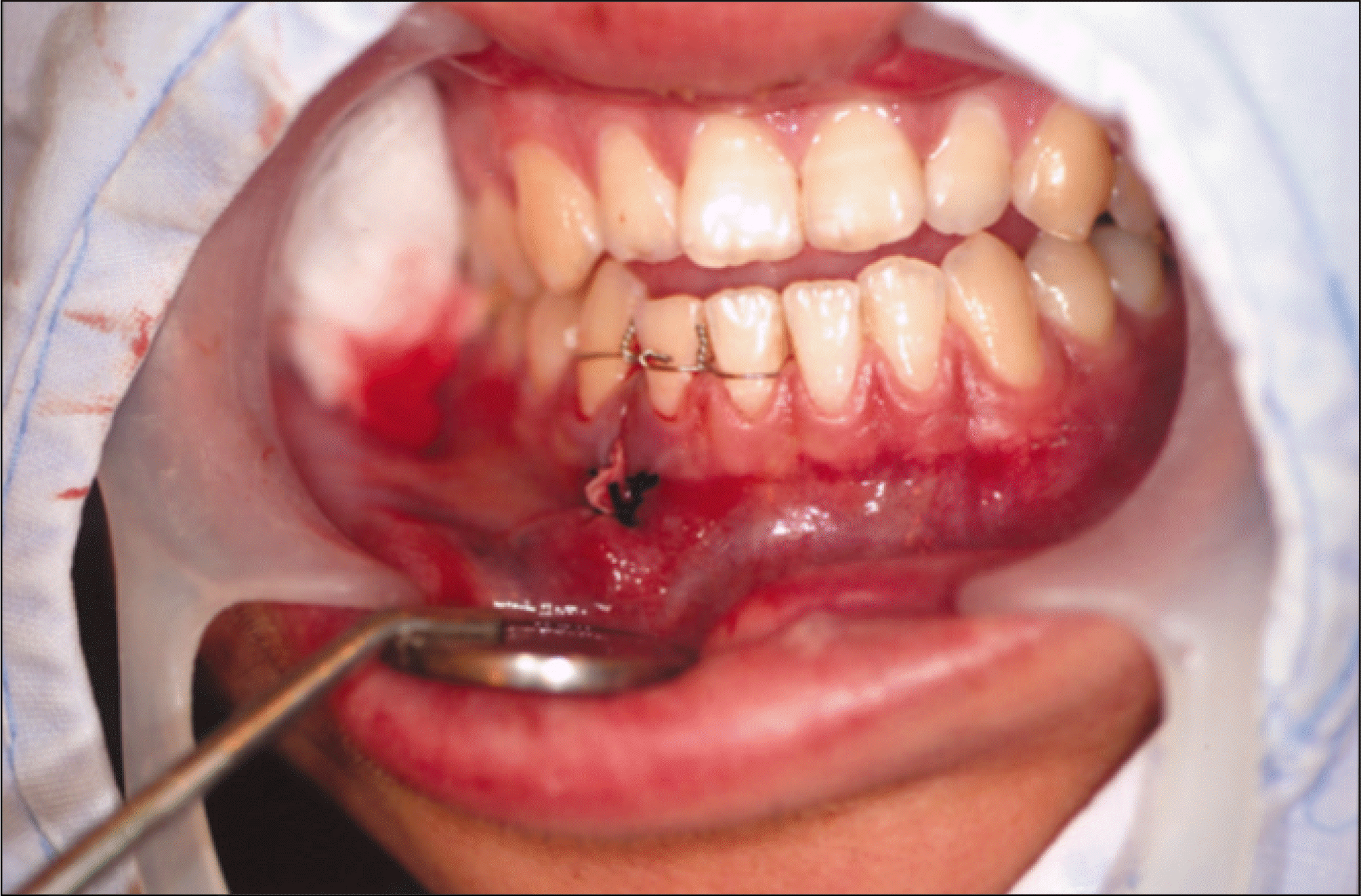 | Fig. 3.Primary wiring and endodontic drainage view of #42 and labial incision and drainage view for the prevention of infection of a mandibular symphysis fracture. |
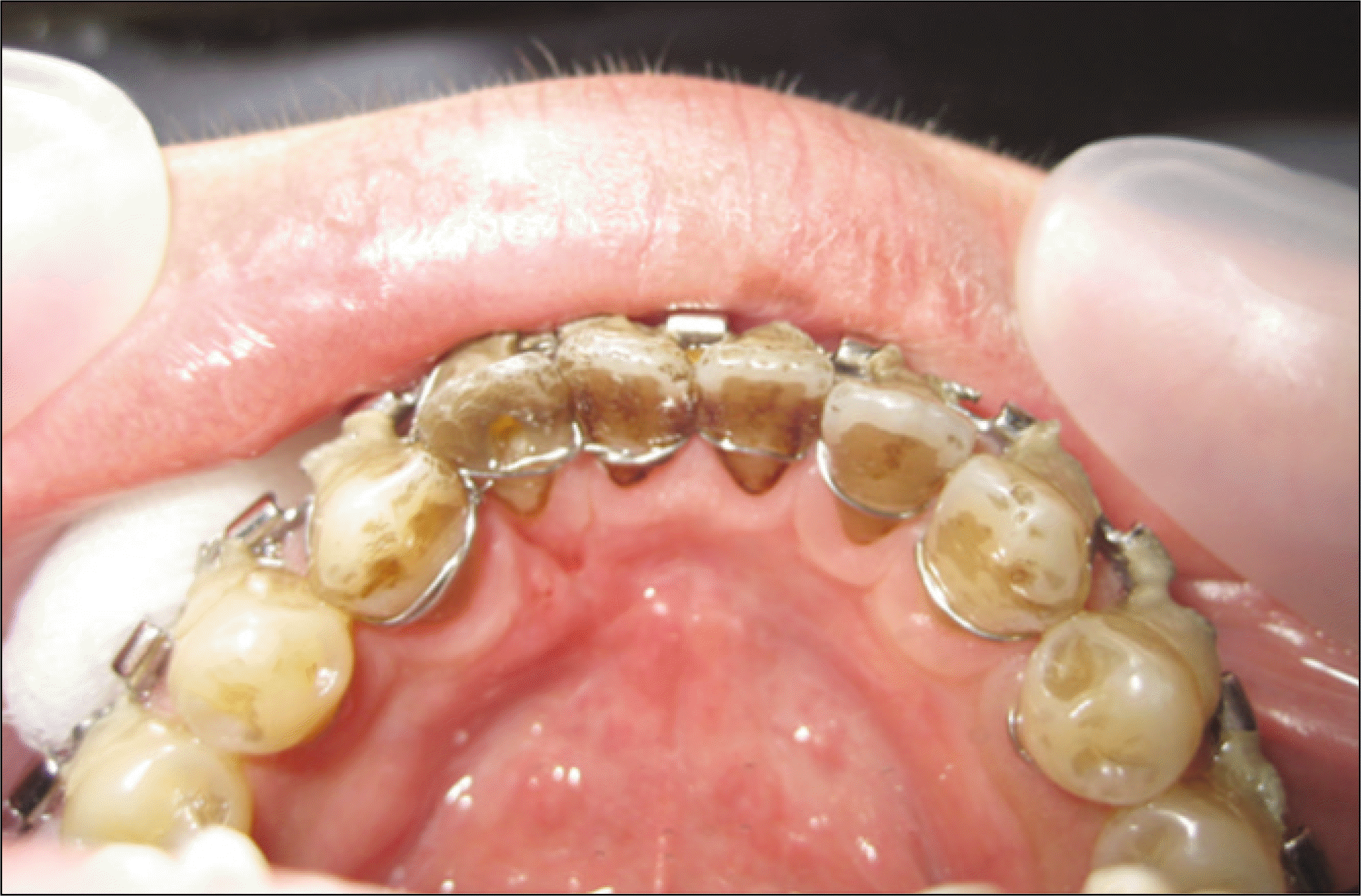
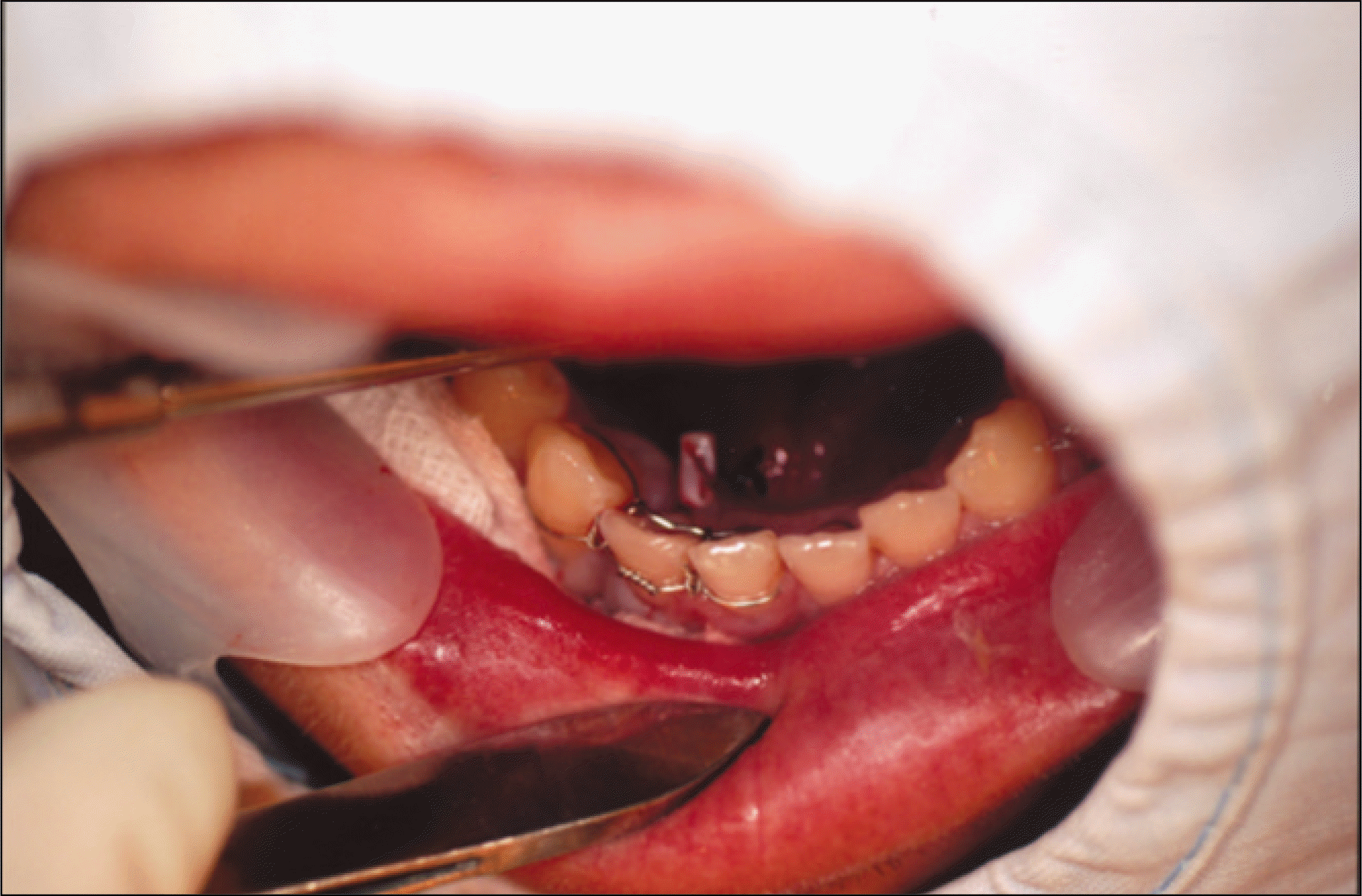 | Fig. 4.Lingual incision and drainage (sutured rubber strip) view for the prevention of infection of a mandibular symphysis fracture. |
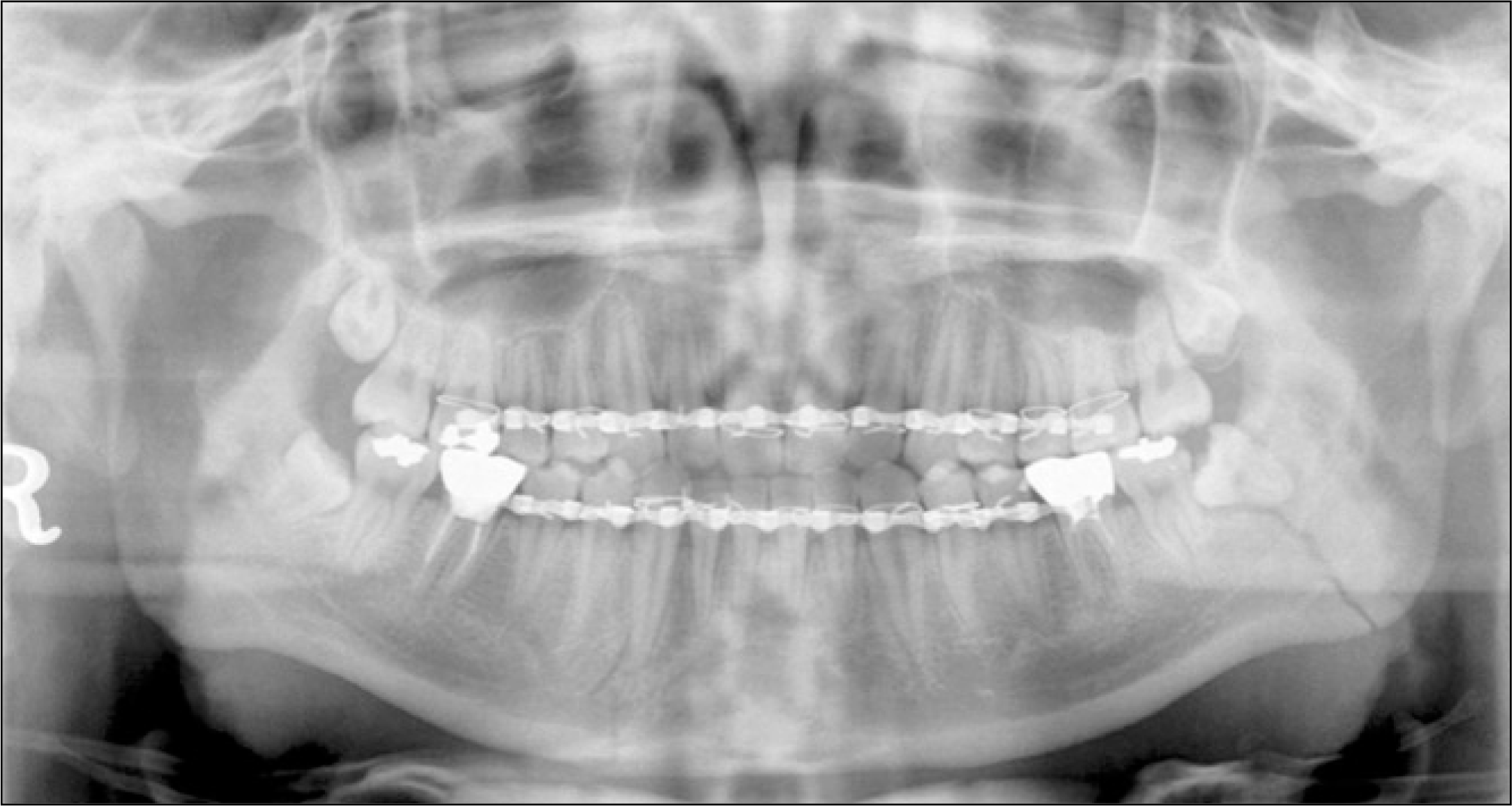
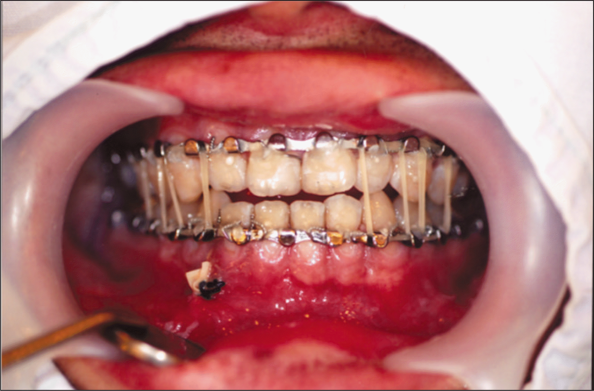 | Fig. 5.Closed reduction view of arch bar application and intermaxillary fixation in a mandibular fracture |
Table 1.
Factors predisposing to infection of mandibular fracture site
Table 2.
Indications and contraindications of extraction on teeth in the line of a mandibular fracture
Table 3.
Contemporary indications of extraction on tooth in the line of a mandibular fracture


 PDF
PDF ePub
ePub Citation
 Print
Print


 XML Download