

 PDF
PDF ePub
ePub Citation
 Print
Print


Abstract
Excessive oral and maxillofacial bleeding causes upper airway obstruction, bronchotracheal and gastric aspiration and hypovolemic shock. Therefore, the rapid and correct bleeding control is very important for saving lives in the emergency room. Despite the conventional bleeding control methods of wiring (jaw fracture, wound suture and direct pressure), continuous bleeding can occur due to the presence of various bleeding disorders. There are five main causes for excessive bleeding disorders in the clinical phase; (1) vascular wall alteration (infection, scurvy etc.), (2) disorders of platelet function (3) thrombocytopenic purpura (4) inherited disorders of coagulation, and (5) acquired disorders of coagulation (liver disease, anticoagulant drug etc.). In particular, infections can alter the structure and function of the vascular wall to a point at which the patient may have a clinical bleeding problem due to vessel engorgement and erosion. Wound infection is a frequent cause of postoperative active bleeding. To prevent postoperative bleeding, early infection control using a wound suture with proper drainage establishment is very important, particularly in the active bleeding sites in a contaminated emergency room. This is a case report of a rational bleeding control method by rapid wiring, wound suture with drainage of a rubber strip & iodoform gauze and wet gauze packing, in a 26-year-old male cerebral palsy patient with active oral and maxillofacial bleeding injuries caused by a traffic accident.
Go to : 
References
1. Conley JJ. Blood vessel complications, In: Conley JJ, ed. Complications of head and neck surgery. 1st ed.Philadelphia: WB Saunders;1979. p. 66–80.
2. Kruger GO. Textbook of oral and maxillofacial surgery. 6th ed.St. Louis: Mosby;1984.
3. Little JW, Falace DA, Miller CS, Rhodus NL. Bleeding disorders. Little JW, Falace DA, Miller CS, Rhodus NL, editors. Dental Management of the Medically Compromised Patient. St. Louis: Mosby;2002. p. 332–64.
4. Dembo JB. Diagnosis and management of oral surgical complications. Falance DA, editor. Emergency dental care: diagnosis and management of urgent dental problems. 1st ed.Baltimore: Lippincott Williams and Wilkins;1995. p. 227–53.
5. Koury ME. Complications in the treatment of mandibular fractures. Kaban LB, Pogrel MA, Perrott DH, editors. Complications in oral and maxillofacial surgery. 1st ed.Philadelphia: WB Saunders;1997. p. 121–63.
6. Goldberg MH. Prevention and control of infection in the surgical patient. Topazian RG, Goldberg MH, editors. Management of infections of the oral and maxillofacial regions. 1st ed.Philadelphia: WB Saunders;1981. p. 329–50.
7. Alling CC 3rd, Alling RD. Bleeding disorders and injuries. Dent Clin North Am. 1982; 26:71–86.
8. Schultz RC. The problems of beginning. Schultz. RC, editor. Facial Injuries. 2nd ed.Chicago: Year Book Medical Publishers;1977. p. 41–64.
9. Min BI. Color atlas of maxillofacial plastic surgery. Seoul: Koon Ja Publishing;1990.
10. Bonn GE, Davis CL. Shock. Fonesca RJ, Walker RV, editors. Oral and maxillofacial trauma. Philadelphia: WB Saunders;1991. p. 58–73.
11. Yoo JH, Kang SH, Kim HS, Kim JB. A clinical study on the emergerncy patients with active oral bleeding. J Korean Assoc Oral Maxillofac Surg. 2002; 28:383–9.
12. Yoo JH, Jung IW. Hypovolemic shock owing to maxillofacial injury-Review of references and report of cases. J Korean Assoc Maxillofac Plast Reconstr Surg. 1988; 10:7–18.
13. Bartlett JG, Gorbach SL. The triple threat of aspiration pneumonia. Chest. 1975; 68:560–5.

14. London PS, Rowe NL, Williams JL. Definitive clinical examination. Rowe NL, Williams JL, editors. Rowe and Williams' maxillofacial injuries. 2nd ed.Edinburgh: Churchill Livingstone;1994. p. 93–148.
15. Douglas WW. Homeostasis: body changes in trauma and surgery. Sabiston DC, editor. Textbook of surgery. Philadelphia: WB Saunders;1986. p. 23–37.
16. Kim GB, Kim CG, Lee YG, Jang ST. Contemporary general surgery. 1st ed.Seoul: Il Cho Gak Publishing;1987. p. 34–51.
17. Assael LA and Ellis III E. Soft tissue and dentoalveolar injuries. Peterson LJ, Ellis III E, Hupp JR, Tucker MR, editors. Contemporary oral and maxillofacial surgery. Saint Louis: CV Mosby;1988. p. 527–55.
18. Kim JB, Chung WG, Noh HJ, Jang SO, Yoo JH, Han SK, et al. A clinical study on the care of odontogenic infections in the patients with major bleeding disorders. J Korean Assoc Oral Maxillofac Surg. 2003; 29:330–7.
19. Hamilton GC. Disorders of hemostasis and polycythemia. Rosen PL. Baker FJ, Barkin RM, editors. Emergency medicine, concepts and clinical practice. 2nd ed.St. Louis: CV Mosby;1987. p. 1647–64.
Go to :
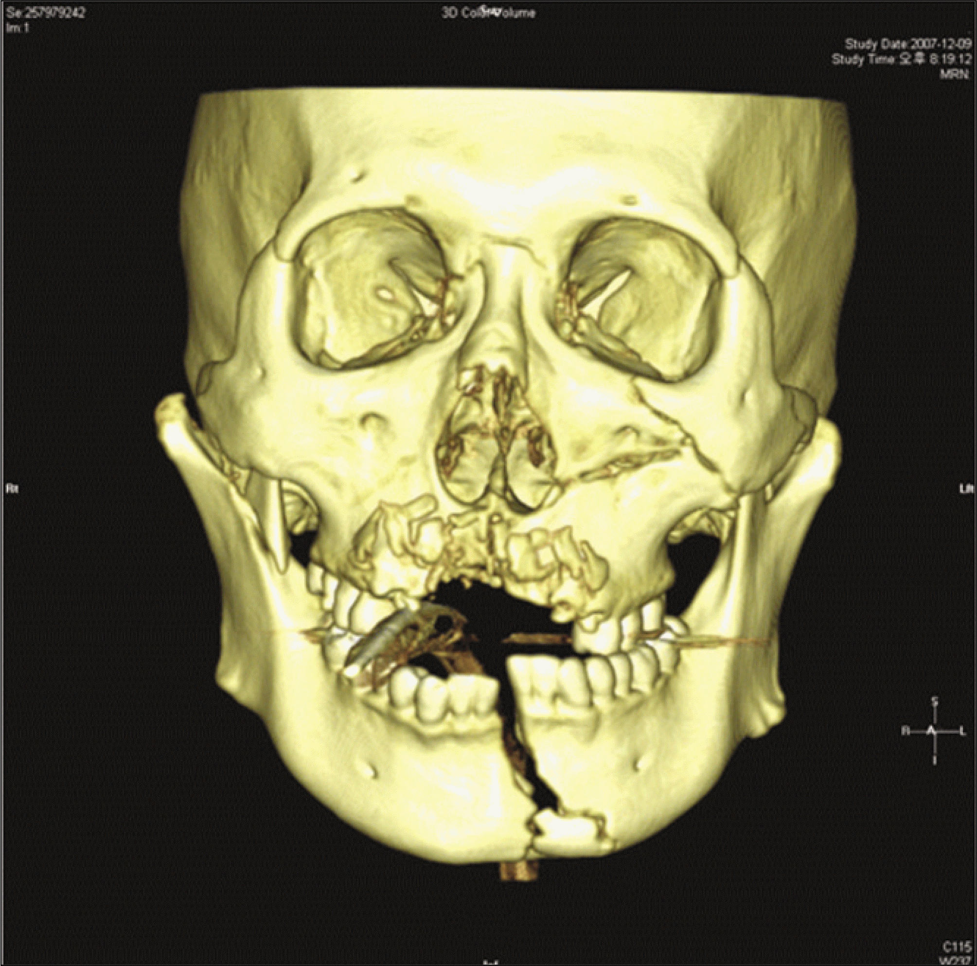 | Fig. 1.Initial 3D-CT view of mandibular and maxillary compound fracture. (3D-CT: 3 dimensional computed tomography) |
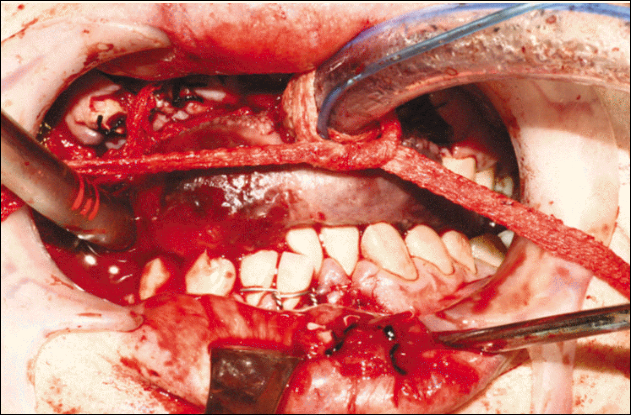
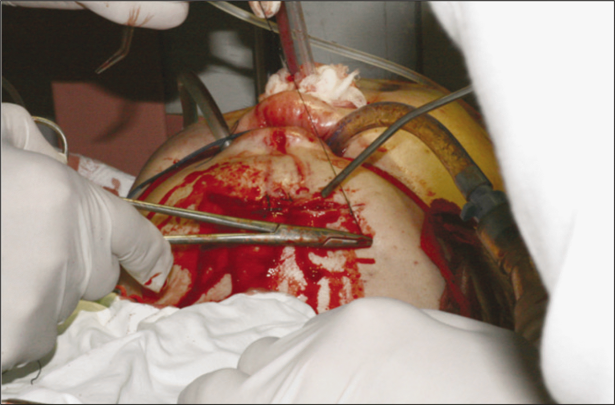
 | Fig. 3.Primary gauze packing and rough wound closure view by use of iodoform gauze, long wet gauze and black silk in the maxillary compound alveolar fracture regions. |
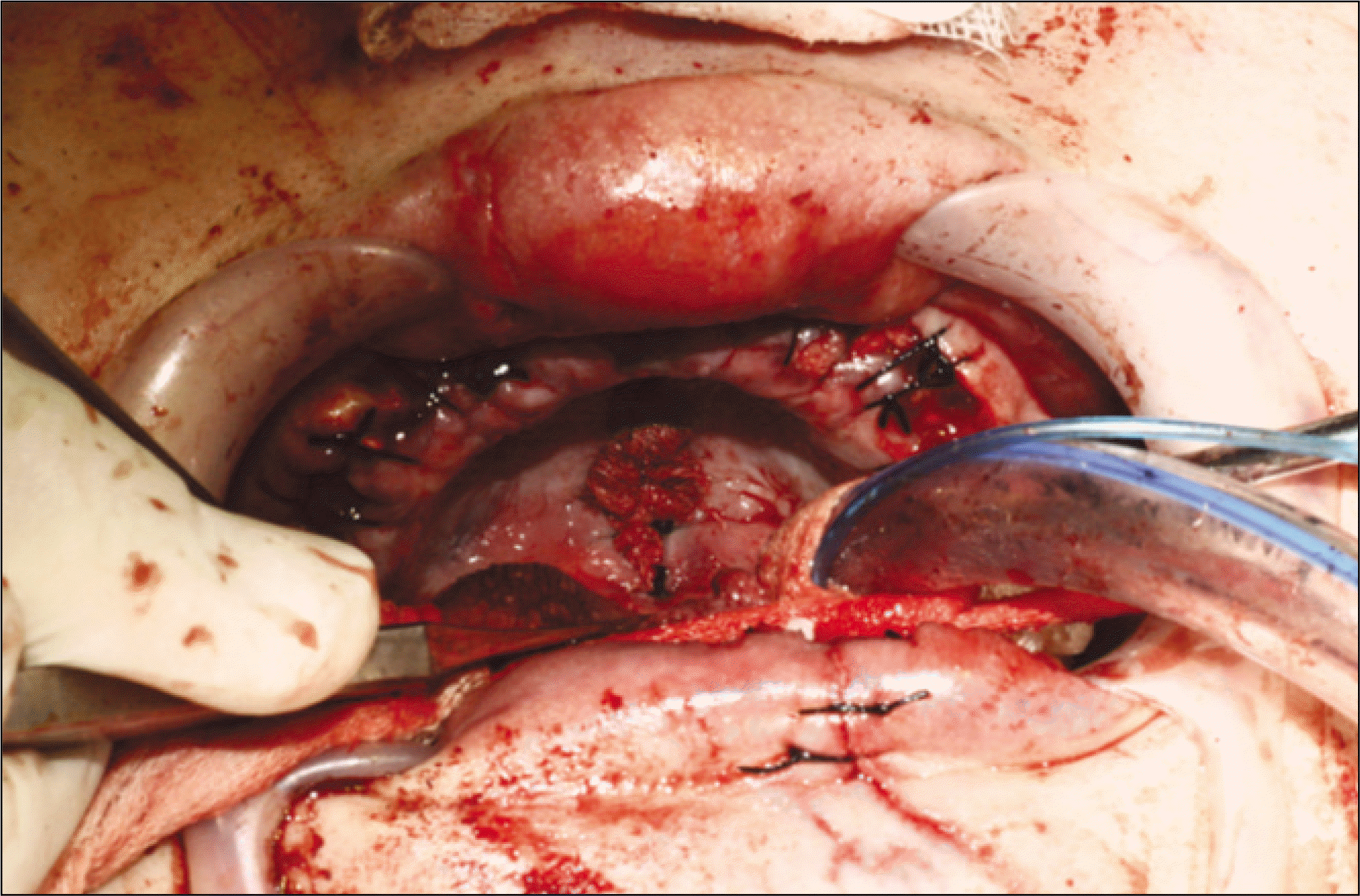
 | Fig. 4.Primary rough closure and rubber strips drainage view in the deep lacerated wounds of sublingual and mouth floor region. |
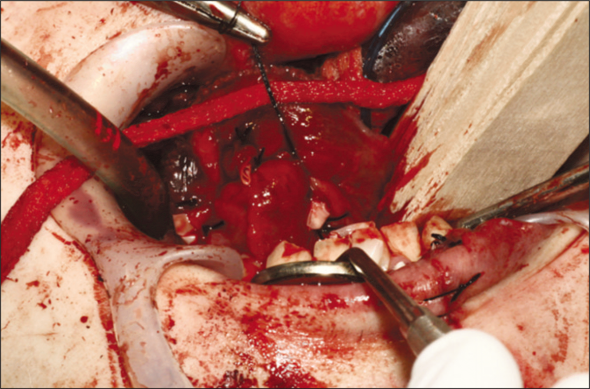
 | Fig. 5.Primary rough wound closure and rubber strips drainage view in the deep lacerated chin wounds. |
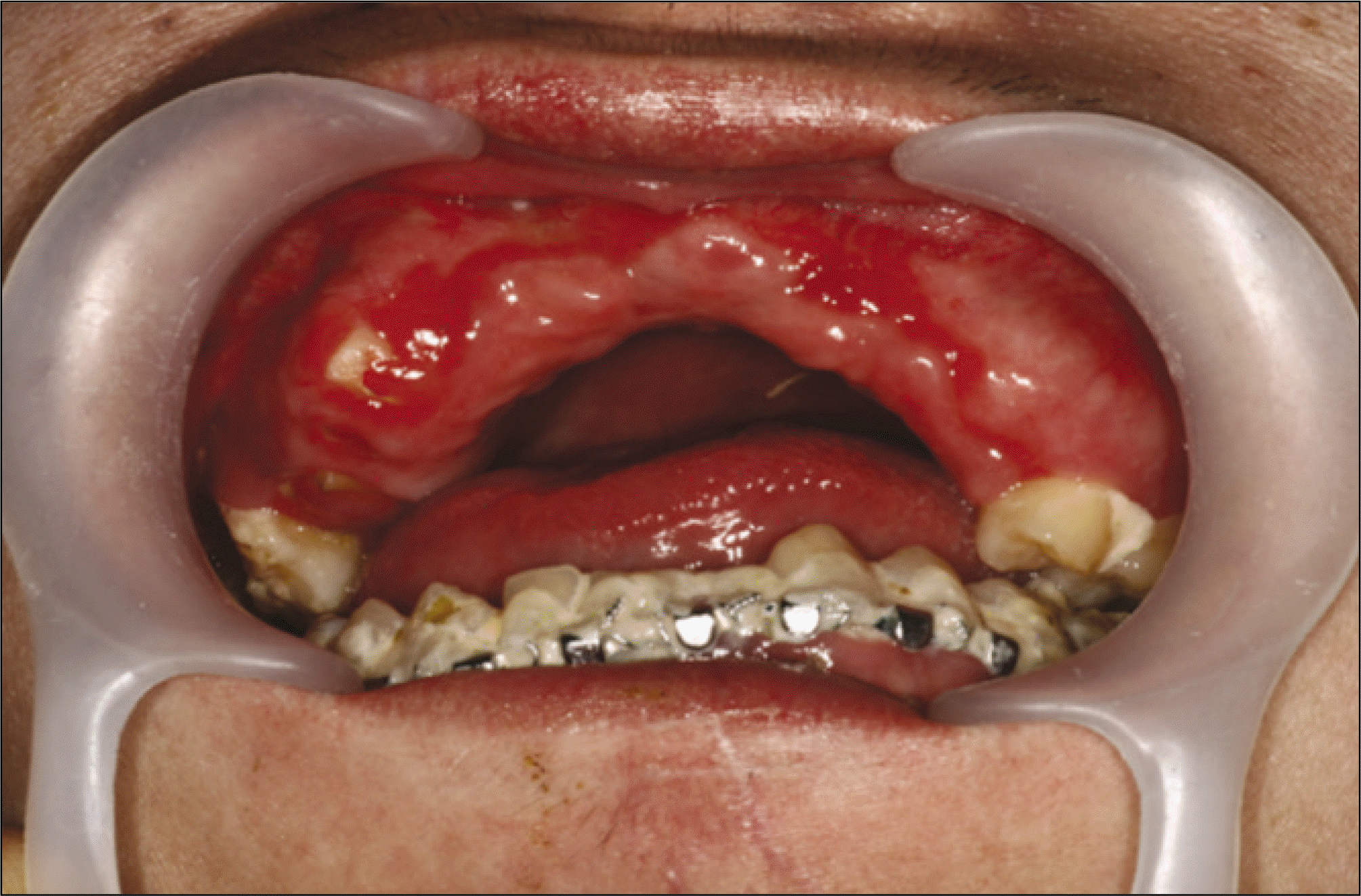 | Fig. 6.Follow-up intraoral view of the mandibular arch bar application and the maxillary residual root rests with good healing of the adjacent soft tissue wounds. |
 XML Download