Abstract
Introduction
To evaluate the skeletal stability after a bilateral sagittal split osteotomy (BSSO) setback of the mandible fixed with a biodegradable internal fixation device or metal internal fixation device.
Materials and Methods
Thirty consecutive patients underwent mandibular setback via BSSO. Fifteen patients were fixed with a biodegradable internal fixation device or metal internal fixation device respectively. Posteroanterior (PA) and lateral cephalograms were taken preoperatively and at two days, 5.5 months and 14.5 months postoperatively. The relevant skeletal points were traced and digitized to evaluate the skeletal changes postoperatively. The relapse rates were analyzed and compared statistically.
Go to : 
References
1. Becktor JP, Rebellato J, Sollenius O, Vedtofte P, Isaksson S. Transverse displacement of the proximal segment after bilateral sagittal osteotomy: a comparison of lag screw fixation versus miniplates with monocortical screw technique. J Oral Maxillofac Surg. 2008; 66:104–11.

2. Joss CU, Vassalli IM. Stability after bilateral sagittal split osteotomy advancement surgery with rigid internal fixation: a systematic review. J Oral Maxillofac Surg. 2009; 67:301–13.

3. Burstone CJ, James RB, Legan H, Murphy GA, Norton LA. Cephalometrics for orthognathic surgery. J Oral Surg. 1978; 36:269–77.
4. Blomqvist JE, Ahlborg G, Isaksson S, Svartz K. A comparison of skeletal stability after mandibular advancement and use of two rigid internal fixation techniques. J Oral Maxillofac Surg. 1997; 55:568–74.

5. Borstlap WA, Stoelinga PJ, Hoppenreijs TJ, van' t Hof MA. Stabilisation of sagittal split advancement osteotomies with miniplates: a prospective, multicentre study with two-year follow-up Part II. Radiographic parameters. Int J Oral Maxillofac Surg. 2004; 33:535–42.
6. Joss CU, Vassalli IM, Thu ¨er UW. Stability of soft tissue profile after mandibular setback in sagittal split osteotomies: a longitudinal and longterm follow-up study. J Oral Maxillofac Surg. 2008; 66:1610–6.

7. Kulkarni RK, Moore EG, Hegyeli AF, Leonard F. Biodegradable poly (lactic acid) polymers. J Biomed Mater Res. 1971; 5:169–81.
8. Suuronen R, Haers PE, Lindqvist C, Sailer HF. Update on bioresorbable plates in maxillofacial surgery. Facial plast Surg. 1999; 15:61–72.

9. Cutright DE, Hunsuck EE, Beasley JD. Fracture reduction using a biodegradable material, polylactic acid. J Oral Surg. 1971; 29:393–7.
10. Cutright DE, Hunsuck EE. The repair of fractures of the orbital floor using biodegradable polylactic acid. Oral Surg Oral Med Oral Pathol. 1972; 33:28–34.

11. Getter L, Cutright DE, Bhaskar SN, Augsburg JK. A biodegradable intraosseus appliance in the treatment of mandibular fractures. J Oral Surg. 1972; 30:344–8.
12. Turvey TA, Bell RB, Phillips C, Proffit WR. Self-reinforced biodegradable screw fixation compared with titanium screw fixation in mandibular advancement. J Oral Maxillofac Surg. 2006; 64:40–6.

13. Suuronen R, Pohjonan T, Hietanen J, Lindqvist C. A 5-year in vitro and in vivo study of the biodegradation of polylactide plates. J Oral Maxillofac Surg. 1998; 56:604–14.

14. Kallella I, Laine P, Suuronen R, Ranta P, Iizuka T, Lindqvist C. Osteotomy site healing following mandibular sagittal split osteotomy and rigid fixation with polylactide biodegradable screws. Int J Oral Maxillofac Surg. 1999; 28:166–70.

15. Turvey TA, Bell RB, Tejera TJ, Proffit WR. The use of self-reinforced biodegradable bone plates and screws in orthognathic surgery. J Oral Maxillofac Surg. 2002; 60:59–65.

16. Ueki K, Nakagawa K, Marukawa K, Takazakura D, Shimada M, Takatsuka S, et al. Changes in condylar long axis and skeletal sta-billity after bilateral sagittal split ramus osteotomy with poly-L-lactic acid or titanium plate fixation. Int J Oral Maxillofac surg. 2005; 34:627–34.
17. Ferretti C, Reyneke JP. Mandibular sagittal split osteotomies fixed with biodegradable or titanium screws: a prospective, comparative study of postoperative stability. Oral Surg Oral Med Oral Pathol Oral Radiol Endod. 2002; 93:534–7.

18. Wood GD. Inion biodegradable plates: the first century. Br J Oral Maxillofac Surg. 2006; 44:38–41.
19. Kim IH, Han CH, Ryu SY. Changes in gonial angle and mandibular width after orthognathic surgery in mandibular prognathic patients. J Korean Assoc Oral Maxillofac Surg. 2006; 32:129–37.
20. Costa F, Robiony M, Politi M. Stability of sagittal split ramus osteotomy used to correct Class III malocclusion: review of the literature. Int J Adult Orthodon Orthognath Surg. 2001; 16:121–9.
21. Joss CU, Vassalli IM. Stability after bilateral sagittal split osteotomy setback surgery with rigid internal fixation: a systematic review. J Oral Maxillofac Surg. 2008; 66:1634–43.

22. Proffit WR, Phillips C, Dann C 4th, Turvey TA. Stability after surgical orthodontic correction of skeletal Class III malocclusion. I. Mandibular setback. Int J Adult Orthodon Orthognath Surg. 1991; 6:7–18.
23. Harada K, Enomoto S. Stability after surgical correction of mandibular prognathism using the sagittal split osteotomy and fixation with poly-L-lactic acid (PLLA) screws. J Oral Maxillofac Surg. 1997; 55:464–8.
Go to : 
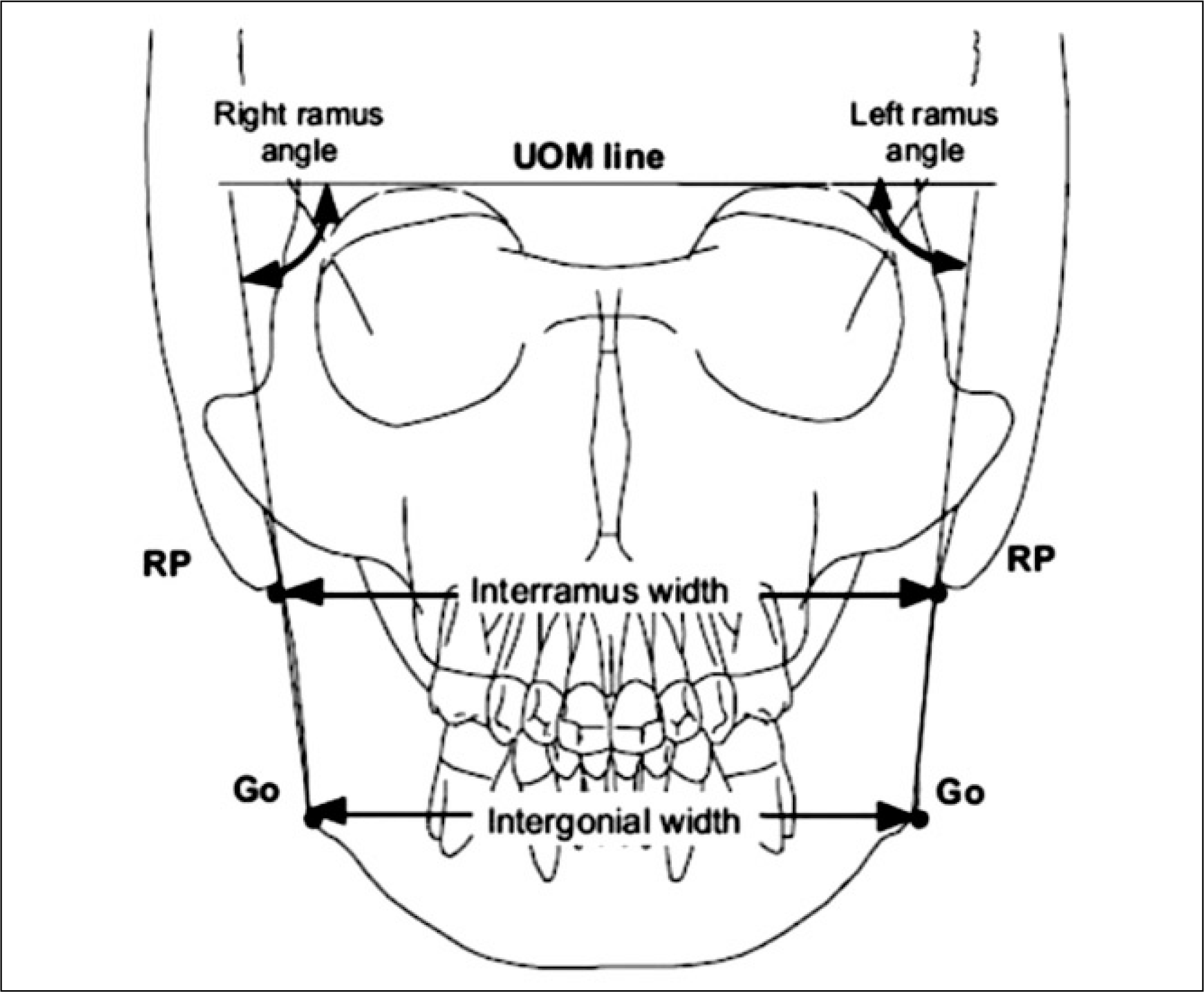
 | Fig. 2.Reference points and lines used in the PA Cephalometric analysis. (UOM: upper orbital margin, RP: ramus point, Go: gonion point, PA: posteroanterior) |
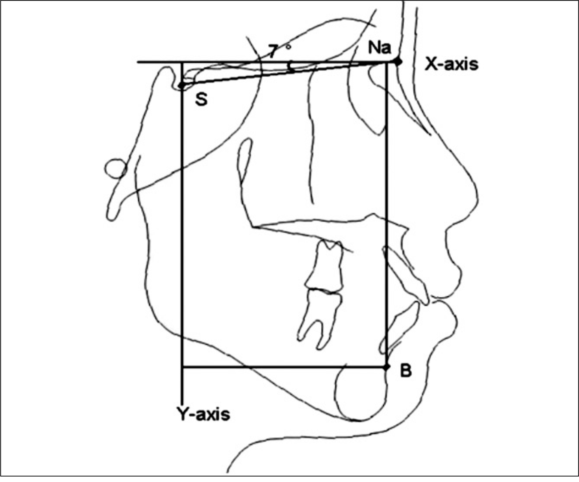
 | Fig. 3.Reference points and lines used in the lateral Cephalometric analysis. (Na: nasion, S: sella, B: B Point) |
Table 1.
Sex and age distributions of the patients
| Age (mean) | Male | Female | Total (%) | |
|---|---|---|---|---|
| Group A | 17–31 (21) | 7 | 8 | 15 (50) |
| Group B | 17–32 (21.8) | 9 | 6 | 15 (50) |
| Total (%) | 16 (53) | 14 (47) | 30 (100) |
Table 2.
Descriptive statistics for horizontal movement at B point and comparison of postoperative changes in horizontal movement at B point between Group A and B
Table 3.
Descriptive statistics for vertical movement at B point and comparison of postoperative changes in vertical movement at B point between Group A and B
Table 4.
Descriptive statistics for intergonial width and comparison of postoperative changes in intergonial width between Group A and B
Table 5.
Descriptive statistics for interramus width and comparison of postoperative changes in interramus width between Group A and B
Table 6.
Descriptive statistics for right ramus angle and comparison of postoperative changes in right ramus angle between Group A and B
Table 7.
Descriptive statistics for left ramus angle and comparison of postoperative changes in left ramus angle between Group A and B


 PDF
PDF ePub
ePub Citation
 Print
Print


 XML Download