

 PDF
PDF ePub
ePub Citation
 Print
Print


Abstract
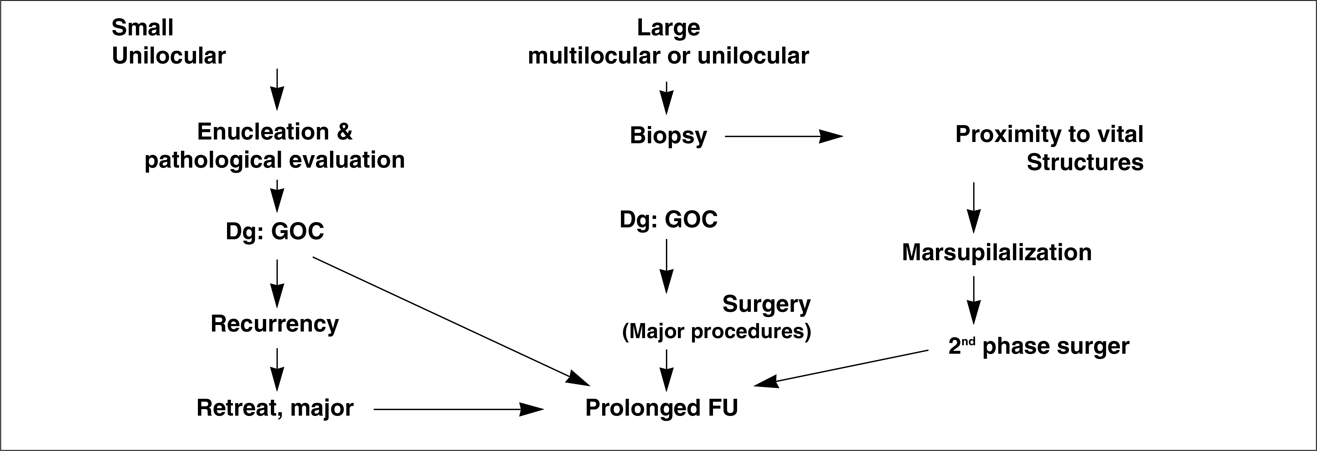
Glandular odontogenic cyst (GOC) is an intraoral cyst originated from serre remnants which has incidence of rare frequency. Only 111 cases have been reported since Gardener first introduced it in 1987. The clinical features are the following components: cortical bone thinning, locally aggressive root resorption, non-painful swelling. The following recurrences rate are 64.3% in conservative treatment, and 0% in wide excision for instance, segmental or marginal mandibulectomy. So, its prognosis is similar to that of odontogenic keratocyst and ameloblastoma. Therefore, periodic recall follow ups are essential to detect disease recurrence. Here, we will report the first case of GOC diagnosed in our department considering with references. And we share this treatment experience because these aggessive lesions may be misjudged for simple dental cyst.
Go to : 
References
1. Krishnamurthy A, Sherlin HJ, Ramalingam K, Natesan A, Premkumar P, Ramani P, et al. Glandular odontogenic cyst: report of two cases and review of literature. Head Neck Pathol. 2009; 3:153–8.

2. Macdonald-Jankowski DS. Glandular odontogenic cyst: systematic review. Dentomaxillofac Radiol. 2010; 39:127–39.
3. Oliveira JX, Santos KC, Nunes FD, Hiraki KR, Sales MA, Cavalcanti MG, et al. Odontogenic glandular cyst: a case report. J Oral Sci. 2009; 51:467–70.
4. Kaplan I, Anavi Y, Hirshberg A. Glandular odontogenic cyst: a challenge in diagnosis and treatment. Oral Dis. 2008; 14:575–81.
5. Kramer IR, Pindborg JJ, Shear M. The WHO histological typing of odontogenic tumours. A commentary on the second edition. Cancer. 1992; 70:2988–94.
6. Kaplan I, Gal G, Anavi Y, Manor R, Calderon S. Glandular odontogenic cyst: treatment and recurrence. J Oral Maxillofac Surg. 2005; 63:435–41.
Go to :
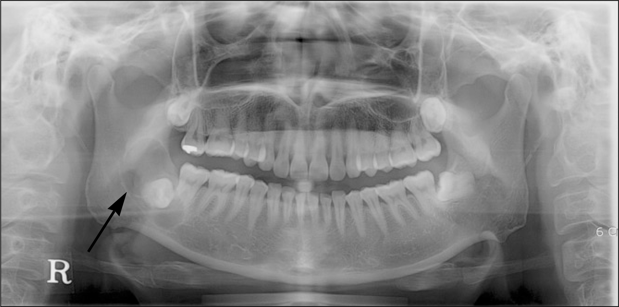 | Fig. 1.Preoperative panoramic view showing well-defined cystic lesion on right ramus area with impacted tooth. |
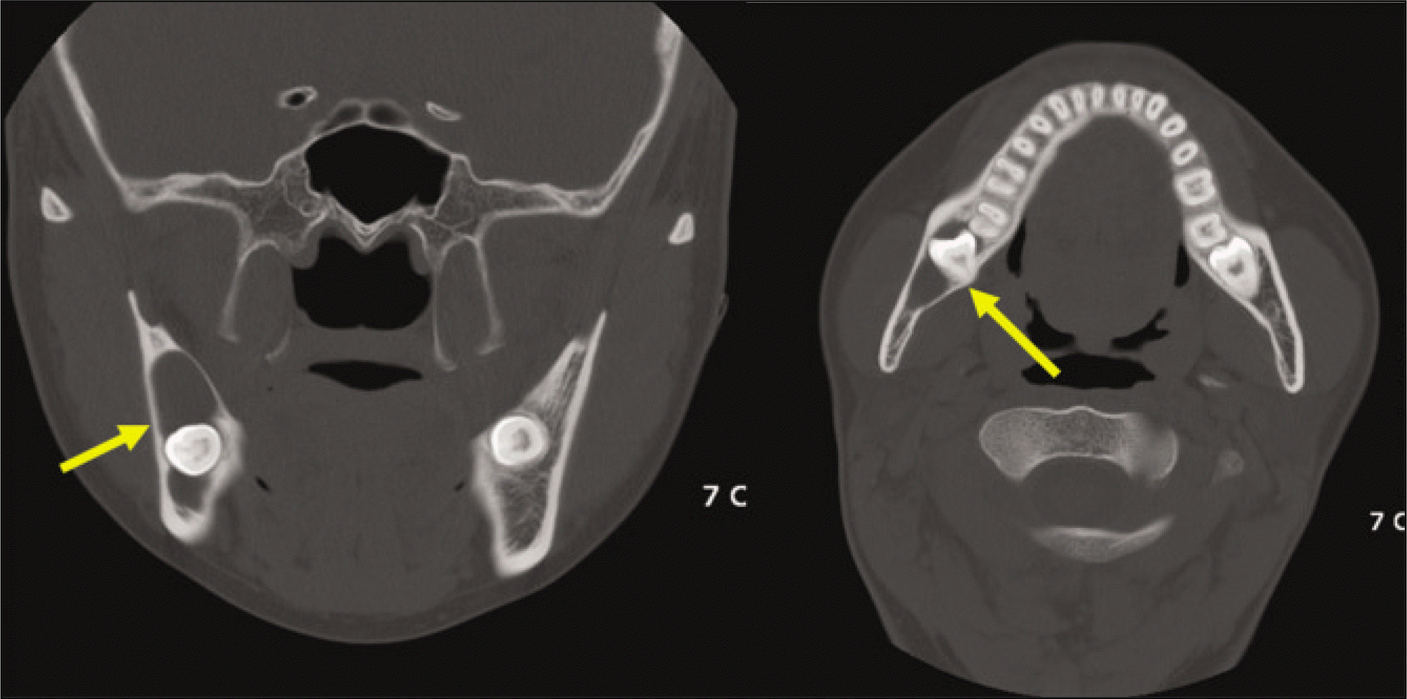 | Fig. 2.Coronal and axial computed tomography scan showing large cystic lesion with cortical thinning. |
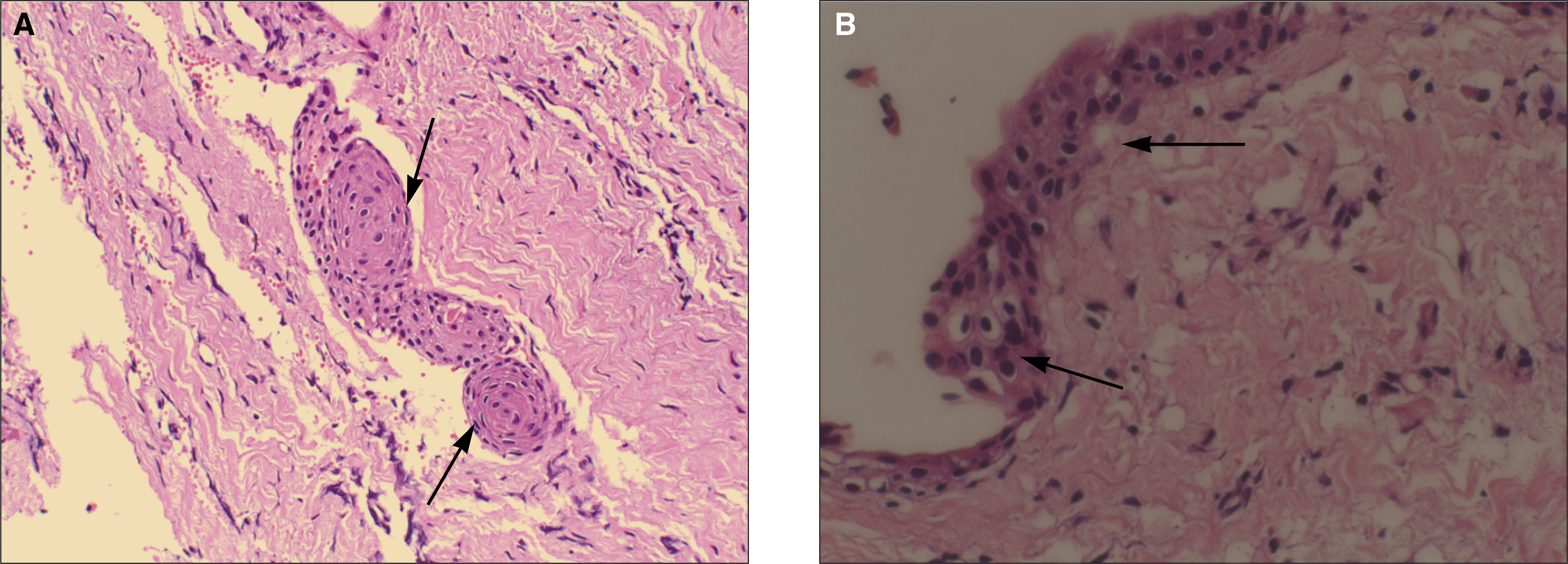 | Fig. 3.A: Histologic section.(H&E staining, original magnification x200) This histologic section showing pseudo-glandular structure in connective tissue. B: Histologic section.(H&E staining, original magnification x400) This histologic section showing squamous epithelial lining, with a flat interface, and intraepithelial goblet cell. |
 XML Download