

 PDF
PDF ePub
ePub Citation
 Print
Print


Abstract
Introduction
The first aim of this study was to isolate the dental tissue-derived stem cells from the dental follicle (DF), dental pulp (DP), and root apical papilla (RAP) of the extracted wisdom teeth. Second was to evaluate their characterization with the expressions of transcription factors and cell surface markers. Finally, their ability of the in vitro multilineage differentiations into osteogenic and adipogenic cells were compared, respectively.
Materials and Methods
Dental tissues, including dental follicle, dental pulp, and root apical papilla, were separated in the extracted wisdom teeth. These three dental tissues were cultured in Dulbecco's modified Eagle's medium (DMEM) with supplements, respectively. After passage 3, the homogeneous shaped dental tissue-derived cells were analyzed the expression of transcription factors (Oct-4, Nanog and Sox-2) and cell surface markers (CD44, CD90 and CD105) with reverse transcription polymerase chain reaction (RT-PCR) and fluorescence-activated cell sorting (FACS) analysis. In order to evaluate in vitro multilineage differentiations, the culture media were changed to the osteogenic and adipogenic induction mediums when the dental tissue-derived cells reached to passage 3. The characteristics of these three dental tissue-derived cells were compared with immunohistochemistry.
Results
During primary culture, heterogenous and colony formatted dental tissue-derived cells were observed in the culture plates. After passage 2 or 3, homogenous spindle-like cells were observed in all culture plates. Transcription factors and mesenchymal stem cell markers were positively observed in all three types of dental tissue-derived cells. However, the quantity of expressed transcription factors was most large in RAP-derived cells. In all three types of dental tissue-derived cells, osteogenic and adipogenic differentiations were observed after treatment of specific induction media. In vitro adipogenic differentiation was similar among these three types of cells. In vitro osteogenic differentiation was most strongly and frequently observed in the RAP-derived cells, whereas rarely osteogenic differentiation was observed in the DP-derived cells.
Conclusion
These findings suggest that three types of human dental tissue-derived cells from extracted wisdom teeth were multipotent mesenchymal stem cells, have the properties of multilineage differentiations. Especially, stem cells from root apical papilla (SCAP) have much advantage in osteogenic differentiation, whereas dental follicle cells (DFCs) have a characteristic of easy adipogenic differentiation.
References
1. Evans MJ, Kaufman MH. Establishment in culture of pluripotential cells from mouse embryos. Nature. 1981; 292:154–6.

2. Wagers AJ, Weissman IL. Plasticity of adult stem cells. Cell. 2004; 116:639–48.
3. Mao JJ, Giannobile WV, Helms JA, Hollister SJ, Krebsbach PH, Longaker MT, et al. Craniofacial tissue engineering by stem cells. J Dent Res. 2006; 85:966–79.
4. Almeida-Porada G, Porada C, Zanjani ED. Adult stem cell plasticity and methods of detection. Rev Clin Exp Hematol. 2001; 5:26–41.
5. Temple S. Stem cell plasticity-building the brain of our dreams. Nat Rev Neurosci. 2001; 2:513–20.
6. Yamada Y, Boo JS, Ozawa R, Nagasaka T, Okazaki Y, Hata K, et al. Bone regeneration following injection of mesenchymal stem cells and fibrin glue with a biodegradable scaffold. J Craniomaxillofac Surg. 2003; 31:27–33.
7. Yamada Y, Ueda M, Naiki T, Nagasaka T. Tissue-engineered injectable bone regeneration for osseointegrated dental implants. Clin Oral Impl Res. 2004; 15:589–97.
8. Fuerst G, Tangl S, Gruber R, Gahleitner A, Sanroman F, Watzek G. Bone formation following sinus grafting with autogenous bone-derived cells and bovine bone mineral in minipigs: preliminary findings. Clin Oral Impl Res. 2004; 15:733–40.
9. Schimming R, Schmelzeisen R. Tissue-engineered bone for maxillary sinus augmentation. J Oral Maxillofac Surg. 2004; 62:724–9.
10. Gronthos S, Mankani M, Brahim J, Robey PG, Shi S. Postnatal human dental pulp stem cells (DPSCs) in vitro and in vivo. Proc Natl Acad Sci USA. 2000; 97:13625–30.
11. Gronthos S, Brahim J, Li W, Fisher LW, Cherman N, Boyde A, et al. Stem cell properties of human dental pulp stem cells. J Dent Res. 2002; 81:531–5.
12. Miura M, Gronthos S, Zhao M, Lu B, Fisher LW, Robey PG, et al. Stem cells from human exfoliated deciduous teeth. Proc Natl Acad Sci USA. 2003; 100:5807–12.
13. Seo BM, Sonoyama W, Yamaza T, Coppe C, Kikuiri T, Akiyama K, et al. SHED repair critical-size calvarial defects in mice. Oral Dis. 2008; 14:428–34.
14. Huang AH, Chen YK, Lin LM, Shieh TY, Chan AW. Isolation and characterization of dental pulp stem cells from a supernumerary tooth. J Oral Pathol Med. 2008; 37:571–4.
15. Morsczeck C, Go ¨tz W, Schierholz J, Zeilhofer F, Ku ¨hn U, Mo ¨hl C, et al. Isolation of precursor cells (PCs) from human dental follicle of wisdom teeth. Matrix Biol. 2005; 24:155–65.
16. Morsczeck C, Moehl C, Go ¨tz W, Heredia A, Scha ¨ffer TE, Eckstein N, et al. In vitro differentiation of human dental follicle cells with dexamethasone and insulin. Cell Biol Int. 2005; 29:567–75.
17. Reynolds AJ, Jahoda CA. Cultured human and rat tooth papilla cells induce hair follicle regeneration and fiber growth. Differentiation. 2004; 72:566–75.
18. Seo BM, Miura M, Gronthos S, Bartold PM, Batouli S, Brahim J, et al. Investigation of multipotent postnatal stem cells from human periodontal ligament. Lancet. 2004; 364:149–55.
19. Sonoyama W, Liu Y, Fang D, Yamaza T, Seo BM, Zhang C, et al. Mesenchymal stem cell-mediated functional tooth regeneration in swine. PLoS ONE. 2006; 1:e79.
20. Morsczeck C, Vo ¨llner F, Saugspier M, Brandl C, Reichert TE, Driemel O, et al. Comparison of human dental follicle cells (DFCs) and stem cells from human exfoliated deciduous teeth (SHED) after neural differentiation in vitro. Clin Oral Investig [in press 2009 Jul 10].
21. Kang EJ, Byun JH, Choi YJ, Maeng GH, Lee SL, Kang DH, et al. In vitro and in vivo osteogenesis of porcine skin-derived mesenchymal stem cell-like cells with a demineralized bone and fibrin scaffold. Tissue Eng Part A. 2010; 16:815–27.
22. Scho ¨ler HR, Ruppert S, Suzuki N, Chowdhury K, Gruss P. New type of POU domain in germ line-specific protein Oct-4. Nature. 1990; 344:435–9.
23. Avilion AA, Nicolis SK, Pevny LH, Perez L, Vivian N, Lovell-Badge R. Multipotent cell lineages in early mouse development depend on SOX2 function. Genes Dev. 2003; 17:126–40.
24. Chambers I, Colby D, Robertson M, Nichols J, Lee S, Tweedie .
Fig. 1.
A. Schematic illustrations of dental pulp (DP), root apical papilla (RAP), and dental follicle (DF) in the extracted impacted wisdom tooth. B. Photograph of the obtained dental tissues after treatment of mechanical dissociation and enzyme (collagenase I) digestion.(Pa: root apical papilla tissue, F: dental follicle tissue, Pu: dental pulp tissue)
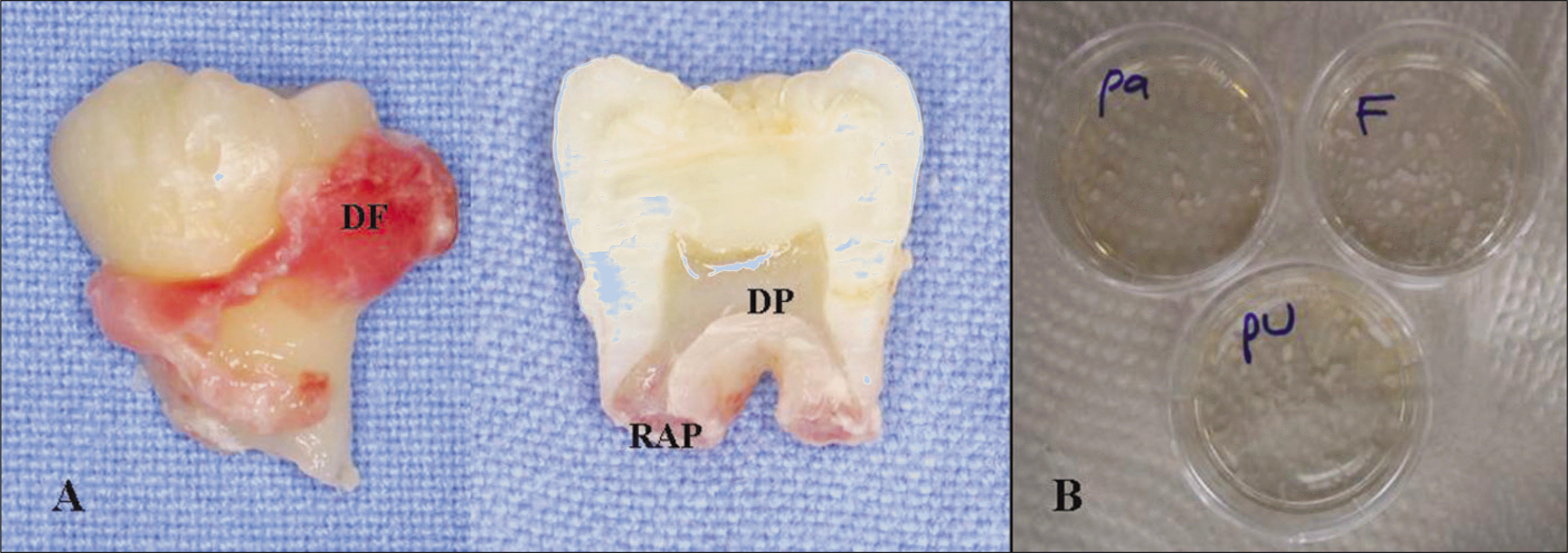
Fig. 2.
Isolation and primary culture of dental tissue-derived cells. Microphotographs of dental follicle-derived cells (DF), root apical papilla-derived cells (RAP), and dental pulp-derived cells (DP) at 1st and 10th days later of primary culture (passage 0). At 1st day of primary culture, round shaped unattached floating primary cells were rarely observed in the culture media. When the culturing cells reached nearly 70–80% confluence of primary culture (10th day of primary culture), remarkably increased cell number and irregular heterogeneous shaped dental tissue-derived cells were observed. In this primary culture period, there are no definitely morphological differences between these three dental tissue-derived cells.

Fig. 3.
Sequential cell proliferations of the third passaged dental tissue-derived cells.(original magnification x100) RAP-derived cells showed the most prominent proliferation rate, especially between 3rd and 6th days of subculture, there is remarkable increase of cell number. However, DF and DP-derived cells showed the remarkable cell number increasing between 6th and 9th days of subculture. (DP: dental pulp, RAP: root apical papilla, DF: dental follicle)

Fig. 4.
Graphs showed the increasing numbers of in vitro cultured cells as time passed. The most prominent cell proliferation rate was observed in the RAP-derived cells. (DP: dental pulp, RAP: root apical papilla, DF: dental follicle)
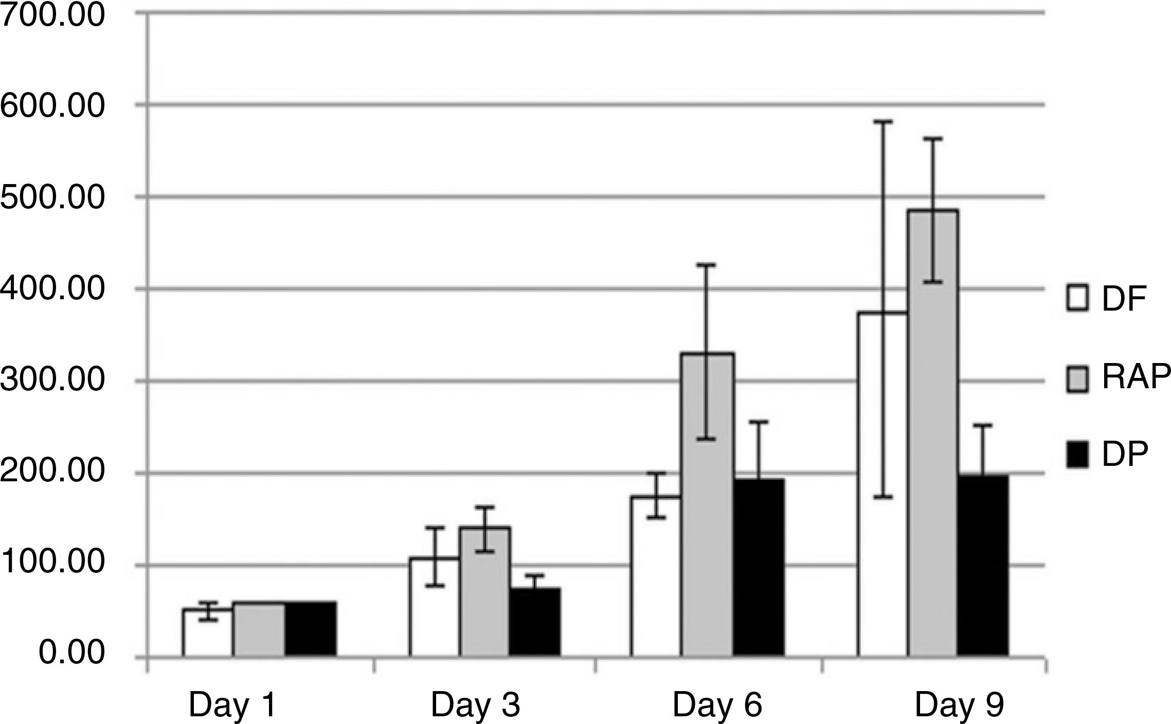
Fig. 5.
Expression of transcription factors, Oct-4, Nanog, and Sox-2 in the cultured dental tissue-derived cells of passage 3 by RT-PCR. A. Although Sox-2 gene was weakly observed, these transcription factors were all detected in the dental tissue-derived cells. B. Quantification of transcription factors expressions. Transcription factors were most strongly detected in the root apical papilla-derived cells.(RAP) (GAPDH: glyceraldehyde-3-phosphate dehydrogenase, DP: dental pulp, RAP: DF: dental follicle, RT-PCR: reverse transcription polymerase chain reaction)
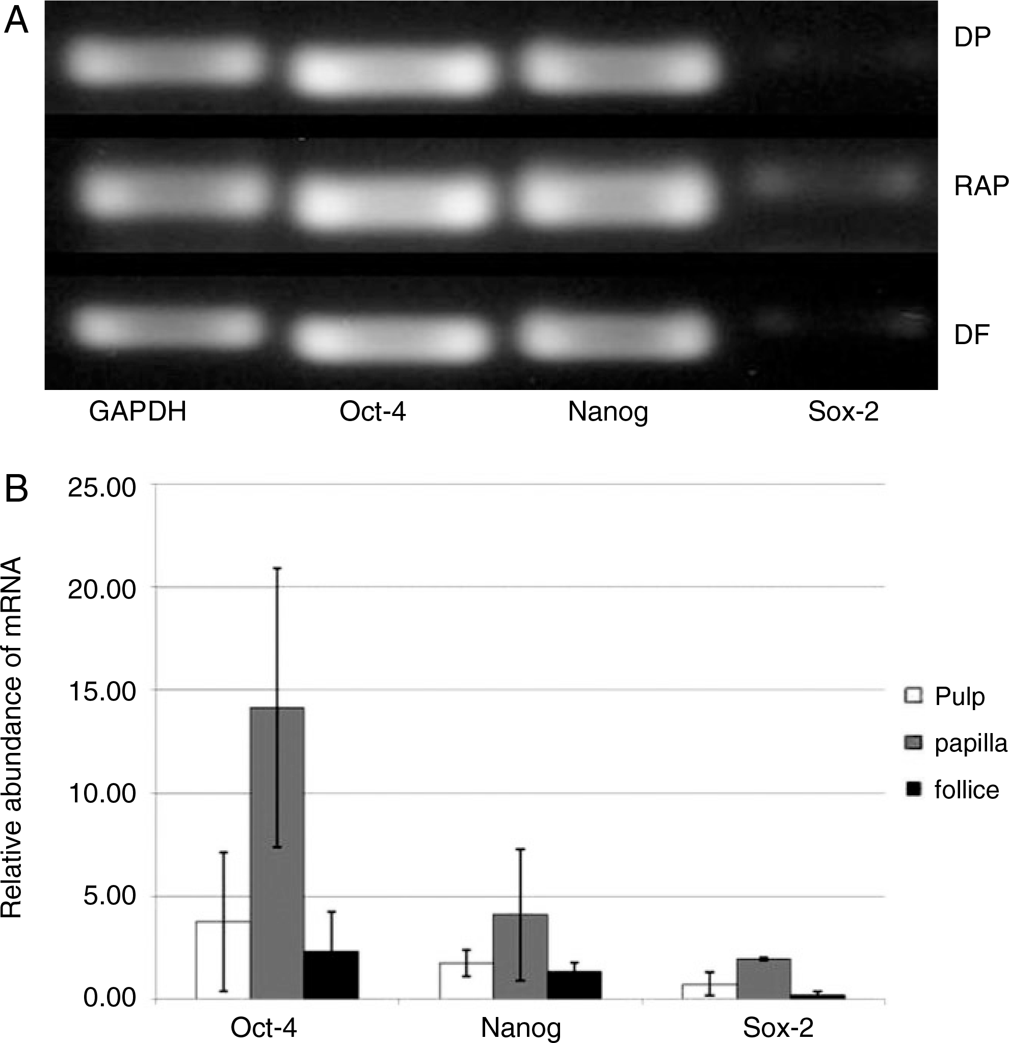
Fig. 6.
Expressions of cell surface markers with FACS analysis. Mesenchymal stem cell marker proteins were detected in the all three types of dental tissue-derived cells. A. Dental follicle cells. B. Dental pulp cells. C. Root apical papilla cells. (FACS: fluorescence-activated cell sorting)
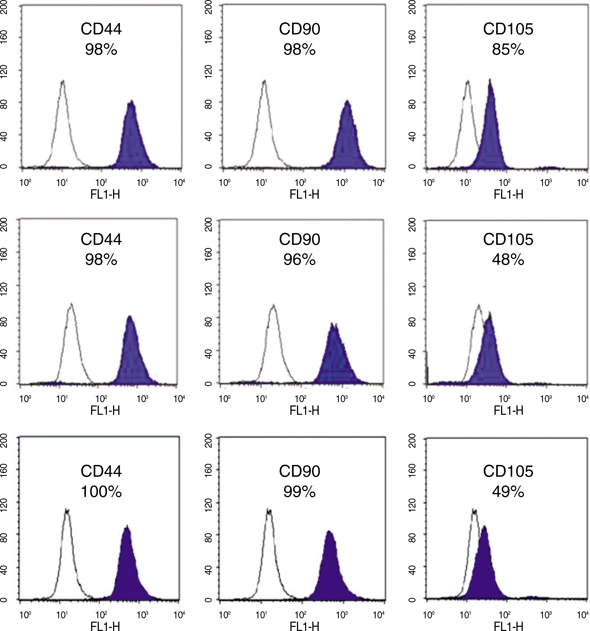
Fig. 7.
Osteogenic differentiations of dental tissue-derived stem cells for 4 weeks. The most abundant bone matrix formation was observed in the RAP cells, whereas it was rarely detected in the DP cells by von Kossa and alizarin red stainings. (DP: dental pulp, RAP: root apical papilla)

Fig. 8.
Adipogenic differentiation of dental tissue-derived stem cells for 4 weeks.(original magnification x100) Small red color stained lipoid nodules were detected in all three types of dental stem cells by oil-red O staining.(arrows) Most actively adipogenic differentiation was showed in the dental follicle-derived cells.

Table 1.
RT-PCR primers used for evaluating transcription factors in dental tissue-derived stem cells
 XML Download