

 PDF
PDF ePub
ePub Citation
 Print
Print


Abstract
Synovial condromatosis of the temporomandibular joint (TMJ) is characterized by the presence of loose bodies (joint mices). It can be confused with temporomandibular disorder clinically, and be with chondrosarcoma histologically. The purpose of this clinical report was to review the clinical, radiological, arthroscopic findings, histological feature and the results of surgical treatment of TMJ synovial chondromatosis. Four patients presented with pain of TMJ and limited mouth opening. The dynamic magnetic resonance imaging (MRI) disclosed a characteristic morphologic changes and displacement of the meniscus with limited translation of the condyle head. Bone scans showed progressive resorptive changes with hot-uptake of the radioisotope. The synovial loose bodies in the joint spaces were removed and sent to pathology for diagnosis as the synovial chondromatosis. The follow-up examination with computed tomography (CT) and MRI revealed no evidence of recurrence and good in function until postoperative 18 months. Diagnostically, the distension of the lateral capsule and fluid findings in the joint on the MRI are very suggestive tool for this synovial chondromatosis, but they are not always detected on the preoperative MRI. Arthroscopic approaches are very useful to inspect the joint spaces and to remove the loose bodies without interruption of the whole synovial membranes.
References
1. Akhtar M, Mahajan S, Kott E. Synovial chondromatosis of the temporomandibular joint. J Bone Joint Surg Am. 1977; 59:266–7.

2. Fee WE Jr, Windhorst P, Wiggins R, Pang L. Synovial chondromatosis of the temporomandibular joint. Otolaryngol Head Neck Surg. 1979; 87:741–8.
3. Ardekian L, Troulis MJ, August M. Synovial chondromatosis of the temporomandibular joint: report and analysis of eleven cases. J Oral Maxillofac Surg. 2005; 63:941–7.
4. Quinn PD, Stanton DC, Foote JW. Synovial chondromatosis with cranial extension. Oral Surg Oral Med Oral Pathol. 1992; 73:398–402.
5. Simon GT, Kendrick RW, Whitlock RI. Osteochondroma of the mandibular condyle. Case report and its management. Oral Surg Oral Med Oral Pathol. 1977; 43:18–24.
6. Koole R, Steenks MH, Witkamp TD, Slootweg PJ, Shaefer J. Osteochondroma of the mandibular condyle. A case report. Int J Oral Maxillofac Surg. 1996; 25:203–5.
7. Ribas Mde O, Martins WD, de Sousa MH, Zanferrari FL, Lanzoni T. Osteochondroma of the mandibular condyle: literature review and report of a case. J Contemp Dent Pract. 2007; 8:52–9.
8. Wolford LM, Mehra P, Franco P. Use of conservative condylectomy for treatment of osteochondroma of the mandibular condyle. J Oral Maxillofac Surg. 2002; 60:262–8.
9. Milgram JW. The classification of loose bodies in human joints. Clin Orthop Relat Res. 1977; 124:282–91.
10. Yu Q, Yang J, Wang P, Shi H, Luo J. CT features of synovial chondromatosis in the temporomandibular joint. Oral Surg Oral Med Oral Pathol Oral Radiol Endod. 2004; 97:524–8.
11. Moses JJ, Hosaka H. Arthroscopic punch for definitive diagnosis of the synovial chondromatosis of the temporomandibular joint. Case report and pathology review. Oral Surg Oral Med Oral Patholol. 1993; 75:12–7.
12. Miyamoto H, Sakashita H, Miyata M, Kurita K. Arthroscopic diagnosis and treatment of temporomandibular joint synovial chondromatosis: report of a case. J Oral Maxillofac Surg. 1996; 54:629–31.
13. Miyamoto H, Sakashita H, Wilson DF, Goss AN. Synovial chondromatosis of the temporomandibular joint. Br J Oral Maxillofac Surg. 2000; 38:205–8.
Table 1.
Demographic data of the patients in the study
| Patient No. | Age Years | Gender Male/Female | Joint Right/Left | Trauma Yes/No | Duration (month) |
|---|---|---|---|---|---|
| 1 | 39 | F | R | N | 19 |
| 2 | 55 | M | L | Y | 13 |
| 3 | 34 | F | L | N | 38 |
| 4 | 47 | F | L | N | 18 |
Table 2.
Clinical findings, preoperative and postoperative
Table 3.
Loose body at radiographic examination and arthroscopic findngs
| No. | Plain | Bonescan | MRI | Arthroscopy | CT |
|---|---|---|---|---|---|
| Radiography Panorama | Preop, Hot spot | Preop | Preop | Postop | |
| 1 | − | + | + | + | − |
| 2 | − | + | + | + | − |
| 3 | − | + | − | + | − |
| 4 | − | + | + | + | − |
Table 4.
Findings at surgery
Fig. 1.
View of arthroscopy of the TMJ where numerous loose bodies can be appreciated involving the superior compartment of the joint. (TMJ: Temporomandibular joint)
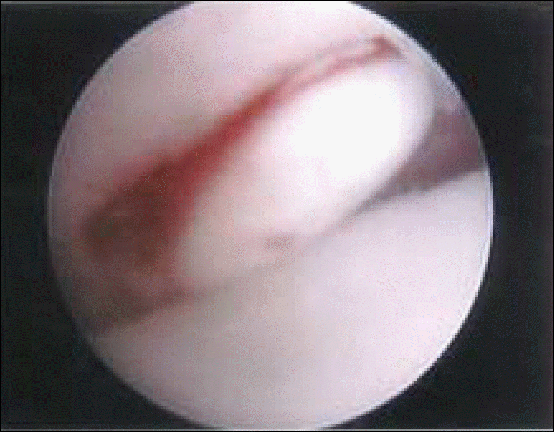
Fig. 2.
View of arthroscopy of the TMJ where numerous loose bodies can be appreciated involving the superior compartment of the joint. (TMJ: Temporomandibular joint)
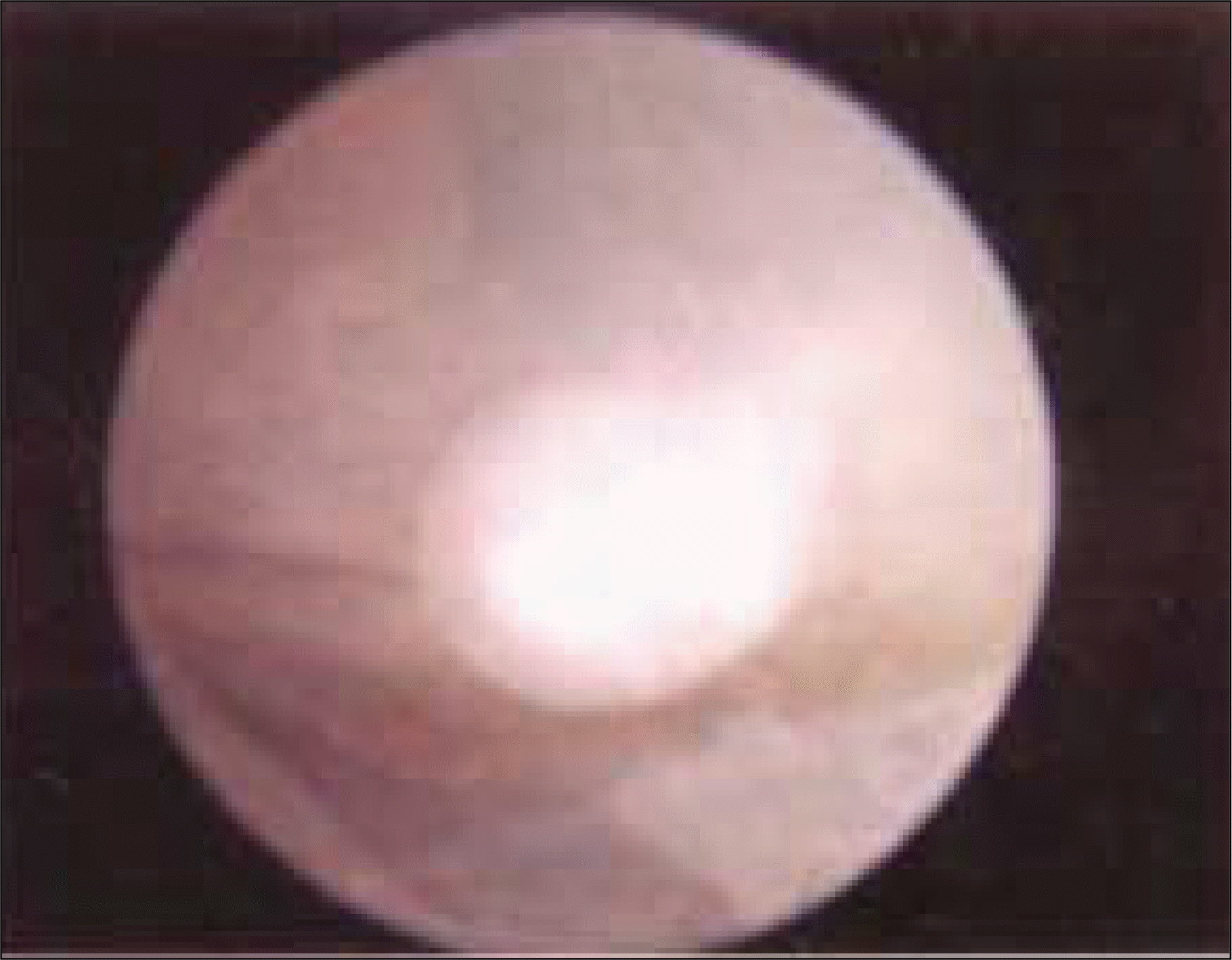
Fig. 3.
View of arthroscopy of the TMJ where numerous loose bodies can be appreciated involving the superior compartment of the joint. (TMJ: Temporomandibular joint)
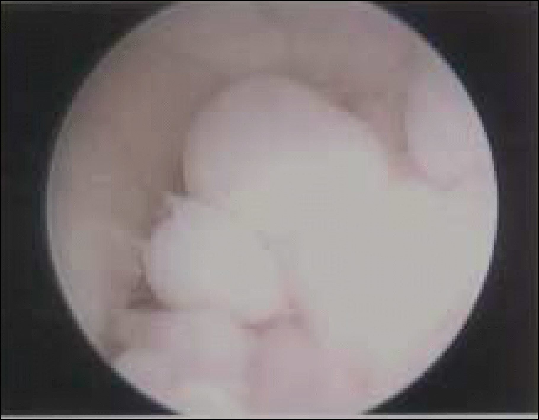
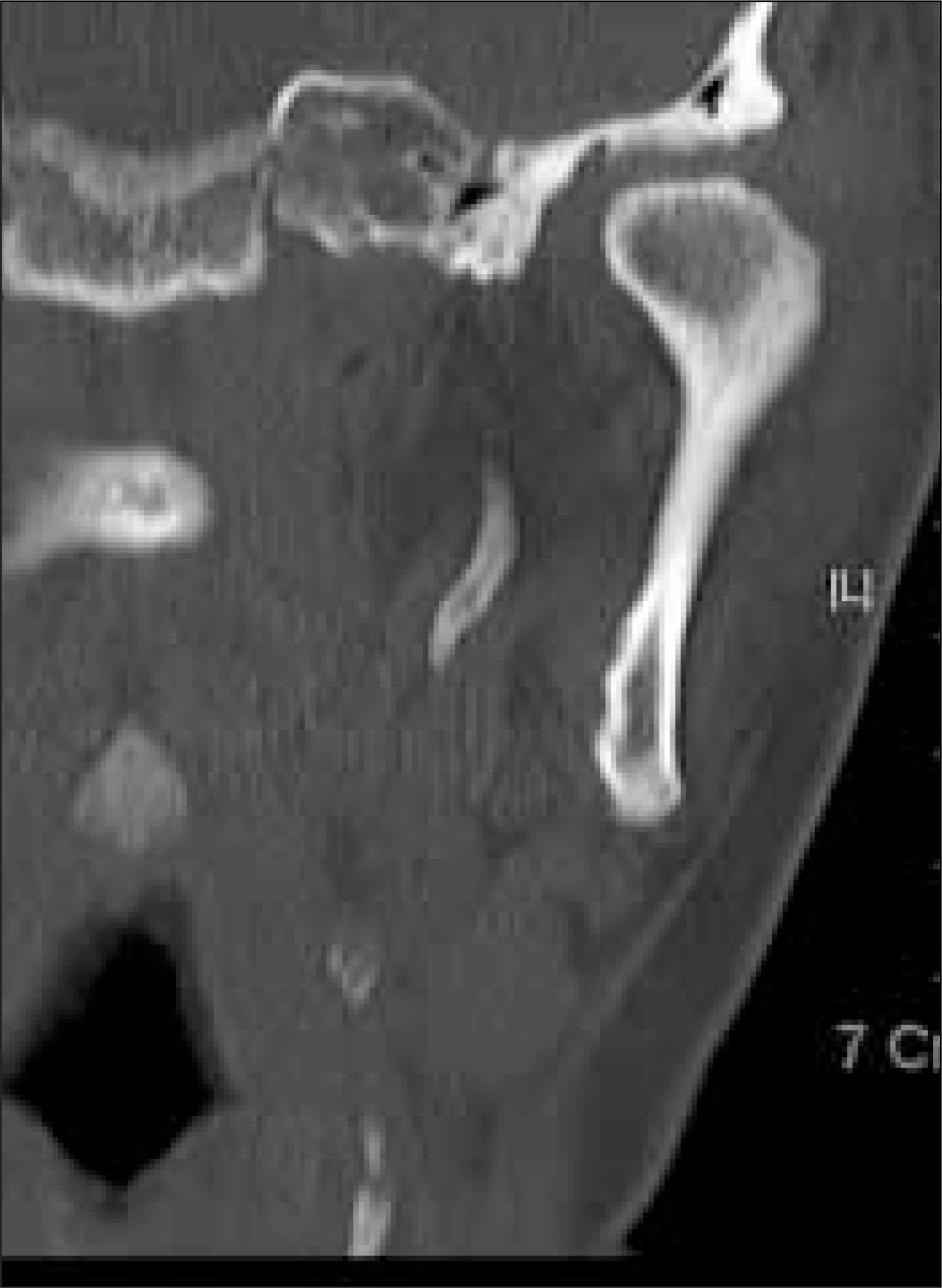
Fig. 4.
A. Bonescan: hot uptake. B, C. Preoperative sagittal and coronal TI-weighted MRI of the left TMJ: loose bodies are examed. D, E. View of arthroscopy of the TMJ where numerous loose bodies can be appreciated involving the superior compartment of the joint. (MRI: magnetic resonance imaging) (TMJ: Temporomandibular joint)

Fig. 5.
The left TMJ is exposed via a preauricular approach. A. Loose bodies escape from the upper compartment after incision of the capsule. B, C. Macroscopic image of over 60 nodules of variables sizes retrieved from the joint. (TMJ: Temporomandibular joint)
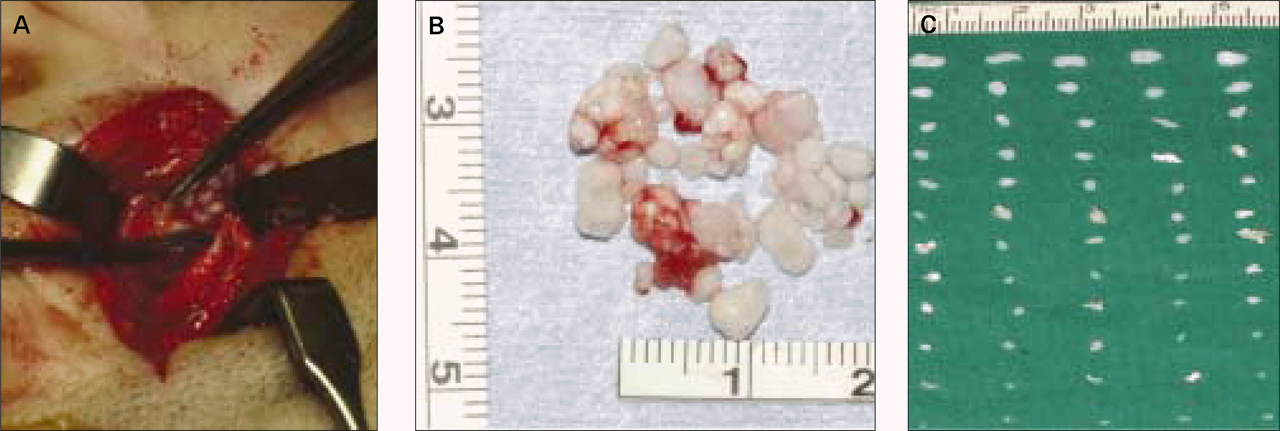
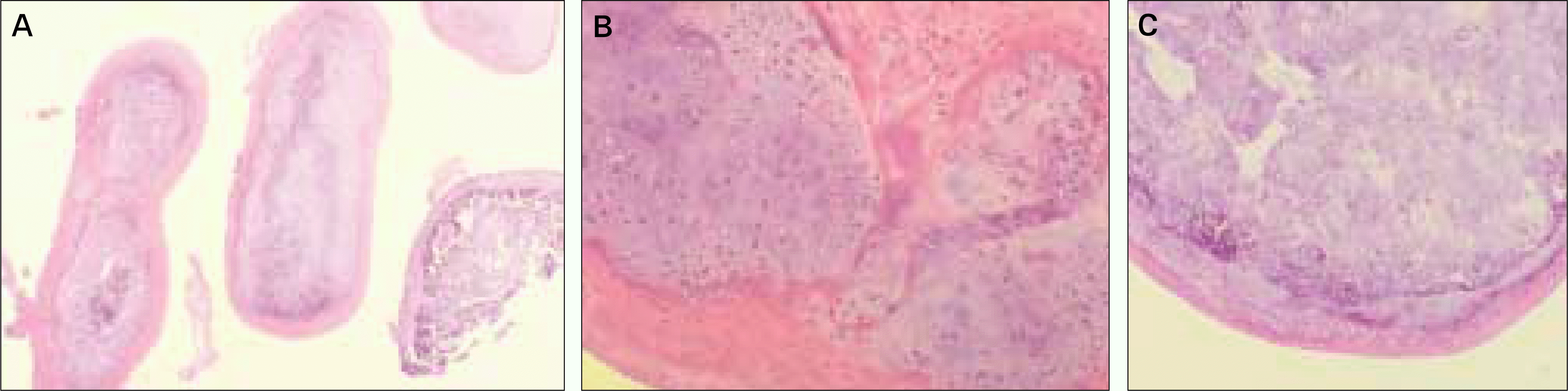
Fig. 6.
A. Microscopic (H&E, x10) appearance of cartilaginous nodules. Nodules of hyaline cartilage with sclerosis, covered with a normal synovium can be seen. B, C. The histopathologic hallmarks are cartilaginous nodules in the synovial membrane. (H&E, x100) Severe inflammation is present. Note lymphocytes and macrophages with some giant cell formation.
 XML Download