

 PDF
PDF ePub
ePub Citation
 Print
Print


INTRODUCTION
18F-AV-1451 is a recently developed tau PET ligand that has high affinity for paired helical filaments of hyperphosphorylated tau in neurofibrillary tangles. Previous studies showed that 18F-AV-1451 uptake was correlated with pathological tau staging.12 However, high AV-1451 retention has also been observed in other conditions unrelated to tau.345 This ‘off-target binding’ includes the substantia nigra, basal ganglia, and choroid plexus. A recent report also showed incidental AV-1451 retention in the regions of meningiomas, vascular malformations, and remote infarcts.3 Nevertheless, there is little knowledge regarding 18F-AV-1451 PET findings in acute or subacute stage of ischemic stroke patients. Herein, we report a case of abnormally increased uptake of AV-1451 which was shown to be a subacute infarction.
CASE REPORT
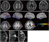
An 83-year-old woman who experienced three years of progressive cognitive decline and apathy visited the Memory Clinic at Samsung Medical Center, Seoul, Korea. She had dyslipidemia and had 9 years of formal education. On neurologic exam, she was alert and showed no focal neurological signs including asymmetric weakness or sensory change. She scored 13 out of 30 on the Mini-Mental State Examination. Neuropsychological testing6 revealed cognitive impairment in multiple domains(language, visuospatial, memory, and frontal-executive dysfunction) which limited her activities of daily living. Brain MRI showed confluent white matter hyperintensities and diffuseatrophy (Fig. 1A). Amyloid PET, assessed with 18F-florbetaben radiotracer, was positive and no asymmetric focal high uptake was observed. Apolipoprotein E genotype was e2/e3. Based on her clinical and imaging findings, we assumed that her dementia was due to mixed etiology: Alzheimer's disease and small vessel disease.
She further underwent 18F-AV-1451 PET to search for distribution of paired helical filament tau. Using cerebellar gray matter as a reference region, we created AV-1451 standardized uptake value ratio images based on mean uptake over 90-110 min post-injection. AV-1451 uptake was observed in the bilateral medial temporal, lateral temporal, and inferior parietal areas, with the overall pattern looking similar to that of Alzheimer's disease. 78 However, a focal hot uptake in the left primary sensory cortex was very unusual (Fig. 1B). This finding was unexpected because there were no relevant symptoms or signs and no structural lesions in that area on brain MRI, taken 20 days before the 18F-AV-1451 PET (Fig. 1A). We re-evaluated the brain MRI 30 days after 18F-AV-1451 PET and found a subacute infarction in the left primary sensory cortex (Fig. 1C, D, and E) with no hemorrhagic transformation.
We obtained written informed consent from the patient. This study was approved by the Institutional Review Board of Samsung Medical Center.
DISCUSSION
We report a case of focal high uptake of AV-1451 which was shown to be a subacute infarction. Although 18F-AV-1451 is a tau PET ligand that has high affinity for paired helical filament tau, various off-target bindings unrelated to tau have been reported. 3459 The underlying substrate of AV-1451 off-target binding in the substantia nigra, basal ganglia, and choroid plexus are not well understood. Previous studies suggested that AV-1451 binds to neuromelanin containing neurons in the substantia nigra.4 Elevated in vivo AV-1451 uptake in the basal ganglia was observed in elderly individuals, which might be due to iron component or different kinetic profile of the tracer in the basal ganglia.5 Another AV-1451 off-target binding is the choroid plexus and the underlying substrate of tracer's uptake is suggested to be leptomeningeal melanocyte or calcifications. 45 AV-1451 uptake in the regions of meningiomas, vascular malformations, and remote infarcts3 has recently been reported, and the authors suggested that increased vascular permeability was responsible for tracer retention.
The reason why AV-1451 uptake was increased in ischemic stroke can be explained in several ways. AV-1451 might bind to tau in the area of the ischemic insult as a result of ischemic neuronal damage, apoptosis, or gliosis, which are reported to be associated with tau hyperphosphorylation.10 Alternatively, increased uptake of AV-1451 might be due to increased tracer leakage related to blood-brain-barrier dysfunction11 as it becomes permeable immediately after hypoxia-ischemia.12
Our present case is in line with a recent report showing two cases of elevated AV-1451 uptake in chronic infarction regions,3 and the author suggested that vascular permeability might have contributed to AV-1451 retention. Similarly, a study of Pittsburgh compound B (PiB) PET showed focal PiB retention in subacute ischemic stroke regions, and the authors suggested leakage of PiB across the blood-brain barrier damaged by the ischemic process and reperfusion injury as a possible mechanism.1314 However, controversies exist. In a larger study of subacute ischemic stroke patients, there was no significant increase of PiB uptake in the infarct area.14
There are several limitations in this case report. First, the interval between AV-1451 and brain MRI was quite long and we might have missed the acute stage of infarction. Second, we lacked pathology data to confirm the exact etiology of the AV-1451 uptake. Therefore, our findings need to be further examined in a group of patients with ischemic stroke along with pathology data.
Nevertheless, we believe that this case is noteworthy because this is the first report to show increased AV-1451 uptake in a recent infarction area. We suggest that one should be cautious in the interpretation of increased AV-1451 uptake and that ischemic stroke should be considered as a possible cause.
 XML Download