

 PDF
PDF ePub
ePub Citation
 Print
Print


INTRODUCTION
Facial nerve paralysis occurs in 7% to 10% of patients with temporal bone fractures.1 There is some dispute in the literature concerning the role, timing, and type of surgery for the management of traumatic facial paralysis.234
A few approaches can be used to decompress facial nerve, including the middle cranial fossa approach or transmastoid approach depending on the site of injury.4 When the suspicious lesion is located only on the tympanic segment, the transmastoid approach is usually performed. For a lesion limited to the tympanic segment, the endoscopic transcanal approach can also provide sufficient exposure for facial nerve decompression, and this approach is minimally invasive compared to the other approaches.
It is now possible to use this approach because of the recently described endoscopic anatomy from the external auditory canal (EAC) to the internal auditory canal (IAC).5 The use of the EAC as a natural surgical corridor allows the facial nerve to be reached from the geniculate ganglion (GG) to the second genu, as well as visualization of its anatomic relationships with the surrounding structures.5678
Go to : 
CASE REPORT
Surgical technique
Using the 0° endoscopic view, an incision of the EAC was performed from 11 o'clock to 7 o'clock. A tympanomeatal flap was endoscopically elevated, anteriorly and inferiorly based to expose the whole scutum. The eardrum was gently detached from the malleus, cutting the adhesion located in the umbus between the eardrum and the handle of the malleus to obtain good exposure of the medial wall of the tympanic cavity. The scutum was partially drilled with caution so as not to injure a chorda tympani, allowing adequate access to the anterior epitympanum. The incus and malleus were removed to expose the facial nerve from the GG to the second genu. The cog and the cochleariform process were identified endoscopically and used as anatomic landmarks for the GG area because of the close anatomic relationship between the GG and these structures. The lateral semi-circular canal was also detected endoscopically, posteriorly, and superiorly with respect to the second genu of the facial nerve, representing the posterior limit of surgical dissection. The cog was gently removed using a micro curette to increase exposure of the GG area. The facial nerve was decompressed from the GG to the second genu. After this procedure, a gelfoam soaked with corticosteroid solution was placed in the surgical field close to the facial nerve. Partial ossicular chain reconstruction with titanium prosthesis and underlay tympanoplasty using tragal cartilage were done. The tympanomeatal flap was repositioned, and the EAC was filled with gelfoam (Fig. 1, Supplementary Video 1, only online).
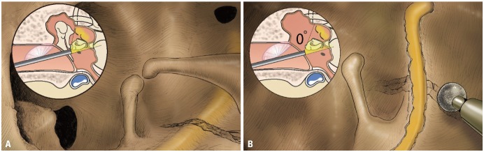 | Fig. 1Schematic drawings of total transcanal endoscopic facial nerve decompression. (A) After tympanomeatal flap elevation, fracture line across the tympanic segment of facial nerve was noted. The disarticulated incudostapedial joint was also found. (B) After removal of malleus and incus, decompression of facial nerve was performed using microdrills. Using this approach, facial nerve can be exposed from geniculate ganglion to second genu.
|
Patients and methods
Between October and December 2016, two patients with traumatic facial nerve palsy underwent total transcanal endoscopic facial nerve decompression from the GG to the second genu. Patients agreed to the use of their medical records and images for specific purpose and informed consents were obtained.
Case 1
A 54-year-old male visited our clinic with left facial palsy and hearing impairment four weeks after a traumatic subdural hematoma and left zygomatic fracture. Otoendoscopic examination revealed left tympanic membrane perforation, and pure tone audiometry showed conductive hearing loss. High-resolution computed tomography (HRCT) of the temporal bone showed fracture line crossed left tympanic segment of facial nerve (Fig. 2A, B, and C).
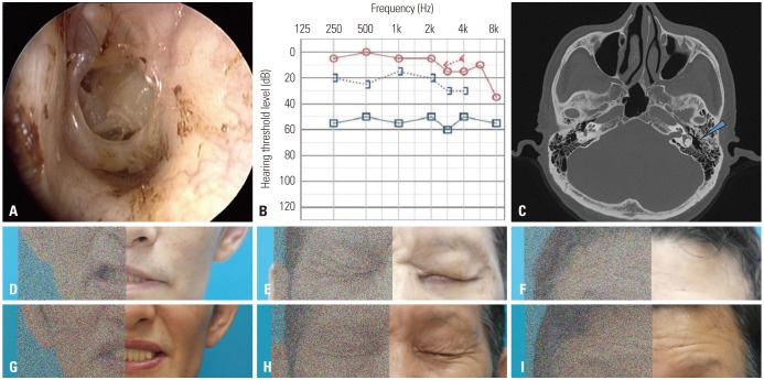 | Fig. 2Otologic manifestations and facial expressions of case 1. (A and B) Traumatic tympanic membrane perforation and conductive hearing loss were noted in the left ear. (C) Pre-operative CT scan, axial view. Note the left temporal bone fracture as shown by the blue arrow. (D-F) Pre-operative and (G-I) six-month post-operative photos of the patient. There was a significant improvement in facial nerve function from House-Brackmann grade IV to II.
|
Intraoperatively, a fracture line across the tympanic segment of facial nerve and disarticulated incudostapedial joint were found. The patient underwent facial nerve decompression from the GG to the second genu through the endoscopic transcanal approach. Six months post-operation, there was significant improvement in facial nerve function (Fig. 2D-I), having from House-Brackmann (H-B) grade IV to grade II.
Case 2
A 39-year-old male who experienced head trauma after falling down 10 days before he visited our clinic with left facial palsy and bilateral hearing impairment. HRCT scan showed fracture line crossed left tympanic segment of facial nerve, and audiological evaluation showed bilateral hearing loss (Fig. 3A and B).
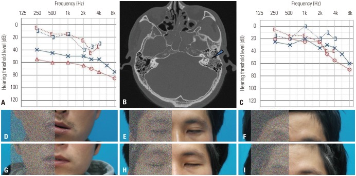 | Fig. 3Otologic manifestations and facial expressions of case 2. (A) Pure tone audiometry showed bilateral conductive hearing loss (average ABG=30 dB). (B) Pre-operative CT scan, axial view. Note the left temporal bone fracture as shown by the blue arrow. (C) Three-month post-operative pure tone audiometry showed improvement. (D-F) Pre-operative and (G-I) five-month post-operative photos of the patient. Patient showed improvement in facial function from House-Brackmann grade V to III.
|
The patient underwent facial nerve decompression from the GG to the second genu by endoscopic transcanal approach. Then, ossiculoplasty was done. Six months after the operation, the patient showed hearing improvement at low frequencies (Fig. 3C), and facial nerve function improved from H-B grade V to III (Fig. 3D-I). This slow recovery was due to difference in degree of injury and personal vulnerability.
Go to :
DISCUSSION
Management of facial nerve palsy secondary to temporal bone fractures has been an issue of dispute among otologic surgeons for years; the role, timing, and type of surgery have been debated. Middle cranial fossa approach and the transmastoid including its modifications, and the two combined together are the approaches currently used for facial nerve decompression depending on the site of injury, which is frequently deduced from the HRCT scan of the temporal bone.123491011
Middle cranial fossa approach is used to expose the IAC and labyrinthine segment of the facial nerve while preserving hearing. The GG and tympanic portion of the nerve can also be decompressed using this approach, especially for longitudinal fractures. When this approach is combined with the transmastoid approach, the whole course of the facial nerve can be exposed and decompressed. However, the middle cranial fossa approach involves invasive procedures such as craniotomy and temporal lobe retraction.
Transmastoid approach is appropriate when the main pathologic lesion is located on the tympanic or mastoid segment. Decompression of the GG or distal portion of the labyrinthine segment is also possible. Drawbacks of this approach, however, include extensive drilling of the mastoid bone, long time, and alteration of normal structures.
Herein, we described total transcanal endoscopic approach for facial nerve decompression as an alternative for lesions limited to the tympanic segment. The workhorse approaches for facial nerve decompression, are still transmastoid or middle cranial fossa craniotomies, however, endoscopic approach is less invasive compared to the other approaches, since it involves neither craniotomy nor extensive mastoid bone drilling. Despite the removal of the incus and malleus to have good exposure of the tympanic segment, which requires ossiculoplasty, this procedure is cosmetically acceptable as there are no external scars or bony depressions due to drilling. The outcome of facial nerve decompression through this approach was assessed using the H-B grading system for facial nerve function, and results were similar to those reported previously for middle cranial fossa, transmastoid, and combined approaches.24911
In conclusion, total transcanal endoscopic facial nerve decompression is minimally invasive alternative for traumatic facial palsy whose lesion is limited to the tympanic segment.
Go to :

 XML Download