

 PDF
PDF ePub
ePub Citation
 Print
Print


INTRODUCTION
The presence of a ureter stone is the most common urologic emergency and is associated with pain, expense, renal obstruction, and urinary tract infection.12 The dilemma facing the urologist is to choose between conservative measures and intervention for ureterolithiasis management. Stone size and location are generally considered the most important factors associated with spontaneous ureter stone passage (SSP).345 According to the current literature, serum C-reactive protein (CRP) concentration, pyuria, hydronephrosis, and helical computed tomography findings of perinephric fat stranding and the tissue-rim sign related to inflammatory changes are predictors associated with SSP.4678
Multiple inflammatory markers, such as CRP concentration and the erythrocyte sedimentation rate (ESR), have generally been used in clinical practice.9 Among the inflammatory markers, the neutrophil-to-lymphocyte ratio (NLR), defined as the ratio of absolute counts of neutrophils and lymphocytes, is a simple and effective marker that reflects an imbalance in inflammatory cells.1011 However, to our knowledge, no data have linked NLR to SSP. The aim of this study was to investigate whether NLR predicts the passage of ureteral stones.
MATERIALS AND METHODS
A retrospective review was performed on 131 patients who were referred to the urology outpatient clinic diagnosed with unilateral ureteral stones at our emergency department between July 2016 and December 2016.
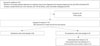
According to the renal colic management protocol of our emergency department (Fig. 1), all patients undergo evaluation using a detailed medical history; physical examination; urinalysis; complete blood count; routine serum chemistry measurements; ESR measurement; CRP measurement; kidneys, ureters and bladder radiography (KUB); and non-contrast-enhanced computed tomography (NCCT). The diagnosis of ureter stones was based on the presence of an unequivocal finding of a stone on NCCT. At the outpatient urology department visit, patients were asked about pain severity or complications and whether they observed any sensation or stone fragments during urination. Plain radiography, urinalysis, and KUB were performed routinely at each follow-up visit. For all patients, our institutional protocol is to perform a NCCT at 3 weeks from the first stone episode if the stone was not spontaneously expelled. For patients who did not experience SSP, whether they continued follow-up for another 2 weeks or underwent intervention was based on their physician's discretion and the patient's preference. Patients who did not receive complete evaluation at initial visit or those who did not complete follow-up at 3 weeks were excluded from analysis.
Predictors of SSP were investigated based on the patient's laboratory and radiographic results evaluated at the emergency department. Stone size was defined by the stone's largest diameter and was stratified into groups: those measuring up to 5 mm and those measuring 5–10 mm. The location of the stone was classified into two groups based on the stone's anatomical position in the upper or lower ureter. Plain radiographic characteristics were used to classify stones as radiopaque or radiolucent.12 Stone density was evaluated based on Hounsfield units (HU) of each stone by a single investigator (K.S.L.).
The study was performed in accordance with the principles of the declaration of Helsinki. Appropriate comparative tests, such as Student's t-test and the χ2 test, were used to compare continuous and categorical variables. Univariate and multivariate analyses were performed according to proportional regression models in order to adjust for potential confounders in predicting SSP. The cut-off values for parameters were determined using the area under the receiver operating characteristic curve. Statistical analysis was performed using SPSS version 23 (SPSS Inc., Chicago, IL, USA). All tests were two-sided, with statistical significance set at p<0.05.
RESULTS
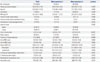
Patient characteristics are shown in Table 1. Eighteen patients (13.7%) did not complete follow-up at 3 weeks. Of 113 (86.3%) patients included for analysis, SSP was observed in 90 (79.6%) patients. Of the 23 (20.4%) patients who did not experience SSP, 11 (47.8%) underwent additional treatment because of failure to expel the stone spontaneously and uncontrollable pain. Ten (90.9%) patients underwent ureteroscopic stone removal, and one (9.9%) patient underwent extracorporeal shock wave lithotripsy.
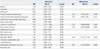
Predictors for SSP are presented in Table 2. In the univariate analysis, SSP was significantly associated with smaller stones (p<0.001), stones located in the lower ureter (p=0.002), previous ureter stone history (p=0.006), previous ureter stone treatment history (p=0.004), percent of neutrophil count (p=0.020), and the NLR (p=0.025).
In the multivariate analysis, stones located in the upper ureter [lower vs. upper: odds ratio (OR), 11.54; 95% confidence interval (CI): 2.889–46.088; p=0.001], size of the stone (≤5 mm vs. 5–10 mm) (OR, 8.16; 95% CI: 2.272–29.285; p=0.001), and NLR (<2.3 vs. ≥2.3) (OR, 9.03; 95% CI: 2.125–38.353; p=0.003) were found to be independent predictors of SSP (Table 2). Unexpectedly, traditional predictors of SSP, namely pyuria, hydronephrosis, and previous ureter stone history, were not associated with SSP in our study. To exclude potential multicollinearity between neutrophil count and NLR, variance inflation factors for these variables were analyzed. There was no harmful collinearity among these variables confirmed by coefficients of variance inflation factors of below 3.6.
DISCUSSION
The present study evaluated the predictors of SSP at 3 weeks for patients with ureter stones ≤10 mm in size. Stone location (lower), stone size (≤5 mm), and NLR (<2.3) were significant positive predictors of SSP. Observation until SSP might cause patients unwanted complications, such as recurrent attacks of renal colic and urinary tract infections. In previous studies, the follow-up strategy for patients with ureter stones varied according to the clinician's preference. Therefore, the 3-week follow-up strategy used in our institution was established for patient monitoring and to help the physician make proper treatment decisions.
The European Association of Urology and the American Uro-logical Association (AUA) guidelines state that the rate of SSP significantly differs according to the stone's location within the ureter. Several studies have examined the role of the stone's location in SSP.131415 Morse and Resnick13 reported that passage rates from the proximal, middle, and distal ureter were 22, 46, and 71%, respectively, from a cohort of 378 patients. In an analysis of 850 patients from six retrospective studies, Hübner, et al.14 reported that the passage rate (without respect to stone size) was 48% from the proximal ureter, 60% from the mid ureter, and 75% from the distal ureter. In our study, the SSP rate was 62.2% from the upper ureter and 88.2% from the lower ureter. Our results are consistent with previous results.
According to the AUA guidelines, 98% of stones <5 mm in size are passed spontaneously with conservative management.16 In a meta-analysis of 224 patients, 68% (95% CI: 46–85%) of patients with stones ≤5 mm passed them spontaneously, and for 104 patients with stones >5 mm but ≤10 mm, 47% (95% CI: 36–59%) passed them spontaneously.12 This study showed that the SSP rate for patients with stones <5 mm was 62.2% and was 88.2% for those with stones 5–10 mm in size. This result for the SSP rate within 3 weeks was not similar to the results presented in previous studies with respect to stone size.
The NLR is a parameter that can be used to evaluate the inflammatory status of a patient. It has proven useful as a prognostic factor in major cardiac events, in several types of cancers, and postoperative complications, as well as a marker of inflammatory or infectious states.1718192021 Our results indicate that NLR could also be utilized for patients with urinary stones as an objective proxy for SSP. Forget, et al.22 reported that the normal NLR values for non-geriatric adults in good health were between 0.78 and 3.53. In a representative sample of 9427 subjects in the United States, the average NLR was 2.15 in the general population.23 The median NLR of the 113 patients in our study was 2.18, and the median NLR in patients who experienced SSP was 2.04. Patients who did not experience SSP had a higher NLR (3.67) than those with SSP, which supports the notion that inflammation plays an important role in the pathophysiology of SSP.
The parameters related to inflammatory changes, including serum CRP concentration, hydronephrosis, and pyuria, and NCCT findings of perinephric fat stranding and the tissue-rim sign were presented as predictive factors for SSP. The median serum CRP concentration in this study was relatively low but within the normal range, and no relation between CRP and SSP was found. Ibrahim, et al.6 reported that conservative treatment was successful when there was no significant pyuria in 125 patients with ureter stones ≤10 mm in size. However, we could not confirm this result in our cohort because of the small number of patients with significant pyuria. Based on a cohort of 66 patients with ureter stones, Takahashi, et al.24 reported that hydronephrosis is associated with a lower likelihood of stone passage. In the current study, all patients had hydronephrosis based on NCCT findings; therefore, we could not analyze wh-ether hydronephrosis was a predictor.
Inflammatory changes in the ureter provoke a reduction in the rate of SSP; therefore, anti-inflammatory drugs, such as NSAIDs and steroids, are generally considered in order to increase SSP rates. In this cohort, all patients received anti-inflammatory drug management to relieve symptoms and promote SSP. With respect to multiple management options for ureter stones, medical expulsive therapy (MET) using alpha bl-ockers has been recommended for muscle relaxation of the lower ureter.25 However, medication for MET, such as calcium channel blockers and alpha-blockers, cannot be routinely used in Korea, because it is not reimbursed for patients with ureter stones. In this study, no patients underwent MET.
A large proportion of patients with a history of ureter stone experiences recurrence within 5 years of the first episode.262728 Clinicians might have difficulty in making treatment decisions for patients who have previously experienced SSP, because previous SSP history might have caused permanent changes in the ureter from inflammation. However, previous SSP history was recently found to be a positive predictive factor for SSP in a prospective clinical study of 251 patients.5 To analyze these conflicting differences, we evaluated previous ureter stone history, previous ureter stone treatment history, and SSP history. SSP history was not found to be a significant parameter in univariate analysis. Previous ureter stone history and previous ureter stone treatment history, including ureteroscopy and extracorporeal shock wave lithotripsy, were not found to be independent predictors of SSP in multivariate analysis.
Our study has an important strength. To our knowledge, it is the first study to investigate NLR as a predictive factor for SSP. Our findings may provide evidence for the development of new therapeutic targets in the management of ureteral stones. However, there were also some limitations. First, the small number of patients might have influenced the results. Further studies with a larger number of patients are required to determine the detailed clinical relevance of our findings in order to aid clinicians in decision-making for selecting patients with ureter stones who should undergo simple observation. Additionally, it is notable that the proportion of patients not enrolled in the study or lost to follow-up was only 13.7%, so a selection bias may have existed. Finally, the level of compliance was difficult to confirm in all patients with respect to fluid intake; patients were asked to consume at least 2 L of fluids daily. Our results were obtained after a relatively short follow-up period for patients under surveillance. The follow-up period should be longer for further evaluation, and multicenter trials are required to clarify whether the newly identified parameters are feasible in this study. In addition, we plan to conduct image an-alysis, such as HU, and the component of stone for the patients who did not experience SSP.
In conclusion, the size and location of ureter stones and low NLR (<2.3) were independent predictors of SSP in patients with ureter stones <1.0 cm in size. Our result supports the notion that ureteral inflammation plays an important role in SSP. Early intervention, rather than expectant management, may be considered for patients presenting with high NLR (≥2.3) at initial stone episode.
 XML Download