

 PDF
PDF ePub
ePub Citation
 Print
Print


INTRODUCTION
Tumor-induced osteomalacia (TIO), also called oncogenic osteomalacia, is a rare paraneoplastic disorder characterized by renal phosphate wasting resulting in hypophosphatemia and impaired bone mineralization. Patients with TIO usually present with musculoskeletal pain or muscle weakness and are susceptible to fractures. Typical biochemical patterns include hypophosphatemia, normocalcemia, normal or low levels of 1,25-dihydroxyvitamin D [1,25(OH)2D], and elevated levels of alkaline phosphatase. TIO is usually caused by a small slowly growing tumor known as the phosphaturic mesenchymal tumor mixed connective tissue variant (PMTMCT).12 Complete surgical resection is required for a cure; however, identification of the causative tumor is often very difficult. Imaging modalities, such as magnetic resonance imaging (MRI), F-18 fluorodeoxyglucose positron emission tomography with computed tomography (FDG-PET/CT), and 111Indium-octreotide scintigraphy are currently used for localization of the tumor.3
Recently, fibroblast growth factor 23 (FGF23) was found to be the main pathophysiological factor for the development of TIO, because FGF23 reduces the expression of type 2a and 2c sodium-phosphate cotransporter (NPT2a and NPT2c) in the renal proximal tubule, thereby leading to increased phosphate excretion.4 Circulating FGF23 levels have been shown to be elevated in patients with TIO and to decrease to normal levels after removal of the tumor.1 Several studies have further shown that systemic venous sampling of FGF23 is helpful for localization of the tumor.5678910 Therefore, our aim was to test clinical utility of systemic venous sampling of FGF23 in patients with suspected TIO in Korean patients.
MATERIALS AND METHODS
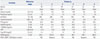
Six patients admitted to our hospital between 2006 and 2015 were analyzed. Symptoms and laboratory findings showing hypophosphatemia and hyperphosphaturia were compatible with TIO. No patients had a family history of hypophosphatemic rickets. In cases 2 and 5 (young-age onset), a genetic test for ph-osphate-regulating endopeptidases homolog, X-linked (PHEX), dentin matrix protein 1 (DMP1), and FGF23 was done for differential diagnosis of hereditary hypophosphatemic disorders; these results were all negative. Laboratory data are summarized in Table 1.
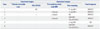
Functional imaging studies of 111Indium-octreotide scintigraphy and FDG-PET/CT were performed first, but revealed negative results in five patients. Subsequent whole body MRI revealed suspicious lesions in three patients. Thus, systemic venous sampling of FGF23 was conducted to detect the blind lesion or to confirm that a suspicious lesion on previous imaging is the culprit tumor. Localized imaging at the region with high FGF23 levels was used next (Table 2).
Systemic venous sampling of FGF23 was performed after obtaining written informed consent. By a single radiologist with expertise in diagnostic intervention radiology, a catheter was inserted through the femoral vein, and blood samples were collected at 10–14 sites, including the bilateral internal jugular veins, bilateral subclavian veins, superior vena cava, inferior vena cava, bilateral common iliac veins, bilateral femoral veins, and bilateral popliteal veins. FGF23 was quantified by means of a commercially available enzyme-linked immunosorbent assay kit (Kainos Laboratories, Tokyo, Japan). The diagnostic sensitivity was 3 pg/mL with a measurement range of 3–800 pg/mL. The inter- and intra-assay coefficients of variation were 4% or less and 3% or less, respectively.
RESULTS
Case 1
A 50-year-old man had lower-limb weakness and pain for 3 years. He had no medical or family history of metabolic bone or muscular diseases. Initially, he visited the neurology department, and neurologic and laboratory examinations yielded normal results, except for hypophosphatemia and elevated levels of alkaline phosphatase. A whole body bone scan showed multifocal radiotracer uptake indicative of a metabolic bone disease. Further evaluation revealed a reduction in maximal tubular reabsorption of phosphate corrected for glomerular filtration rate (Tmp/GFR), suggestive of a diagnosis of TIO. However, 111Indium-octreotide scintigraphy and whole body MRI were not helpful for localization of the tumor, and we decided to perform venous sampling of FGF23.
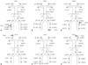
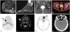
The patient's serum FGF23 level in a peripheral vein was 375.1 pg/mL (reference range: 10–50 pg/mL) and was the highest in the right profunda femoris vein (408.6 pg/mL, 1.2-fold higher than the peripheral concentration) (Fig. 1A). These findings were indicative of the presence of a FGF23-producing tumor adjacent to the right upper leg. MRI of the right leg revealed a 2.7-cm mass in the gluteus maximus muscle (Fig. 2A). The patient underwent surgical treatment, and diagnosis of PMTMCT was made. Serum phosphate and FGF23 levels normalized.
Case 2
A 17-year-old boy had pain in both knees and ankles for 4 years. He had a history of a femur trochanter fracture after falling and underwent recurrent surgical treatment due to nonunion at age 15 and a distal humerus fracture after arm-wrestling at age 16. Laboratory data showed low levels of serum phosphate and upregulated alkaline phosphatase. Bone mineral density (BMD) measured by dual-energy X-ray absorptiometry revealed severely low bone mass in the lumbar spine (Z-score −3.7) and total hip (Z-score −5.0). Mutations of PHEX, DMP1, and FGF23 were absent, suggestive of TIO. 111Indium-octreotide scintigraphy and FDG-PET/CT did not show a suspected tumor, and whole body MRI identified a 1-cm focal lesion in the right mi-dtibia, raising suspicions of a hemangioma or osteomalacic lesion. He underwent a surgical intervention, but histopathological examination revealed woven bone formation. There was also no improvement in symptoms. Therefore, we performed systemic venous sampling of FGF23.
The serum FGF23 level in a peripheral vein was 1231.6 pg/mL and was the highest in the left femoral vein (1435.6 pg/mL), 1.3-fold higher than the peripheral concentration (Fig. 1B). MRI of the left leg was done, and we found a 4.2-cm mass in the posterior aspect of the ankle abutting the peroneal artery and vein (Fig. 2B). By a two-stage operation, the tumor was completely resected, and histopathological analysis revealed PMTMCT. Serum phosphate and FGF23 rapidly normalized. The lumbar spine and total hip BMD increased by 40% (lumbar spine Z score −1.2, total hip Z score −1.7) after 5 months and finally normalized after 17 months.
Case 3
A 48-year-old man was referred to us with complaints of hip pain and decreased height (by 5 cm) for 1 year. Biochemical analysis showed hypophosphatemia, decreased Tmp/GFR, and 24-hour urinary glucose at 15057 mg (reference range: 500–1500 mg/day) without diabetes. We suspected both acquired Fanconi syndrome and TIO. However, characteristics of typical Fanconi syndrome, including metabolic acidosis, proteinuria, or hypouricemia, were all negative. 111Indium-octreotide scintigraphy yielded negative results. However, FDG-PET/CT and MRI identified a 0.8-cm round lesion in the left femoral head (Fig. 2C and D). Thus, systemic venous sampling of FGF23 was conducted.
The serum FGF23 level in a peripheral vein was 204.4 pg/mL and was the highest in the left common iliac vein (380.4 pg/mL, 1.9-fold higher than the peripheral concentration) (Fig. 1C). These results strongly suggested that the tumor in his left femoral head was responsible for the TIO. After left total hip replacement for removal of the tumor, serum FGF23 decreased to an undetectable level, and symptoms improved significantly. The pathologic diagnosis was PMTMCT.
Case 4
A 33-year old man was admitted with aggravating back pain. He began to experience the pain and stiffness at the age of 20 years, and symptoms continued to worsen. Laboratory examination showed hypophosphatemia and decreased Tmp/GFR. Imaging by 111Indium-octreotide scintigraphy and FDG-PET/CT yielded negative results, while whole body MRI revealed a small mass in his right tibia with decreased trabeculation. He underwent surgical intervention, and histopathological examination revealed an intraosseous lipoma. Postoperatively, he was treated with calcium, vitamin D, and oral phosphate. At 5-year follow-up, he complained of lower-leg pain again, and follow-up MRI revealed a newly developed 2.8-cm enhancing mass in the right proximal tibia, suggestive of an intraosseous lipoma. To verify that the mass was responsible for TIO, we performed systemic venous sampling of FGF23.
The serum FGF23 level in a peripheral vein was 86.7 pg/mL and lower than average in the right femoral vein (78.7 pg/mL) (Fig. 1D). These results suggested that the mass found on MRI was a lipoma, rather than an FGF23-producing tumor. Because the FGF23 concentration in both jugular veins were respectively 125.5 and 124.1 pg/mL, 1.3-fold higher than average (the highest), secondary venous sampling was focused on head and neck veins, including the bilateral subclavian vein, internal jugular vein, sigmoid sinus vein, transverse sinus vein, and superior and inferior sagittal sinus veins. FGF23 levels were highest in the right internal jugular vein (144.3 pg/mL), although the difference in comparison to the other veins was minimal. MRI of the head and neck did not identify a suspicious lesion either. Accordingly, he received medical supplementation with calcium, active vitamin D, and oral phosphate.
At 3-year follow up, we conducted 68Ga-DOTATOC PET/CT. We noted increased gallium uptake in the right mandible (Fig. 2E). Subsequent neck MRI demonstrated focal enhancement in the right mandible and adjacent buccal mucosa (Fig. 2F). As he had a history of tooth extraction (#46, 47) of that area with chronic periodontitis, an excisional surgery was performed. Histopathologic examination revealed giant cell granuloma with unmineralized osteoid formation, an entity that may be included in the PMTMCT.11 His serum phosphate and FGF23 levels normalized after the removal without medication. We planned to carefully follow up laboratory and clinical signs.
Case 5
A 20-year-old man was referred to us with pain in both wrists lasting for 10 years. A laboratory investigation revealed hypophosphatemia and markedly decreased Tmp/GFR. Because of suspected hypophosphatemic rickets, a genetic test was performed, although the results were all negative. Various imaging modalities, including 111Indium-octreotide scintigraphy, FDG-PET/CT and whole body MRI, were conducted to detect the causative lesion; however, all yielded negative results. We decided to perform systemic venous sampling of FGF23.
The serum FGF23 level in a peripheral vein was 136.7 pg/mL, with a peak in his left peroneal vein (221.3 pg/mL, 1.4-fold higher than the peripheral level) (Fig. 1E). However, subsequent localized MRI failed to detect the responsible tumor. He is still on medical replacement of calcium, active vitamin D, and oral phosphate.
Case 6
A 52-year-old man presented with generalized bone pain lasting for 6 months. He had a history of surgical treatment of a vertebral fracture at age 50 years, and his laboratory findings were compatible with TIO. 111Indium-octreotide scintigraphy, FDG-PET/CT, and whole body MRI were performed, but with negative results. Thus, we conducted systemic venous sampling of FGF23.
The serum FGF23 level in a peripheral vein was 492.3 pg/mL and was highest in the right jugular vein (576.3 pg/mL), 1.2-fold higher than the peripheral level (Fig. 1F). However, MRI of the head and neck did not reveal a suspicious tumor. Accordingly, he was treated with medical replacement of calcium, active vitamin D, and oral phosphate.
At 3-year follow-up, we performed 68Ga-DOTATOC PET/CT. There was increased gallium uptake in the left ethmoid and nasal cavity (Fig. 2G). Subsequent CT confirmed a 1.8-cm, soft-tissue lesion in the left ethmoid sinus (Fig. 2H). He underwent surgical treatment, and PMTMCT was diagnosed. Because the resection was incomplete, postoperative radiation therapy was administered. After completion of the radiation therapy, serum phosphate levels normalized, and MRI showed no residual visible tumor.
In summary, five patients out of six achieved a complete cure after surgical resection, and only one patient is still on medical supplementation. Diagnostic modalities helpful for detection of the tumor were venous sampling of FGF23 (83%, 5/6), followed by post-sampling localized MRI (cases 1, 2, and 3) and 68Ga-DOTATOC PET/CT (case 4, 6). In case 5, an approximate location was suggested by venous sampling of FGF23; however, MRI of the suspicious site could not identify the lesion. Both 111Indium-octreotide scintigraphy (0%, 0/6) and FDG-PET/CT (20%, 1/5) were not helpful in our cases.
DISCUSSION
TIO was first reported in 1947 by McCance12 who described a 15-year-old girl with symptoms of TIO and resistance to vitamin D treatment. While her symptoms were resolved after tumor resection, the relationship between the tumor and bone disease was not clearly recognized.12 In 1959, Prader, et al.13 recognized that a “rachitogenic” substance secreted by the tumor was the cause of TIO. The tumor responsible for TIO is small and grows slowly, and is usually located in bone, a soft tissue, or the head or neck. Thus, this lesion is difficult to identify by conventional imaging methods.14 This situation delays diagnosis often by several years: it takes up to 2.5 years to recognize TIO after patients experience symptoms and usually more than 5 years to localize the causative tumor.1516
111Indium or 99mTechnetium octreotide scintigraphy has been used for the detection of TIO, because a mesenchymal tumor causing TIO often expresses somatostatin receptors (SSTRs), which enable visualization of the tumor by octreotide scintigraphy.17 In studies by Jiang, et al.18 46 of 94 patients (48%), in whom the tumor was not localized successfully by other imaging modalities, showed a tumor on octreotide scintigraphy. Forty of them underwent surgical treatment, and in 37 patients, the diagnosis was histologically confirmed.
FDG-PET/CT has shown good sensitivity in various reports. 192021 In the largest survey, which was conducted by Jagtap, et al.20 FDG-PET/CT localized tumors in seven of eight patients (87%). TIO was pathologically confirmed in four patients, was false positive in one patient, and histopathological confirmation was not performed in two patients. In a recent trial by Agrawal, et al.21 FDG-PET/CT detected a tumor in two out of four patients (50%), and TIO was confirmed by histopathology. Meanwhile, in the present study, only one of five patients (20%, case 3) showed increased FDG uptake in FDG-PET/CT, far below what was expected. This could be explained as follows: the tumor grows very slowly and, thus, is less hypermetabolic or vascular than can be detected by FDG-PET/CT.
FGF23 is a well-known phosphaturic factor, “phosphatonin,” secreted by the tumors causing TIO.22 Other known potential phosphatonins include secreted frizzled related protein 4 and matrix extracellular phosphoglycoprotein. They play a major role in the pathophysiology of TIO, autosomal dominant hypophosphatemic rickets, and X-linked hypophosphatemia.23 FGF23 is a polypeptide hormone belonging to the FGF19 superfamily. It is produced by bone and exerts effects in the kidneys. By binding to target cells forming the Klotho-FGF receptor complex, it transduces signals to inhibit NPT2a and NPT2c transporters in the proximal renal tubule, thereby activating renal phosphate excretion. FGF23 also plays a role in 1,25(OH)2D metabolism by inhibiting 1α-hydroxylase, leading to a decrease in 1,25(OH)2D levels.13 FGF23 concentration is elevated in patients with TIO and normalizes after surgical resection of the tumor.24
Venous sampling of FGF23 was first used for the diagnosis of TIO in 2004,6 and additional case reports followed.578910 Of a total of 28 patients, researchers were able to localize tumors in 19 patients (67%) by FGF23 sampling, and the patients achieved a complete cure after surgical treatment. In the present study, FGF23 sampling was useful in five of six patients (83%). They (cases 1, 2, 3, 4, and 6) had an especially high FGF23 level in a certain vein, and the relevant anatomical lesion was found by subsequent additional imaging studies. In contrast, one patient (cases 5) had elevated FGF23 levels in a focal area but no plausible lesion on subsequent imaging. This result suggests that venous sampling of FGF23 is helpful for estimating the location of the tumor, as well as confirming the diagnosis in the presence of the related lesion on other imaging modalities.
At present, SSTR imaging can be performed with better resolution by means of 68Ga-DOTATOC, 68Ga-DOTANOC, or 68Ga-DOTATATE PET/CT. This approach combines the specificity of octreotide scintigraphy and sensitivity of PET/CT, thus emerging as a new diagnostic tool.2125 In one study comparing 111Indium-octreotide scintigraphy and 68Ga-DOTATATE PET/CT in five patients, octreotide scintigraphy detected tumors in only one patient, while 68Ga-DOTATATE PET/CT identified tumors in all five patients.26 This finding can be explained by a higher affinity for SSTR2 in 68Ga-DOTA conjugated PET/CT, which allows for visualization of a previously undetectable tumor.27 In our study, octreotide scintigraphy was not helpful for diagnosis in all six patients, while 68Ga-DOTATOC PET/CT helped to detect the tumor in cases 4 and 6.
Nevertheless, this does not mean that FGF23 sampling has low clinical value. In cases 2 and 4, patients underwent surgical treatment according to the assumption that the mass detected by MRI was responsible for TIO. However, we later found that this mass was misidentified. There is a chance that MRI will detect a lesion false positively; moreover, it is impossible to determine whether a mass is responsible for TIO by means of imaging only. In such circumstances, venous sampling of FGF23 may be helpful by demonstrating the location of the FGF23-producing tumor. In case 4, the patient was able to avoid an unnecessary surgical procedure, because the location of elevated FGF23 was not relevant to the mass visible on MRI.
In terms of threshold values, the highest level of FGF23 was 1.2- to 1.5-fold higher than the peripheral level, lower than that in other case reports in which a 1.5- to 3.0-fold elevation was recorded.510 Only a 20% increase can be useful for detecting tumors when there is an overall rise in FGF23 levels.
This study has some limitations: we did not perform mutation testing in all patients; therefore, genetic causes of hypophosphatemia could not completely ruled out. In addition, we analyzed only a small patient population (in whom we conducted systemic venous sampling of FGF23), because TIO is a very rare disease. Despite these limitations, we showed the value of venous sampling of FGF23 for predicting the location of the tumor in Korean patients for the first time. This approach can be more powerful in the presence of a related lesion on subsequent imaging studies.
 XML Download