Journal List > Yonsei Med J > v.58(4) > 1032209

Lim, Uhm, and Pak: Focal Atrial Tachycardia Arising from the Inferior Vena Cava

Abstract

The inferior vena cava (IVC) is a rare site of focal atrial tachycardia (AT). Here, we report a 20-year-old woman who underwent catheter ablation for anti-arrhythmic drug-resistant AT originating from the IVC. She had undergone open-heart surgery for patch closure of an atrial septal defect 17 years previously and permanent pacemaker implantation for sinus node dysfunction 6 years previously. The AT focus was at the anterolateral aspect of the IVC-right atrial junction, and it was successfully ablated under three-dimensional electroanatomical-mapping guidance. We suspect that the mechanism of this tachycardia was associated with previous IVC cannulation for open-heart surgery.

INTRODUCTION

The inferior vena cava (IVC) is a rare site of focal atrial tachycardia (AT), and AT can occur long time after open heart surgery.1 Although three-dimensional (3D)-electroanatomical mapping provides detailed electrophysiology, a review of surgical record is important to understand the mechanism of tachycardia.2 Here, we report a case of a successfully ablated focal AT originating from the IVC-right atrial (RA) junction, confirmed by 3D electroanatomical mapping 17 years after surgery for atrial septal defect (ASD).

CASE REPORT

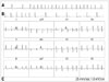
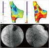
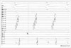
A 20-year-old woman was admitted to our hospital because of recurrent episodes of palpitation. She had a history of ASD patch closure surgery at the age of 3 years and permanent pacemaker implantation (DDDR type) for sick sinus syndrome at the age of 13 years. For ASD closure, the surgeon utilized an autologous pericardial patch and performed venous cannulations at the superior vena cava (28 Fr) and the IVC (30 Fr) for cardiopulmonary bypass. Computed tomography and transthoracic echocardiography revealed normal left ventricular function, and there was no evidence of residual ASD shunt or other structural heart diseases. Over the past 2 years, she experienced highly symptomatic AT, and the frequency and duration showed an increasing tendency. Tachycardia was induced and terminated abruptly (Fig. 1A and B). On electrocardiography, P-wave morphology was biphasic in V1; negative in II, III, and aVF; and positive in I and aVL. AT was characterized by P-waves separated by an isoelectric interval, suggesting a focal mechanism of AT (Fig. 1C). Because this tachycardia was only partially responsive to flecainide and β-blockers, we decided to perform catheter ablation. Although focal ectopic AT was suspected as the mechanism, we mapped AT with 3D electroanatomical mapping (NavX, St. Jude Medical, St. Paul, MN, USA) because the patient had a history of cardiac surgery. The tachycardia was found to originate from the anterolateral aspect of the IVC-RA junction, and it showed centrifugal activation with some irregularity during mapping (Fig. 2). There were low-amplitude, fractionated, and double potentials at the earliest activation site (Fig. 3). AT was successfully terminated 5 seconds after a single radiofrequency application (5-mm Blazer II Catheter, EP Technologies Inc., San Jose, CA, USA; 50 W and 60℃), and thereafter, it was not inducible with pacing with or without isoproterenol infusion. The procedure was completed without any complications.

DISCUSSION

AT in patients with surgically corrected congenital heart disease is generally caused by a macro-reentry mechanism.13 However, we described herein a case of focal AT originating from the IVC-RA junction, which was a cannulation site for cardiopulmonary bypass during ASD surgery performed 17 years earlier. The IVC is a rare site of focal atrial arrhythmia.45 Kato, et al.6 reported a case of IVC tachycardia late after cardiopulmonary bypass with IVC cannulation. In the present case, we mapped a similar IVC tachycardia with 3D electroanatomical mapping and ablated it under 3D electroanatomical-mapping guidance. Although there were three case reports regarding focal AT arising from the IVC so far,678 this was the first case report to demonstrate the mechanism of AT originating from IVC using 3D mapping. 3D electroanatomical mapping allows reconstruction of AT mechanisms and represents an advance in the precise localization and ablation of the arrhythmogenic substrates of post-surgical AT.238 Due to overlapping in electrophysiologic characteristics, the question of whether the mechanism of this AT was enhanced automaticity or micro-reentry is unclear. Low-amplitude, fractionated, and double potentials around the ablation target site, as well as reproducible AT induction with critical range of pacing cycle length, favor reentrant mechanisms. However, cycle length variations during tachycardia suggest non-reentrant mechanisms. AT was terminated with a single application of radiofrequency energy.
Although Murphy, et al.9 demonstrated that the development of late AT after surgical ASD repair is low in patients operated during childhood, an arrhythmogenic substrate can be produced with cardiopulmonary bypass cannulation, creating arrhythmogenic muscular sleeves with an electrical connection between the IVC and RA. These findings suggest that patients who undergo cardiopulmonary bypass using vena cava cannulation can present with IVC tachycardia.10
In conclusion, we reported herein a case of IVC tachycardia, presumably related to earlier cardiopulmonary bypass cannulation performed for open-heart surgery. The tachycardia was successfully ablated under 3D electroanatomical-mapping guidance.

Figures and Tables

Fig. 1

Electrocardiography (ECG) rhythm strip showing the initiation (A) and termination (B) of clinical atrial tachycardia (AT). The initiation of AT occurs with atrial extrastimuli and terminates abruptly. The patient's basic self-rhythm is junctional rhythm because of sinus node dysfunction after cardiac surgery. A 12-lead ECG of the clinical AT (C).

ymj-58-884-g001
Fig. 2

Three-dimensional (3D) activation map (A). The focal pattern of activation is demonstrated using electroanatomical mapping. The earliest activation site of the tachycardia near the junction between the inferior vena cava (IVC) and right atrium (RA) is shown on the anatomical reconstruction of the RA. The area in white represents the earliest activation region during initiation of the propagation sequence. 3D voltage map (B). Endocardial voltage mapping in the RA is demonstrated. The voltage amplitudes of the sample points (small yellow dots) are assessed, and the points are set to a color scale as indicated in the figure. Large, irregular regions of extremely low voltage (red) consistent with myocardial scarring can be detected over the entire RA. Fluoroscopic projections showing the intracardiac positions of the ablation catheter (C). The catheter ablation site located in the IVC-RA junction is presented in the left anterior oblique view (LAO) and right anterior oblique view (RAO).

ymj-58-884-g002
Fig. 3

Intracardiac electrograms and ablation signals at the inferior vena cava, where the local electrogram recorded from the distal electrodes of the ablation catheter during tachycardia preceded the onset of the earliest atrial activation (H9, 10) by 40 ms. ABL, ablation; HRA, high right atrium; HIS, His bundle; H, duodecapolar catheter.

ymj-58-884-g003

Notes

The authors have no financial conflicts of interest.

References

1. de Groot NM, Zeppenfeld K, Wijffels MC, Chan WK, Blom NA, Van der Wall EE, et al. Ablation of focal atrial arrhythmia in patients with congenital heart defects after surgery: role of circumscribed areas with heterogeneous conduction. Heart Rhythm. 2006; 3:526–535.
crossref
2. Magnin-Poull I, De Chillou C, Miljoen H, Andronache M, Aliot E. Mechanisms of right atrial tachycardia occurring late after surgical closure of atrial septal defects. J Cardiovasc Electrophysiol. 2005; 16:681–687.
crossref
3. Lukac P, Pedersen AK, Mortensen PT, Jensen HK, Hjortdal V, Hansen PS. Ablation of atrial tachycardia after surgery for congenital and acquired heart disease using an electroanatomic mapping system: Which circuits to expect in which substrate? Heart Rhythm. 2005; 2:64–72.
crossref
4. Scavée C, Jaïs P, Weerasooriya R, Haïssaguerre M. The inferior vena cava: an exceptional source of atrial fibrillation. J Cardiovasc Electrophysiol. 2003; 14:659–662.
5. Yamane T, Miyazaki H, Inada K, Matsuo S, Miyanaga S, Date T, et al. Focal source of atrial fibrillation arising from the ostium of the inferior vena cava. Circ J. 2005; 69:756–759.
crossref
6. Kato Y, Horigome H, Takahashi-Igari M, Aonuma K. Focal atrial tachycardia originating from inside the inferior vena cava late after surgical repair of congenital heart defects. Pediatr Cardiol. 2011; 32:846–848.
crossref
7. Higa S, Tai CT, Lin YJ, Liu TY, Lee PC, Huang JL, et al. Focal atrial tachycardia: new insight from noncontact mapping and catheter ablation. Circulation. 2004; 109:84–91.
crossref
8. Katsivas AG, Manolis AG, Vassilopoulos C, Ioanidis P, Giotopoulou A, Kyriakides Z. Electroanatomical mapping of a right atrial tachycardia originating within the inferior vena cava. Hellenic J Cardiol. 2004; 45:187–190.
9. Murphy JG, Gersh BJ, McGoon MD, Mair DD, Porter CJ, Ilstrup DM, et al. Long-term outcome after surgical repair of isolated atrial septal defect. Follow-up at 27 to 32 years. N Engl J Med. 1990; 323:1645–1650.
crossref
10. Pap R, Kohári M, Makai A, Bencsik G, Traykov VB, Gallardo R, et al. Surgical technique and the mechanism of atrial tachycardia late after open heart surgery. J Interv Card Electrophysiol. 2012; 35:127–135.
crossref
TOOLS
Similar articles