

 PDF
PDF ePub
ePub Citation
 Print
Print


INTRODUCTION
Sex hormone-binding globulin (SHBG) is a serum glycoprotein produced predominantly in hepatocyts.12 It controls the transport of sex steroid hormones in the blood circulation to their target tissues.13 Many previous studies have revealed that low levels of serum SHBG are associated with metabolic syndrome, type II diabetes, and cardiovascular disease.45 Nonalcoholic fatty liver disease (NAFLD) grade is also known to be inversely related to serum SHBG levels:67 as the grade of hepatic steatosis increases, serum SHBG level decreases.27 Hepatocytes are the primary sites of SHBG synthesis, and therefore the synthesis of SHBG could be associated with liver function.1 Various studies have investigated the relationship between SHBG and biomarkers of liver function.8 However, despite several epidemiological studies, it is still controversial as to whether serum alanine aminotransferase (ATL) is associated with SBHG. Flechtner-Mors, et al.2 found no relationship between ALT and SHBG, while Ayala, et al.9 found that SHBG and ALT were inversely related with each other.1 Whereas ALT is known to reflect hepatocellular injury or death, it is still unclear whether ALT, a liver function marker, modulates the relationship between SHBG and hepatic steatosis.10 Furthermore, ALT is a predictive marker of insulin resistance1112 and several studies have suggested that hyperinsulinemia is associated with decreased expression of SHBG.13
This study aimed to investigate the association between SHBG and hepatocyte damage represented by ALT among Korean male patients with hepatic steatosis, enrolled in a health examination program.
MATERIALS AND METHODS
Study population
This retrospective study included 2837 men and women aged >19 years old who visited the Health Promotion Center of Gangnam Severance Hospital, Yonsei University College of Medicine in Seoul, Korea. Subjects underwent routine health examination between January 2007 and July 2010. Of them, all female 1118 subjects were excluded because most female subjects did not undergo tests for serum SHBG level. Also, an additional 478 male subjects were excluded because of missing values. Subjects who were positive for hepatitis B surface antigen or hepatitis C antibody were also excluded. In addition, we excluded 143 subjects with a history of hepatocellular carcinoma and 155 subjects with alcohol consumption ≥140 g/week. Ultimately, a total of 922 men were included in this study.
Measurements
Examinations were performed by medical staff according to a standard protocol. Demographic, anthropometric, and laboratory data were gathered for each participant. The participants were asked whether they were undergoing or had recently undergone treatment for any disease. If the patient was under treatment, they were questioned for the date of diagnosis and a list of current medications. Trained staff reviewed the completed questionnaires and entered responses into the database. Participants were classified as non-smokers, ex-smokers or current smokers. Subjects were categorized by alcohol intake as a heavy drinker (alcohol ingestion ≥140 g/week) or light drinker (alcohol ingestion <140 g/week). Body weight and height were measured in light indoor clothing and no shoes to the nearest 0.1 kg and 0.1 cm, respectively. Body mass index (BMI) was calculated as the ratio of weight (kg)/height (m2). Blood pressure measurement was obtained with the participant in the sitting position, after 5 min of rest using an automated device (TM-2665P, A&D Co., LTD., Tokyo, Japan).
The diagnosis of a fatty liver was based on abdominal ultrasonography with a 3.5-MHz transducer (HDI 5000, Philips, Bothell, WA, USA).14 Ultrasonography was performed by an experienced radiologist. Participants were diagnosed with fatty liver if at least two of the following three findings were present: increased liver echogenicity, deep attenuation, and vascular blurring.
Venous blood sampling was performed after a fasting period of 12 h. White blood cell (WBC) counts were quantified by an automated blood cell counter (ADVIA 120, Bayer, Tarrytown, NY, USA). Fasting plasma glucose, total cholesterol, high density lipoprotein (HDL) cholesterol, triglycerides (TGs), and ALT were measured using a Hitachi7600-110 chemistry autoanalyzer (Hitachi, Tokyo, Japan). T3, free T4, and SHBG were measured via electrochemiluminescent methods using Modular E170 (Roche Diagnostics, Mannheim, Germany).
Statistical analysis
The distribution of SHBG was markedly skewed and log-transformed in the analyses where normality was required. ALT elevation was defined as ALT ≥40 U/L.1516 We divided the study participants into tertiles of SHBG levels with cut-points of 28.60 and 48.90 nmol/L, and defined the lowest tertile of SHBG as low SHBG.17
Multivariable logistic regression was used to assess the association of ALT elevation with risk of low SHBG. We adjusted for age, BMI, systolic blood pressure (SBP), diastolic blood pressure (DBP), WBC, glucose, T3, total cholesterol, HDL, and TG. To investigate the interrelationships among hepatic steatosis, elevated ALT levels, and risk of low SHBG, we divided our participants into the following three groups: a reference group of participants who had no hepatic steatosis and no ALT elevation, the hepatic steatosis group without ALT elevation, and the hepatic steatosis with ALT elevation group. The effect of interactions between hepatic steatosis and ALT on the risk of lower SHBG level was examined by multivariate logistic regression. Results are expressed as odds ratios (ORs) with 95% confidence intervals (CIs). All analyses were performed using SPSS software (version 18.0, SPSS Inc., Chicago, IL, USA). All statistical tests were two-sided and significance was determined at a p-value<0.05.
RESULTS
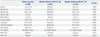
Baseline characteristics of each group are described in Table 1. In hepatic steatosis with ALT ≥40 group, SHBG was 32.5±13.7, which was significantly lower than that of the reference group (p<0.001). The subjects in the hepatic steatosis with ALT ≥40 group had higher BMI, SBP, DBP, glucose, and TG. They were more likely to have a history of diabetes. The subjects in the hepatic steatosis with ALT ≥40 group were also younger and had lower HDL cholesterol levels.
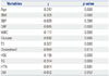
Table 2 shows the correlation analysis between SHBG and several factors known to be associated with SHBG. BMI, DBP, WBC, glucose, cholesterol, and TG were negatively correlated with SHBG, while age, T3 and HDL were positively associated with SHBG.
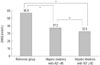
Fig. 1 shows the mean values of SHBG for the study population between the reference group, hepatic steatosis group without ALT elevation, and hepatic steatosis with ALT elevation group after adjustment for several factors. In the hepatic steatosis with ALT elevation group, the mean SHBG level was lower than all other groups.
Multivariate logistic regression models were used to evaluate the relationship between hepatic steatosis and elevated ALT levels with the risk of low SHBG levels (Table 3). In multivariable regression analysis model 1, adjusting for age and BMI, the OR for lower SHBG was 3.287 (95% CI 2.007–5.385) in the hepatic steatosis group with ALT ≥40, compared with the reference group. Even after progressive adjustment for several factors that were previously found to be associated with SHBG, the subjects in the hepatic steatosis group with ALT ≥40 were still more likely to have a lower SHBG level (OR: 2.729; 95% CI 1.591–4.681). Before we divided men into three groups, we conducted a logistic regression analysis to confirm if there is an interaction between hepatic steatosis and ALT and the risk of lower SHBG level. We found that there was a significant interaction between hepatic steatosis and ALT in men (p=0.014).
DISCUSSION
In this cross-sectional study, we examined the joint effects of hepatic steatosis and ALT on SHBG concentration. A previous observational study showed that hepatic steatosis was independently related with a low SHBG level.1 In addition , the severity of hepatic steatosis was proportionally correlated with the risk of a low SHBG level.2 A previous study also reported that serum ALT level was inversely associated with SHBG level.3 Taken together, hepatic steatosis and serum ALT may be associated with a high risk of a low SHBG concentration. However, the joint effect of hepatic steatosis and ALT on serum SHBG level has not been investigated. In multivariate logistic regression analysis, we observed that the hepatic metabolic disturbance jointly affected low SHBG concentrations independent of BMI and metabolic profiles. This is the first report showing that increased ALT could be marker of low SHBG in patients with hepatic steatosis.
Non-alcoholic fatty liver comprises a spectrum of liver condition ranging from simple steatosis to steatohepatitis and cirrhosis. Although liver biopsy is regarded as the gold standard for the assessment of the severity of fatty liver, it has limited applicability in clinical settings, because of the risk related to the technique and uncertainty of the distribution of fatty infiltration. Previous studies have reported an association between serum ALT and the grade of hepatic steatosis as confirmed by liver biopsy,18 suggesting that patients with fatty liver and higher ALT might be at increased risk for a lower SHBG level compared to patients with hepatic steatosis and a lower ALT level. Regarding these results, severity of hepatic steatosis may be closely associated with lower SHBG levels. Ayala, et al.9 also showed that serum SHBG levels were inversely associated with ALT serum level.
Level of SHBG are used in clinical practice to determine amounts of free estradiol and testosterone. However, recent studies have reported that SHBG levels are altered in several diseases, including obesity, thyroid hormone disorder, poly cystic ovarian syndrome, and Cushing's disease.1 In addition, several epidemiologic studies have shown that low SHBG levels are associated with a higher risk for developing metabolic syndrome and type 2 diabetes. Moreover, these relationships are dose-dependent and observed even if the SHBG level are within normal range.1920 Although, the potential roles of SHBG the pathogenesis of the metabolic disease remains to be elucidated, regardless of being within normal range or not, the levels of SHBG have clinical meaning.
There are some potential mechanisms by which elevated ALT levels affect serum SHBG levels. First, hepatocytes are the primary synthesis sites of SHBG, even though SHBG is also expressed in testicular germ cells. Thyroid hormone, insulin, and cytokines regulate the expression SHBG in hepatocytes.121 These hormones interact with their receptors and activate various signal cascades, resulting in alterations of hepatocyte nuclear factor 4 alpha protein levels, which regulate SHBG expression.1 Also, metabolic syndrome, sex hormone, dietary, and liver fat influence serum SHBG levels. Hua, et al.22 reported that serum SHBG level was inversely associated with NAFLD and correlated with lipid profiles. They observed a negative relationship between serum SHBG and TG and a positive relationship between HDL. In a study by Chubb, et al.23 lower SHBG was also shown to be associated with increased serum levels of smaller, denser low density lipoproteins. The relationship between serum SHBG and lipid profiles was discussed in the study of Desmeules, et al.24 focusing on post-heparin hepatic lipase and lipoprotein lipase activity. Previous studies have shown that participants with elevated lipoprotein lipase activity had a lower TG and higher HDL levels, whereas participants with higher hepatic lipase activity had a lower HDL levels. Desmeules, et al.24 observed a negative relationship between serum SHBG and hepatic lipase activity, where as a positive relationship was found between serum SHBG and lipoprotein lipase activity. This implied that correlation between lipid profiles and activity of hepatic lipase and lipoprotein lipase could affect serum SHBG levels. In the study by Miksztowicz, et al.25 hepatic lipase activity was significantly increased in patients with hepatic steatosis grade 3, compared to those with grade 1. Serum SHBG levels were associated with the high-grade NAFLD in the study by Shin, et al.7 Correlations of lipid profiles, hepatic lipase, lipoprotein lipase, NAFLD, and SHBG were noted with NAFLD and elevated liver enzymes. In the study by Ekstedt, et al.26 serum ALT levels were elevated in follow-up of NAFLD patients with progressive fibrosis. Second, almost all ALT is found in the cytosol of hepatocytes. ALT activity in the liver is about 3000 times greater than that in the serum. Thus, in cases of hepatocellular injury or death, release of ALT from damaged liver cells increases serum ALT levels.27 Therefore, elevated ALT levels could reflect hepatocellular injury or death, and thus could also be associated with a decrease of serum SHBG level. Third, insulin resistance could be another possible mechanism explaining this association between ALT and SHBG. Previous study has suggested that an increased ALT level is associated with insulin resistance.2228 A recent study proposed that glucose-induced lipogenesis may affect hepatic production of SHBG, and glucose also could directly decrease the expression of SHBG.29 In the present study, we observed an inverse relationship between serum glucose and SHBG concentration. Therefore, this finding suggested that there could be interactions among ALT, glucose and SHBG.
Our study has several limitations. First, it followed a retrospective cross-sectional method, from which it is difficult to conclude a causal relationship. Therefore, a cause-and-effect relationship among ALT and SHBG cannot be inferred. Second, although a liver biopsy is the gold standard for the diagnosis of a hepatic steatosis, a biopsy-proven NAFLD was not assessed in the present study. Despite some limitations, ultrasonography is a non-invasive and preferred modality for mass screening for hepatic steatosis with a reasonable sensitivity and specificity. Third, our study did not include sex hormone levels, which is another factor that influences SHBG synthesis. However, despite with these limitations, the present study includes other meaningful factors, particularly T3 levels which could influence SHBG levels. Fourth, exclusion criteria of this study did not comprise autoimmune hepatitis. However, in Korea, the prevalence of autoimmune hepatitis seems to be lower than that in Western countries.30 Therefore, the effect of this criterion might be small in this study.
In male patients with hepatic steatosis, elevated serum ALT levels were associated with lower serum SHBG levels. Further investigation of the association between SHBG and ALT should be pursued in future longitudinal studies.

 XML Download