Journal List > Yonsei Med J > v.58(4) > 1032182

Gwag, Chun, Hwang, Park, On, Kim, and Park: Comparison of De Novo versus Upgrade Cardiac Resynchronization Therapy; Focused on the Upgrade for Pacing-Induced Cardiomyopathy

Abstract

Purpose

This study aimed to determine whether upgrade cardiac resynchronization therapy (CRT) shows better outcomes than de novo CRT. To do so, we compared the efficacy of CRT between de novo and upgrade groups, focusing particularly on the effect of upgrade CRT on patients with pacing-induced cardiomyopathy (PiCM).

Materials and Methods

PiCM was defined as new-onset dilated cardiomyopathy following pacemaker implantation in patients with baseline normal ejection fraction ≥50%. Electro-mechanical reverse remodeling and clinical outcomes were compared among the de novo (n=62), PiCM upgrade (n=7), and non-PiCM upgrade (n=8) CRT groups.

Results

The PiCM upgrade group showed significantly greater electro-mechanical reverse remodeling than the de novo CRT or non-PiCM upgrade groups at 6-month follow-up. The rate of super-responders was significantly higher in the PiCM upgrade group than the other CRT groups. The group factor of the PiCM upgrade was identified as an independent predictor of super-responder in multivariate analysis (odds ratio 10.4, 95% confidential interval 1.08–99.4, p=0.043). During the median follow-up of 15.8 months, the PiCM upgrade group showed the lowest rate of composite clinical outcomes, including cardiac death, heart transplantation, and heart failure-related rehospitalization (p=0.059).

Conclusion

The upgrade CRT for PiCM patients showed better performance in terms of electro-mechanical reverse remodeling than de novo implantation or upgrade CRT in non-PiCM patients.

INTRODUCTION

Chronic right ventricular (RV) pacing could deteriorate left ventricular (LV) systolic function and increase mortality via pacing-induced electrical and mechanical dyssynchrony,12345 particularly in patients with pre-existing LV systolic dysfunction (LVSD).167 However, even in patients without underlying LVSD, chronic RV pacing can cause new-onset LVSD, which can be defined as pacing-induced cardiomyopathy (PiCM).38
Cardiac resynchronization therapy (CRT) corrects electro-mechanical dyssynchrony and is now established as a standard treatment for advanced heart failure (HF) patients with a pacemaker (upgrade CRT) or without a pacemaker (de novo CRT).91011 However, it remains unclear whether upgrade CRT shows better outcomes than de novo CRT,12131415 and no previous studies have investigated underlying LV function before pacemaker implantation in patients receiving upgrade CRT.12131415
We hypothesized that the efficacy of CRT would be greater in patients with PiCM (upgrade CRT for PiCM) than in those without PiCM (upgrade CRT for non-PiCM) or those treated by de novo implantation (de novo CRT). Therefore, this study aimed to compare the effect of CRT between the upgrade and de novo CRT groups, as well as between the PiCM upgrade and non-PiCM upgrade groups.

MATERIALS AND METHODS

Patients and baseline assessment

We enrolled consecutive patients who received CRT at our center from March 2012 to July 2015. Patients with 1) drug-refractory advanced HF with New York Heart Association (NYHA) class III or ambulatory IV, 2) LV ejection fraction (LVEF) <35%, and 3) QRS duration >120 ms were included. Exclusion criteria included 1) switching off the CRT device within 3 months or 2) a follow-up duration of less than 3 months after implantation.
PiCM was defined when all of the following criteria were met: 1) LVEF prior to permanent pacemaker (PPM) insertion ≥50%, 2) new-onset LVSD (LVEF <35%) in pacing-dependent (>90% RV pacing)16 patients, and 3) the absence of other causes of HF, including previous myocardial infarction, valvular heart disease, tachycardia-induced cardiomyopathy, toxic (alcohol or chemotherapy) cardiomyopathy, hypertensive cardiomyopathy, myocarditis, or other infiltrative diseases. All CRT upgrade patients who did not fulfil the criteria for PiCM were classified into non-PiCM. We categorized the patients into three groups: PiCM-upgrade, non-PiCM upgrade, and de novo CRT (Fig. 1).
Baseline information, such as NYHA functional class and echocardiographic and electrocardiographic (ECG) parameters, was collected in all patients. Echocardiographic parameters included LVEF, end-diastolic dimension (LVEDD), end-systolic dimension (LVESD), end-diastolic volume (LVEDV), and end-systolic volume (LVESV). Additionally, pre-PPM LVEF was investigated in the upgrade CRT groups. ECG parameters encompassed the duration and morphology of the QRS complex. The baseline QRS morphology was categorized as left bundle branch block (LBBB) when all of the following criteria were met: 1) QRS duration >120 ms, 2) QS or rS in lead V1, and 3) monophasic R wave with no Q wave in lead V6 and I; paced QRS complexes in the upgrade groups and native QRS complexes in the de novo group were used for this classification. We also investigated HF duration, defined as the time interval between the first time a patient received diuretics or vasodilator/inotropic therapy for HF symptoms of congestion and/or low cardiac output and CRT implantation. The Institutional Review Board at Samsung Medical Center approved the study protocol and waived the requirement for informed consent.

Device therapy

CRT implantation was performed under local anesthesia using a transvenous approach. The LV lead was preferably placed into the anterolateral, lateral cardiac veins, or posterolateral ventricular veins. An epicardial LV lead was screwed into the lateral mid-LV segment via thoracoscopic surgery unless the transvenous approach via the coronary sinus was successful. The position of the RV or LV lead was confirmed by left and right anterior oblique fluoroscopic images. RV lead positions were classified as apical or septal. LV lead positions were classified as basal, mid, or apical in the right anterior oblique view and as anterior, anterolateral, lateral, posterolateral, or posterior in the left anterior oblique view.17 After implantation, atrioventricular and ventriculo-ventricular delays were determined showing the greatest stroke volume (Doppler-guided) or narrowest QRS duration (ECG-based) before discharge.

Follow-up assessment and outcomes

The primary outcome was electrical and mechanical reverse remodeling evaluated at 6 months after CRT implantation. For assessment of electrical reverse remodeling, absolute [(pre-CRT-post-CRT)] and relative [(pre-CRT-post-CRT)/pre-CRT×100, %] change in the QRS duration was calculated. In a similar way, absolute and relative changes in LVESD, LVEDD, LVESV, LVEDV, and LVEF were calculated for mechanical reverse remodeling.
Secondary outcomes included improvement in symptoms assessed by change in NYHA class, responder rate, and long-term clinical outcomes. CRT responders were defined as follows: 1) ‘clinical responder’ for patients with any improvement in NYHA class, 2) ‘echocardiographic responder’ for patients with a relative decrease in LVESV by ≥15%, and 3) ‘super-responder’ for those with any improvement in NYHA class and a relative decrease in LVESV by ≥30%. Super-response rates were also assessed according to LVEF criteria as follows: 1) LVEF increase ≥fourth quartile or 2) LVEF ≥45% at 6 months after CRT.
Long-term clinical outcomes were assessed at the last follow-up in terms of all-cause death, cardiac death, heart transplantation, HF-related rehospitalization, and major adverse cardiac events (MACEs), encompassing cardiac death, heart transplantation, and HF-related rehospitalization.

Statistical analysis

Continuous variables are presented as medians with interquartile ranges and categorical variables are presented as numbers with percentages. Comparisons between groups were performed using chi-square tests, Mann-Whitney U tests, or Kruskal-Wallis tests, as appropriate. Survival free from adverse clinical outcomes was estimated by Kaplan-Meier curve and compared by log rank test. A Cox proportional hazard model was used to assess the risk factors for clinical composite events, and the logistic regression method was used to determine the predictors of a super-responder. Variables for which the p value was <0.2 were included in multivariate analysis. Statistical analyses were performed with the IBM SPSS Statistics version 23 (IBM Corporation, Armonk, NY, USA). All tests were two-tailed, and a p value<0.05 was considered statistically significant.

RESULTS

Patients and baseline characteristics

We identified 82 patients who underwent a CRT procedure during the enrollment period. Five of these patients were excluded because of the following reasons: two patients (one patient in the non-PiCM upgrade and one in the de novo group) were followed for less than 3 months, one patient (de novo group) died after the CRT procedure during the admission period, and two patients (de novo group) had their CRT devices turned off due to worsening HF symptoms. Of the remaining 77 patients, 62 underwent de novo implantation of CRT devices (de novo group), and 15 had their old PPM replaced by a CRT device (upgrade group). Indications for a pacemaker and RV pacing burden in CRT upgrade patients are summarized in Supplementary Table 1 (only online). Among the 15 patients in the upgrade group, seven met the criteria for PiCM, showing normal LVEF before PPM insertion and no definite cause of LVSD before CRT implantation (the PiCM upgrade group), while the remaining eight had reduced LVEF prior to PPM insertion (the non-PiCM upgrade group) (Fig. 1).
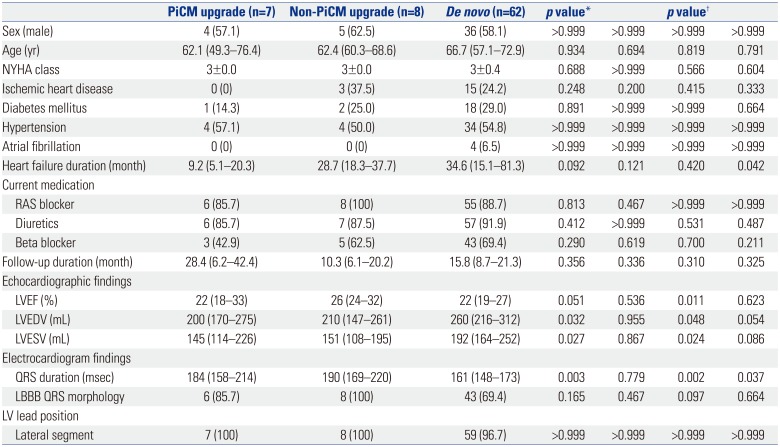
There were no differences in baseline demographic characteristics among the groups. For echocardiographic findings, the non-PiCM upgrade group showed the highest median LVEF, while LV volumes were greatest in the de novo group. QRS duration in both upgrade groups was significantly longer than that in the de novo group, and LBBB morphology tended to be more frequent in the upgrade groups. Most patients in all three groups had their LV leads in lateral (anterolateral, lateral, or posterolateral) LV segments (Table 1).

Primary outcomes

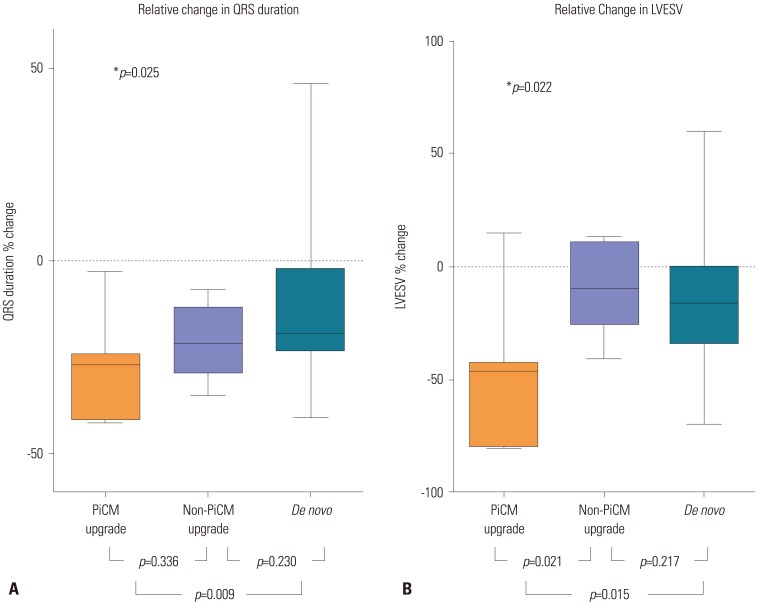
Fig. 2 and the Supplementary Table 2 (only online) show the primary outcomes. The percent decrease in QRS duration was greatest in the PiCM upgrade group (26.8% vs. 21.4% vs. 19.0%, p=0.025). The percent reduction in LVESV was significantly greater in the PiCM upgrade group than in the other two groups at 6-month follow-up (46.2% vs. 9.4% vs. 18.1%, p=0.022).

Secondary outcomes

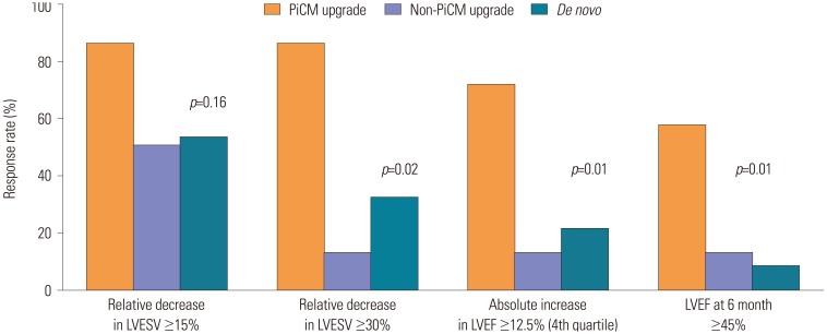
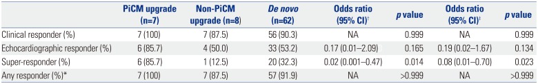
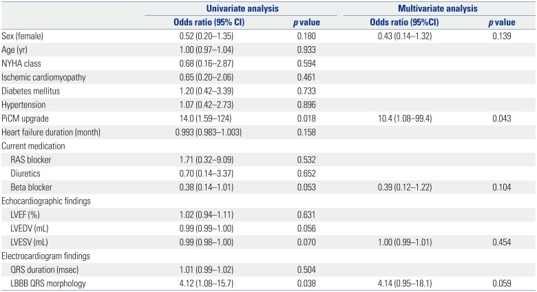
The PiCM upgrade group showed the highest responder rates, although the clinical and echocardiographic responder rates at 6 month did not reach statistical significance (Table 2, Fig. 3). Especially, the super-responder rate was remarkably higher in the PiCM upgrade than the other groups (6.9 and 2.6 times higher than in the non-PiCM upgrade and de novo group, respectively). The super-response rates according to LVEF criteria were also highest in the PiCM upgrade group (Fig. 3). On univariate and multivariate analyses, the PiCM upgrade group showed a significant association with super-responders (odds ratio 10.4, 95% confidence interval 1.08–99.4, p=0.043) (Table 3).
Patients underwent follow-up for a median of 15.8 (7.4–21.6) months, with no significant differences in follow-up duration among the groups. However, the PiCM upgrade group showed a tendency toward more favorable outcomes in terms of MACEs, compared to the other patient groups (p=0.059) (Fig. 4). More detailed clinical outcomes are summarized in Supplementary Table 3 and Supplementary Fig. 1 (only online).

DISCUSSION

In this study, PiCM upgrade was associated with more favorable outcomes with regard to electrical and mechanical reverse remodeling. Furthermore, PiCM upgrade was an independent predictor of CRT super-response. Survival analysis showed that clinical outcomes tended to be better in the PiCM upgrade group than the other groups.
Previous studies comparing the efficacy of CRT between upgrade and de novo groups showed conflicting results.12131415 In a prospective observational study including 135 CRT cases, the authors showed that upgrade CRT had more favorable outcomes than de novo CRT over a period of 4 years in terms of death, heart transplantation, or LV assist devices.15 However, no significant differences were observed in functional class change, QRS narrowing, and mortality at 1-year follow-up in the European CRT Survey, which compared 692 cases of upgrade CRT against 1675 of de novo CRT.14 This conflicting data might result from the fact none of the previous researchers specified the efficacy of upgrade CRT according to initial pre-PPM LV systolic function. In other words, patients with preserved pre-PPM LVEF (PiCM upgrade) and those with reduced pre-PPM LVEF (non-PiCM upgrade) were all incorporated into the same group, increasing the heterogeneity of the upgrade group. On the contrary, we made an extra effort to break down the upgrade CRT group into the PiCM and non-PiCM upgrade subgroups; we investigated LVEF prior to PPM implantation and the presence of other causes of LVSD by a comprehensive review of patient medical records, including clinical information, echocardiography, ECG and other imaging and laboratory findings. We hypothesized that patients with PiCM would be better CRT responders than those in the other CRT groups, because, by definition, most of them had non-ischemic substrate and LBBB pattern, which are well-known predictors for favorable CRT response.18192021 PiCM patients have other favorable features as well, including a greater degree of QRS narrowing after CRT upgrade.192022 and a shorter duration of HF than those treated by de novo CRT implantation.222324
In our data, almost half of patients (46.7%) in the upgrade CRT group were classified as PiCM, according to the definition mentioned above in the Methods section. As compatible with our hypothesis, the PiCM upgrade group showed the greatest electro-mechanical reverse remodeling (p<0.05) and the lowest rate of adverse events, including cardiac death, heart transplantation, and HF-related rehospitalization (p=0.059). We believe statistical significance could also be achieved for long-term clinical outcomes if a greater number of patients were followed for a longer period of time.
Although patients in the PiCM upgrade and non-PiCM upgrade groups shared similar manifestations of LV dilation, systolic dysfunction, and PPM dependency, clinical scenarios might have been quite different. In the PiCM upgrade group, conduction disorder requiring artificial pacing came first, and then LV dilatation and systolic dysfunction followed chronic RV pacing. However, in the non-PiCM upgrade group, pre-existing LV dilation and systolic dysfunction likely brought about a subsequent conduction disorder. Therefore, CRT could be a fundamental treatment for PiCM as it corrects the primary cause of LVSD.
There were several limitations of our study. As a single-center study of a small number of patients, there are concerns regarding selection bias. Especially, this study is underpowered to perform multivariate analysis for predictors of super-responder due to small patient number in upgrade groups. Furthermore, there was no standardized follow-up protocol, and retrospective data were collected for a relatively short follow-up duration. To overcome these limitations, we are also conducting analyses using nationwide, multicenter data.
The PiCM upgrade group showed better CRT response, compared to the non-PiCM upgrade and de novo groups, in terms of electrical and mechanical reverse remodeling. Therefore, CRT upgrade should be considered a fundamental therapy for patients with unexplained ongoing LVSD after PPM implantation.

Notes

The authors have no financial conflicts of interest.

References

1. Sweeney MO, Hellkamp AS. Heart failure during cardiac pacing. Circulation. 2006; 113:2082–2088. PMID: 16636167.
crossref
2. Sweeney MO, Hellkamp AS, Ellenbogen KA, Greenspon AJ, Freedman RA, Lee KL, et al. Adverse effect of ventricular pacing on heart failure and atrial fibrillation among patients with normal baseline QRS duration in a clinical trial of pacemaker therapy for sinus node dysfunction. Circulation. 2003; 107:2932–2937. PMID: 12782566.
crossref
3. Cho EJ, Park SJ, Park KM, On YK, Kim JS. Paced QT interval as a risk factor for new-onset left ventricular systolic dysfunction and cardiac death after permanent pacemaker implantation. Int J Cardiol. 2016; 203:158–163. PMID: 26523358.
crossref
4. Thackray SD, Witte KK, Nikitin NP, Clark AL, Kaye GC, Cleland JG. The prevalence of heart failure and asymptomatic left ventricular systolic dysfunction in a typical regional pacemaker population. Eur Heart J. 2003; 24:1143–1152. PMID: 12804929.
crossref
5. Yang JH, Kim JY, Kim SH, Oh YS. Reversal of pacing-induced cardiomyopathy by normal QRS axis pacing. Korean Circ J. 2016; 46:421–424. PMID: 27275181.
crossref
6. Mazza A, Bendini MG, Leggio M, Riva U, Ciardiello C, Valsecchi S, et al. Incidence and predictors of heart failure hospitalization and death in permanent pacemaker patients: a single-centre experience over medium-term follow-up. Europace. 2013; 15:1267–1272. PMID: 23444421.
crossref
7. O'Keefe JH Jr, Abuissa H, Jones PG, Thompson RC, Bateman TM, McGhie AI, et al. Effect of chronic right ventricular apical pacing on left ventricular function. Am J Cardiol. 2005; 95:771–773. PMID: 15757609.
8. Zhang XH, Chen H, Siu CW, Yiu KH, Chan WS, Lee KL, et al. New-onset heart failure after permanent right ventricular apical pacing in patients with acquired high-grade atrioventricular block and normal left ventricular function. J Cardiovasc Electrophysiol. 2008; 19:136–141. PMID: 18005026.
crossref
9. Cleland JG, Daubert JC, Erdmann E, Freemantle N, Gras D, Kappenberger L, et al. The effect of cardiac resynchronization on morbidity and mortality in heart failure. N Engl J Med. 2005; 352:1539–1549. PMID: 15753115.
crossref
10. Moss AJ, Hall WJ, Cannom DS, Klein H, Brown MW, Daubert JP, et al. Cardiac-resynchronization therapy for the prevention of heart-failure events. N Engl J Med. 2009; 361:1329–1338. PMID: 19723701.
crossref
11. European Society of Cardiology (ESC). European Heart Rhythm Association (EHRA). Brignole M, Auricchio A, Baron-Esquivias G, Bordachar P, et al. 2013 ESC guidelines on cardiac pacing and cardiac resynchronization therapy: the task force on cardiac pacing and resynchronization therapy of the European Society of Cardiology (ESC). Developed in collaboration with the European Heart Rhythm Association (EHRA). Europace. 2013; 15:1070–1118. PMID: 23801827.
12. Foley PW, Muhyaldeen SA, Chalil S, Smith RE, Sanderson JE, Leyva F. Long-term effects of upgrading from right ventricular pacing to cardiac resynchronization therapy in patients with heart failure. Europace. 2009; 11:495–501. PMID: 19307283.
crossref
13. Kabutoya T, Mitsuhashi T, Hata Y, Hashimoto T, Nakagami R, Osada J, et al. Beneficial effects of upgrading from right ventricular pacing to cardiac resynchronization therapy in patients with heart failure compared to de novo cardiac resynchronization therapy. J Arrhythmia. 2010; 26:16–20.
crossref
14. Bogale N, Witte K, Priori S, Cleland J, Auricchio A, Gadler F, et al. The European Cardiac Resynchronization Therapy Survey: comparison of outcomes between de novo cardiac resynchronization therapy implantations and upgrades. Eur J Heart Fail. 2011; 13:974–983. PMID: 21771823.
crossref
15. Tayal B, Gorcsan J 3rd, Delgado-Montero A, Goda A, Ryo K, Saba S, et al. Comparative long-term outcomes after cardiac resynchronization therapy in right ventricular paced patients versus native wide left bundle branch block patients. Heart Rhythm. 2016; 13:511–518. PMID: 26545939.
crossref
16. Adelstein E, Schwartzman D, Gorcsan J 3rd, Saba S. Predicting hyperresponse among pacemaker-dependent nonischemic cardiomyopathy patients upgraded to cardiac resynchronization. J Cardiovasc Electrophysiol. 2011; 22:905–911. PMID: 21332868.
crossref
17. Rad MM, Blaauw Y, Dinh T, Pison L, Crijns HJ, Prinzen FW, et al. Left ventricular lead placement in the latest activated region guided by coronary venous electroanatomic mapping. Europace. 2015; 17:84–93. PMID: 25186457.
crossref
18. Solomon SD, Foster E, Bourgoun M, Shah A, Viloria E, Brown MW, et al. Effect of cardiac resynchronization therapy on reverse remodeling and relation to outcome: multicenter automatic defibrillator implantation trial: cardiac resynchronization therapy. Circulation. 2010; 122:985–992. PMID: 20733097.
19. Çelebi Ö, Knaus T, Blaschke F, Habedank D, Döhner W, Nitardy A, et al. Extraordinarily favorable left ventricular reverse remodeling through long-term cardiac resynchronization: super-response to cardiac resynchronization. Pacing Clin Electrophysiol. 2012; 35:870–876. PMID: 22553930.
crossref
20. Verhaert D, Grimm RA, Puntawangkoon C, Wolski K, De S, Wilkoff BL, et al. Long-term reverse remodeling with cardiac resynchronization therapy: results of extended echocardiographic follow-up. J Am Coll Cardiol. 2010; 55:1788–1795. PMID: 20413027.
21. Killu AM, Grupper A, Friedman PA, Powell BD, Asirvatham SJ, Espinosa RE, et al. Predictors and outcomes of “super-response” to cardiac resynchronization therapy. J Card Fail. 2014; 20:379–386. PMID: 24632340.
crossref
22. Steffel J, Milosevic G, Hürlimann A, Krasniqi N, Namdar M, Ruschitzka F, et al. Characteristics and long-term outcome of echocardiographic super-responders to cardiac resynchronisation therapy: ‘real world’ experience from a single tertiary care centre. Heart. 2011; 97:1668–1674. PMID: 21821856.
crossref
23. António N, Teixeira R, Coelho L, Lourenço C, Monteiro P, Ventura M, et al. Identification of ‘super-responders’ to cardiac resynchronization therapy: the importance of symptom duration and left ventricular geometry. Europace. 2009; 11:343–349. PMID: 19240109.
crossref
24. Verbrugge FH, Dupont M, Vercammen J, Jacobs L, Verhaert D, Vandervoort P, et al. Time from emerging heart failure symptoms to cardiac resynchronisation therapy: impact on clinical response. Heart. 2013; 99:314–319. PMID: 23038789.
crossref

SUPPLEMENTARY MATERIALS

Supplementary Table 1

Indication for Pacemaker and Right Ventricular Pacing Burden in CRT Upgrade Patients
ymj-58-703-s001.pdf

Supplementary Table 2

Changes in NYHA Class and Electrocardiographic and Echocardiographic Values after 6 Months
ymj-58-703-s002.pdf

Supplementary Table 3

Clinical Outcomes
ymj-58-703-s003.pdf

Supplementary Fig. 1

Change in New York Heart Association (NYHA) class between before cardiac resynchronization therapy (CRT) and 6 months after CRT. PiCM, pacing-induced cardiomyopathy.
ymj-58-703-s004.pdf
Fig. 1

Study population. CHF, chronic heart failure; CRT, cardiac resynchronization therapy; PiCM, pacing-induced cardiomyopathy.

ymj-58-703-g001
Fig. 2

Percent narrowing of QRS duration (A) and percent reduction in left ventricular end-systolic volume (B) 6 months after cardiac resynchronization therapy. *p value refers to the difference among the three groups by Kruskal-Wallis test. LVESV, left ventricular end-systolic volume; PiCM, pacing-induced cardiomyopathy.

ymj-58-703-g002
Fig. 3

Echocardiographic responder and super-responder rates according to various echocardiographic criteria at 6 months after cardiac resynchronization therapy. LVEF, left ventricular ejection fraction; LVESV, left ventricular end-systolic volume; PiCM, pacing-induced cardiomyopathy.

ymj-58-703-g003
Fig. 4

Comparison of survival curves for MACEs. MACEs, major adverse cardiovascular events; PiCM, pacing-induced cardiomyopathy.

ymj-58-703-g004
Table 1

Baseline Characteristics of Study Population

ymj-58-703-i001
PiCM upgrade (n=7) Non-PiCM upgrade (n=8) De novo (n=62) p value* p value
Sex (male) 4 (57.1) 5 (62.5) 36 (58.1) >0.999 >0.999 >0.999 >0.999
Age (yr) 62.1 (49.3–76.4) 62.4 (60.3–68.6) 66.7 (57.1–72.9) 0.934 0.694 0.819 0.791
NYHA class 3±0.0 3±0.0 3±0.4 0.688 >0.999 0.566 0.604
Ischemic heart disease 0 (0) 3 (37.5) 15 (24.2) 0.248 0.200 0.415 0.333
Diabetes mellitus 1 (14.3) 2 (25.0) 18 (29.0) 0.891 >0.999 >0.999 0.664
Hypertension 4 (57.1) 4 (50.0) 34 (54.8) >0.999 >0.999 >0.999 >0.999
Atrial fibrillation 0 (0) 0 (0) 4 (6.5) >0.999 >0.999 >0.999 >0.999
Heart failure duration (month) 9.2 (5.1–20.3) 28.7 (18.3–37.7) 34.6 (15.1–81.3) 0.092 0.121 0.420 0.042
Current medication
 RAS blocker 6 (85.7) 8 (100) 55 (88.7) 0.813 0.467 >0.999 >0.999
 Diuretics 6 (85.7) 7 (87.5) 57 (91.9) 0.412 >0.999 0.531 0.487
 Beta blocker 3 (42.9) 5 (62.5) 43 (69.4) 0.290 0.619 0.700 0.211
Follow-up duration (month) 28.4 (6.2–42.4) 10.3 (6.1–20.2) 15.8 (8.7–21.3) 0.356 0.336 0.310 0.325
Echocardiographic findings
 LVEF (%) 22 (18–33) 26 (24–32) 22 (19–27) 0.051 0.536 0.011 0.623
 LVEDV (mL) 200 (170–275) 210 (147–261) 260 (216–312) 0.032 0.955 0.048 0.054
 LVESV (mL) 145 (114–226) 151 (108–195) 192 (164–252) 0.027 0.867 0.024 0.086
Electrocardiogram findings
 QRS duration (msec) 184 (158–214) 190 (169–220) 161 (148–173) 0.003 0.779 0.002 0.037
 LBBB QRS morphology 6 (85.7) 8 (100) 43 (69.4) 0.165 0.467 0.097 0.664
LV lead position
 Lateral segment 7 (100) 8 (100) 59 (96.7) >0.999 >0.999 >0.999 >0.999

PiCM, pacing-induced cardiomyopathy; NYHA, New York Heart Association; RAS, renin-angiotensin system; LV, left ventricular; LVEF, LV ejection fraction; LVEDV, LV end-diastolic volume; LVESV, LV end-systolic volume; LBBB, left bundle branch block.

Values are presented as median (interquartile range) or number of patients (%), except for NYHA class, which is presented as mean±standard deviation.

*p value refers to the difference among the three groups by Kruskal-Wallis test, p value refers to the difference between two groups by Mann-Whitney U test; PiCM versus non-PiCM upgrade, non-PiCM upgrade versus de novo, PiCM versus de novo group, in that order. Lateral segment of LV lead position refers to anterolateral, lateral, or posterolateral coronary vein or lateral mid-LV epicardium.

Table 2

Major Responder Rates and Response Rates According to LVEF Criteria 6 Months after Cardiac Resynchronization Therapy

ymj-58-703-i002
PiCM upgrade (n=7) Non-PiCM upgrade (n=8) De novo (n=62) Odds ratio (95% CI) p value Odds ratio (95% CI) p value
Clinical responder (%) 7 (100) 7 (87.5) 56 (90.3) NA 0.999 NA 0.999
Echocardiographic responder (%) 6 (85.7) 4 (50.0) 33 (53.2) 0.17 (0.01–2.09)0 0.165 0.19 (0.02–1.67) 0.134
Super-responder (%) 6 (85.7) 1 (12.5) 20 (32.3) 0.02 (0.001–0.47) 0.014 0.08 (0.01–0.70) 0.023
Any responder (%)* 7 (100) 7 (87.5) 57 (91.9) NA >0.999 NA >0.999

LVEF, left ventricular ejection fraction; PiCM, pacing-induced cardiomyopathy; CI, confidence interval.

Values are presented as number of patients (%).

*Any responder indicates the patients who meet the criteria of either clinical or echocardiographic responder, Indicates the value between the PiCM upgrade and non-PiCM upgrade group, indicates the value between the PiCM upgrade and the de novo group.

Table 3

Predictors for Super-Responder

ymj-58-703-i003
Univariate analysis Multivariate analysis
Odds ratio (95% CI) p value Odds ratio (95%CI) p value
Sex (female) 0.52 (0.20–1.35) 0.180 0.43 (0.14–1.32) 0.139
Age (yr) 1.00 (0.97–1.04) 0.933
NYHA class 0.68 (0.16–2.87) 0.594
Ischemic cardiomyopathy 0.65 (0.20–2.06) 0.461
Diabetes mellitus 1.20 (0.42–3.39) 0.733
Hypertension 1.07 (0.42–2.73) 0.896
PiCM upgrade 14.0 (1.59–124) 0.018 10.4 (1.08–99.4) 0.043
Heart failure duration (month) 0.993 (0.983–1.003) 0.158
Current medication
 RAS blocker 1.71 (0.32–9.09) 0.532
 Diuretics 0.70 (0.14–3.37) 0.652
 Beta blocker 0.38 (0.14–1.01) 0.053 0.39 (0.12–1.22) 0.104
Echocardiographic findings
 LVEF (%) 1.02 (0.94–1.11) 0.631
 LVEDV (mL) 0.99 (0.99–1.00) 0.056
 LVESV (mL) 0.99 (0.98–1.00) 0.070 1.00 (0.99–1.01) 0.454
Electrocardiogram findings
 QRS duration (msec) 1.01 (0.99–1.02) 0.504
 LBBB QRS morphology 4.12 (1.08–15.7) 0.038 4.14 (0.95–18.1) 0.059

NYHA, New York Heart Association; PiCM, pacing-induced cardiomyopathy; RAS, renin-angiotensin system; LVEF, left ventricular ejection fraction; LVEDV, left ventricular end-diastolic volume; LVESV, left ventricular end-systolic volume; LBBB, left bundle branch block.

Adjusted covariates included sex, PiCM, use of beta blocker, and QRS morphology.

TOOLS
Similar articles