

 PDF
PDF ePub
ePub Citation
 Print
Print


INTRODUCTION
Congenital erythrocytosis (CE) can be classified as primary or secondary. Primary CE or primary familial and congenital polycythemia (PFCP) is caused by mutations of the erythropoietin receptor gene (EPOR),1 whereas secondary CE is caused by high oxygen affinity hemoglobin (Hb) variants, 2,3-bisphosphoglycerate (2,3-BPG) deficiency, and dysregulation of hypoxia-sensing pathway.1 More than 90 high oxygen affinity Hb variants have been reported in CE (Database of Human Hemoglobin Variants and Thalassemia mutations; http://globin.bx.psu.edu/hbvar/menu.html). The prognosis and treatment of CE and myeloproliferative neoplasm, Polycythemia vera (P. vera), are different.234 Therefore, differentiation of CE from P. vera is important.
Herein, we describe the Hb variant, Hb Heathrow [β103(G5)Phe→Leu], found in a Korean patient with isolated erythrocytosis.
CASE REPORT
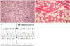
A 25-year-old man was referred from a local hospital for evaluation of high levels of Hb and management of gout. There was no hepatosplenomegaly on physical examination. A complete blood count showed the following results: Hb 20.4 g/dL, hematocrit 58%, reticulocyte count 2.90%, white blood cell count 6.83×10⁹/L, and platelet count 195×109/L. Bone marrow biopsy indicated no definite evidence of myeloproliferative features (Fig. 1A and B). Mutations were not found in JAK2 V617 or exon 12, MPL W515K/L, CALR exon 9. Serum EPO level was within the normal range, 7.3 mIU/mL (reference range: 4.3–29.0 mIU/mL). No abnormalities were found on Hb electrophoresis. P50 (partial pressure at which Hb is half saturated with oxygen), which is an indicator of left-shift of oxygen dissociation curve (high oxygen affinity state) was calculated to be 14.3 mm Hg (reference value 22.6–29.4 mm Hg), using venous blood gas analysis (recommended by international federation of clinical chemistry).15 Congenital (familial) erythrocytosis was suspected. We performed alpha 1-globin gene (HBA1)/alpha 2-globin gene (HBA2), beta-globin gene (HBB), and Von Hippel Lindau (VHL) gene mutation analysis according to a previous study.6 All coding sequences and flanking intronic regions of HBA1/HBA2, HBB, and VHL were analyzed. Sequencing was performed by using the BigDye Terminator Cycle Sequencing Ready Reaction Kit on the ABI Prism 3130 Genetic Analyzer (Applied Biosystems, Foster City, CA, USA). Sequence variation was described according to guidelines of the Human Genome Variation Society (http://www.hgvs.org/mutnomen). In case of HBB, the conventional numbering system was also used (Database of Human Hemoglobin Variants and Thalassemia mutations).
DISCUSSION
More than 90 high oxygen affinity Hb variants have been reported until now (Database of Human Hemoglobin Variants and Thalassemia mutations). High oxygen affinity Hb variants are inherited in an autosomal dominant pattern. The HBB mutation is more prevalent than HBA1/HBA2 mutation.8 High oxygen affinity Hb variants are derived mostly via three mechanisms; mutation affecting transition of R-state (relaxed binding structure, high oxygen affinity) to T-state (tight binding structure, low oxygen affinity), mutation in the 2,3-BPG binding site, and mutation in the heme pocket.1 Hb Heathrow [β103 (G5)Phe→Leu] and Hb Saint Nazaire [β103(G5)Phe→Ile] are examples of mutation in the heme pocket.79 White, et al.7 reported a first case of Hb Heathrow in English family. Patients with Hb Heathrow showed high Hb level (15.7–21.0 g/dL), normal platelet count, normal white blood cell count and low P50.710
The diagnostic strategies for CE have been suggested in several studies. The acquired secondary (pulmonary, renal, cardiac, etc.) and acquired primary erythrocytosis (P. vera; JAK2 mutation with low serum EPO level) are excluded.1 Low serum EPO level with negative JAK2 mutation is suggestive of PFCP.8 In cases of low P50 (<22.6 mm Hg) (high oxygen affinity state), HBA1/HBA2, HBB, and BPGM gene sequencing analyses are recommended.1 VHL, EPAS1, and EGLN1 gene sequencing analyses must be performed in patients with normal serum EPO level.14 However, the clinician must keep in mind that the incidence of erythrocytosis without a known genetic cause (idiopathic erythrocytosis) is surprisingly high.8 For example, Bento, et al.8 reported that only 25 of 70 patients (36%) had a known causative gene defect. Similarly, only 13.2 % of Korean patients with erythrocytosis had a known causative gene mutation.6 Therefore, further studies are needed to identify the new genetic cause of erythrocytosis.
In conclusion, we reported first case of Hb Heathrow in Asian. High oxygen affinity Hb variant is a common cause of CE. Therefore, the presence of a high oxygen affinity Hb variant must be ruled out, especially in a case of erythrocytosis with normal/high serum EPO level and low P50.
 XML Download