

 PDF
PDF ePub
ePub Citation
 Print
Print


INTRODUCTION
The retrievable type of inferior vena cava (IVC) filter has been widely used to prevent pulmonary thromboembolism (PTE) in patients with deep vein thrombosis (DVT) and contraindication of anticoagulation. According to their clinical experience, the manufacturer's recommendation and US Food and Drug Administration (FDA) safety advisory,1 physicians tend to remove the IVC filter within 3 weeks. However, the optimal retrieval time has not yet been clearly established. In some instance, forced retrieval of filter might cause vascular injury and related after effects such as PTE.
CASE REPORT
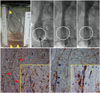
A 75-year-old man, involved in a traffic accident, was admitted to the orthopedic department due to multiple trauma including pelvic bone fractures and hemoperitoneum. He underwent surgery and was ordered by doctor to take absolute bed rest in the intensive care unit. On the 6th post-operation day, he developed dyspnea with blood tinged sputum and left calf swelling. The ultrasonography revealed DVT in the left calf vein, and the computed tomography of chest showed pulmonary hemorrhage without evidence of PTE. He should have had an installation of retrievable IVC filter (OptEase™; Cordis Europa, Roden, the Netherlands) to prevent PTE because he could not take any kind of anticoagulant. Fortunately, there was no embolic event at that period. Eighteen days later, there was no evidence of active bleeding. Thereafter, we tried to remove the filter according to the manufacturer's recommendation. However, we barely managed to retrieve it against our expectations because the filter won't come into the catheter (Supplementary Video 1 S1, S2, and S3, only online). There were several pieces of trapped tissue in the retrieved filter (arrow heads in Fig. 1A). Post-retrieval cavography demonstrated mobile rugged filling defects at the filter-detached vascular wall side suggesting a significant vessel wall injury (circles in Fig. 1B) (Supplementary Video 1 S4, only online). Pathological examinations, such as immunohistochemical staining of the tissue, were positive for smooth muscleactin and CD34, and confirmed that they were parts of vessel including smooth muscle cells (red arrows and rectangle in Fig. 1C) and endothelial cells (blue arrows and rectangle in Fig. 1D), respectively.
DISCUSSION
The US FDA issued and updated a safety advisory, urging that implanting clinicians responsible for the ongoing care of patients with retrievable IVC filters should remove the filter as soon as protection from pulmonary embolism is not needed because prolonged dwell times in patients with retrievable filters who have no longer an indication for the device could cause serious complications such as caval thrombosis, filter migration and erosion through the caval wall.12 In our case, we tried to remove the filter at 18 days after installation. However, there were some difficulties in the retrieval of filter because of neointimalization on the filter struts. In some case of longer dwelled filter (5.6 months in this cited literature), forced removal could cause another disaster including severe caval injury and associated potential thromboembolic event.34 Our present case indicates that the forced retrieval of cava filter has a potential unwanted venous injury and its related pulmonary embolic event even if the cava filter was retrieved on time.
 XML Download