

 PDF
PDF ePub
ePub Citation
 Print
Print


INTRODUCTION
Patients with glaucoma often have coexisting vitreoretinal diseases, such as proliferative diabetic retinopathy, epiretinal membrane, or uveitis, which may require vitrectomy. Additionally, occasional occurrences of intraocular lens (IOL) complications also necessitate vitrectomy. When vitrectomy is performed in eyes that have undergone glaucoma surgery, there may be damage to the filtering bleb or glaucoma drainage implant (GDI), which affects intraocular pressure (IOP) control.
In a standard pars plana vitrectomy, the infusion cannula used during vitrectomy is generally inserted infratemporally, while the second and third incisions are made supranasally and supratemporally, respectively, just above the 180° plane. Although this technique is applicable to most cases, certain circumstances compel the surgeon to create alternative approaches to achieve better intraoperative access to the posterior segment.
A suitable position of the surgeon relative to the patient's eye and appropriate sclerotomy locations are essential to perform effective surgical maneuvers during vitrectomy. Furthermore, the incisions made during these procedures should be located so as to avoid conjunctival scars, scleral thinning, filtering blebs, and regions of abnormal pars plana.1
Here, we describe a modified technique using a supratemporal surgeon position and altered sclerotomy sites for microincisional sutureless vitrectomy (MSV) in patients with a preexisting GDI or filtering bleb, and report early clinical results.
SURGICAL TECHNIQUE AND CASE SERIES
We retrospectively reviewed all medical records of patients who underwent MSV using our modified position-shift technique in an eye with a GDI or filtering bleb between August 2010 and September 2015. This study was approved by the Institutional Review Board of Ajou University Hospital, Suwon, Republic of Korea.
Surgeries were performed in one operating room by a single surgeon (JHS). The surgeon was right-handed. Five patients previously underwent GDI implantation in the supratemporal quadrant of the same eye using an Ahmed FP-7 (New World Medical Inc., Rancho Cucamonga, CA, USA). The tube tip of the GDI was located in the anterior chamber in all five cases. Two patients had undergone trabeculectomy and the filtering bleb had been located in the supratemporal quadrant.
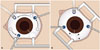
Surgery was conducted using retrobulbar anesthesia (via 2 mL of a lidocaine/bupivacaine hydrochloride mixture) under monitored anesthesia care. To preserve the supratemporally placed GDI or filtering bleb, we decided to perform the surgery by shifting the surgeon's position and altering the conventional sclerotomy sites. A caliper was used to mark the sclera 3–3.5 mm posterior to the limbus. Three sclerotomy incisions were created using a single-step Alcon (Alcon Laboratories Inc., Fort Worth, TX, USA) 23-gauge or 25-gauge trocar-cannula. The cannula for the infusion line was positioned inferonasally, while the other two sclerotomy incisions were made at positions 45° counterclockwise away from the original site (Fig. 1A). In cases where the vitrectomy was performed in combination with cataract extraction, phacoemulsification with a standard clear corneal incision and IOL implantation in the capsular bag were performed after insertion of the trocar cannulas. In the case of IOL repositioning with scleral fixation, conjunctival dissection was performed to bury the 10-0 polypropylene fixation suture in the supranasal and infratemporal conjunctiva, away from the GDI or filtering bleb.
The surgeon, the operating microscope, and the foot pedals were then adjusted to a supratemporal orientation. The surgeon was seated supratemporally, and the assistant was seated supranasally (Fig. 1B). The infranasal cannula was used to place the infusion, and the superonasal and inferotemporal cannulas were used for the light pipe and the vitreous cutter, respectively.
A non-contact wide-field viewing system (BIOM; Oculus Inc., Wetzlar, Germany) was primarily used for fundus visualization. A total vitrectomy was performed in all cases, that is, we tried to remove as much vitreous as possible with the induction of posterior vitreous detachment using 0.2–0.4 mL of triamcinolone (10 mg/mL) for vitreous staining. Additional surgical techniques were used as needed to address the pathology of the case. In the case of scleral fixation of an IOL, the sclerotomy sites and dissected conjunctiva were sutured with 8-0 polyglactin 910 sutures after cannula removal. Sclerotomy sites were not sutured in any other case (Supplemental Video 1, only online).
We used our modified technique to treat seven consecutive patients with GDIs or filtering blebs who required vitrectomy. The patients' demographics and surgical results are summarized in Table 1. Visual acuity improved in three patients and was unchanged in the other four patients. The IOPs of all patients were within the normal range (11–21 mm Hg), although three patients (patients 1, 3, and 6) required more glaucoma medication with a longer follow-up. In patient 5, normal IOP was sustained using a lower dosage of glaucoma medication after vitrectomy than before surgery. In one case of vitrectomy due to a GDI occluded by vitreous incarceration (patient 2), vitreous fibers around the tube tip were sufficiently removed, but the IOP was 40 mm Hg the next day. The glaucoma specialist performed glaucoma surgery to implant another GDI in the supranasal quadrant in this patient. We used tamponade in only one case (patient 6). Patient 6 had trabeculectomy-related hypotony maculopathy that was finally accompanied by retinal folding and a decline in visual acuity to 20/100. Six months after vitrectomy using our modified technique and 12% C3F8 gas tamponade, retinal anatomy normalized and vision improved to 20/25 with good IOP control.
In all seven cases, GDIs and filtering blebs were well preserved without surgical trauma.
DISCUSSION
A suitable position of the surgeon relative to the patient's eye and appropriate sclerotomy locations are essential to perform effective surgical maneuvers during vitrectomy. Here, we described a modified technique using a supratemporal surgeon position and altered sclerotomy sites for MSV in patients with a GDI or filtering bleb and early clinical results.
The sclerotomy sites for vitrectomy and the position of the filtering bleb or GDI often overlap. Patients whose eyes were treated previously with a glaucoma filtering bleb or GDI and require additional surgery are at risk of losing appropriate IOP control if an incision is required over a functioning bleb or GDI.2 In a previous study of 25-gauge microincision vitrectomies to treat vitreoretinal disease in glaucomatous eyes after trabeculectomy, the insertion point of the cannula was shifted so as to avoid disturbing the conjunctiva adjacent to the filtering bleb.3 A few previous reports have described the utility of a temporal approach during vitrectomy. In these studies, vitrectomy was performed with the surgeon seated temporally along with an appropriate change in sclerotomy sites. The infusion cannula incision was made in an infranasal position, while the both second and third sclerotomy incisions were made temporally. 45 However, shifting superiorly positioned sclerotomies to a temporal position may cause injury to the posterior long ciliary nerve or ciliary vessels, and also result in difficult maneuvering for the surgeon during the operation. Therefore, we proposed modified positions for the surgeon as well as the sclerotomy incision sites to avoid trauma to both important anatomic structures and ocular surface morbidities, such as GDIs or filtering blebs, and to make surgical maneuvers easier. The supratemporal approach enables the separation of incision sites and the GDI or filtering bleb, thereby reducing the risk of injury to the supratemporal conjunctiva and fibrous tissue overlying the GDI or filtering bleb. Transposition of the infusion line from the infratemporal to the infranasal quadrant and counterclockwise rotation of the other two incision sites by 45° allow free movement of the surgeon's hands.
Despite the successful outcomes of this procedure, this study has some limitations, including its retrospective nature, the small number of patients, and relatively short follow-up period. Another shortcoming of this study is that vitrectomy was performed on the left eye in only two cases. Because this is a retrospective case series that included consecutive patients who underwent glaucoma surgery and then vitrectomy on the same eye, an uneven distribution in the laterality of the study eyes ensued. Most cases included in this study did not need tamponade, except for patient 6 who had a filtering bleb. Although all GDI-implanted eyes had their tube tips in the anterior chamber and did not need tamponade, the role of tamponade in GDI failure should be considered when vitrectomy is performed in eyes with tube implantation. When an eye with an implanted tube undergoes vitrectomy and tamponade, there is a possibility of dysfunction or blockage of the tube. Some techniques have been reported to avoid this complication, including repositioning the tube tip from the pars plana into the anterior chamber of the pseudophakic eye.6
Three out of seven eyes in this study needed more anti-glaucoma medications after vitrectomy at the last follow-up. One possible explanation is that postoperative intraocular inflammation might have affected the GDIs and filtering blebs despite cautious control of postoperative inflammation. Another possibility is that the function of the GDI and filtering bleb might have deteriorated from gradual blockage of the tube or subconjunctival fibrosis, even without any surgical trauma, in the long-term follow-up of 6–62 months.
Although our technique is not an entirely new approach, to the best of our knowledge, there have been no previous reports. This is the first report that illustrates the technique in detail and presents a series of cases with early results. Further investigations may be needed to evaluate changes in IOP and complications in the late postoperative period.
In conclusion, our technique of modifying the surgeon's position and sclerotomy incision sites is a simple, safe, and well-tolerated approach for performing vitrectomy in patients who previously underwent glaucoma surgery while not disturbing the GDI or filtering bleb and maintaining IOP control. This quarter-shifted MSV directed from a supratemporal position may provide advantages when performing vitrectomy in eyes with GDIs and filtering blebs as well as with other forms of ocular surface morbidity in the same region.


 XML Download