

 PDF
PDF ePub
ePub Citation
 Print
Print


INTRODUCTION
A number of factors can limit a joint's range of motion, including tightness of soft-tissue structures such as muscle, tendon, ligament, and joint capsule. When connective tissue is not stretched, the collagen component gradually shortens. As a result, the periarticular collagen and the connective tissue of the muscle shorten. Furthermore, immobilization of a muscle in a shortened position also causes a decrease in the muscle length through a decrease in the number of sarcomeres in the muscle.1 The tightness of hamstring muscles is one of the main factors hindering performance in daily and sporting activities.2 Tightness of the hamstrings has been reported to be associated with the occurrence of back pain in adults.3
Recently, extracorporeal shock wave therapy (ESWT), which has proven effective in musculoskeletal injuries, has been also proposed for treatment of spastic muscles.4567 ESWT was introduced in the 1990's and now is used for the treatment of musculoskeletal conditions, such as calcific tendinopathies of the shoulder, lateral epicondylitis of the elbow, and plantar fasciitis.4 A shock wave is a sonic pulse characterized by an initial rapid rise of a high peak pressure in less than 10 nanoseconds, followed by a low tensile amplitude, a short life cycle of approximately 10 microseconds.5 The effects of shockwaves in tendon pathologies can be through a biological mechanism called mechanotransduction, by which the tissues exposed to shockwaves convert the mechanical stimulation of the shockwaves into biochemical signals through the release of growth factors involved in neoangiogenesis, tendon proliferation, and collagen synthesis.6 However, the mechanisms by which ESWT affects spasticity remain unclear. Obviously, it is not enough to explain the effects of ESWT on spasticity by the therapeutic mechanism on tendon pathologies. The reduction in spasticity after ESWT might be due to its direct action on fibrous areas by altering the rheological properties of chronically hypertonic muscles and by reducing intramuscular connective tissue stiffness.6
We speculated that ESWT would be effective in improving range of motion in normal adults with muscle tightness if the therapeutic effect of ESWT on spasticity is due to direct action on muscle or intramuscular connective tissues. To the best of our knowledge, no study has determined the changes of muscle stiffness in normal adults after ESWT. Therefore, the objective of this study was to compare the effect of ESWT for hamstring muscle tightness in normal adults with stretching exercise and to assess the safety and the feasibility of a larger, ensuing hypothesis testing study.
MATERIALS AND METHODS
Participants
We recruited healthy volunteers by posting flyers at our hospital. The inclusion criteria were age 20–69 years and finger-to-floor distance exceeding 5 cm. Participants were excluded from the study if they had a history of receiving ESWT or performed stretching exercise of hamstring muscle within three months before screening; were taking medications that could have an impact on the study or on the response to ESWT, including muscle relaxants, non-steroidal anti-inflammatory drugs, anticoagulants, or botulinum toxin treatment; had coagulopathy disorder; presented with hamstring spasticity caused by central nervous system lesion; were cognitively impaired; had a skin disorder including skin ulcer or open wound; or displayed hamstring muscle spasm caused by specific diseases like fracture or back pain. Twenty-nine healthy adults (25 men and 4 women) were enrolled between September 2014 and August 2015. Approval from our Institutional Review Board was obtained before conducting the study. Written informed consent was obtained from all participants after they were briefed about the purpose of the study and the examination procedures.
Study design
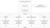
The was a prospective, randomized, single-blinded, clinical pilot trial designed to compare the benefit of ESWT and stretching exercise in the relief of hamstring tightness. The selected participants were divided randomly into four groups by the investigator (N.Y.K). We used a random permuted block design, where envelopes were sealed by persons not associated with the study. Group 1 received ESWT. Group 2 performed stretching exercises. Group 3 received ESWT and performed stretching exercises. Group 4 was not treated (control group). The same physiatrist examined all participants for eligibility. Two investigators who evaluated the outcome measures were blinded to the group allocation throughout the study, although the physiatrist who performed all interventions and the participants were not blinded. The investigators conducted the finger-to-floor test and measured the popliteal angle to evaluate the hamstring tightness of participants. Clinically, hamstring muscle tightness is commonly measured indirectly by these two methods. These tests are easy to perform and are reliable.891011 The study design is illustrated in Fig. 1.
Extracorporeal shock wave therapy
Participants receiving ESWT only and ESWT with stretching exercises received three ESWT sessions each week on both hamstring muscles while in the prone position. A model AR2 electromagnetic ESWT (Dornier MedTech, Kennesaw, GA, USA) was used. A total of 2000 impulses were applied to each hamstring muscle at the biceps femoris long head (n=1000), semitendinosus (n=500), and semimembranosus (n=500). All impulses were applied in the middle of the respective muscle belly. The applied energy flux density (EFD) was 0.074 mJ/mm2; the energy did not require the use of anesthesia or analgesic drugs. The repetition frequency of shock wave was 5 Hz.
Stretching exercise
Participants who performed stretching exercises only and who received ESWT in addition to stretching exercises stretched for 12 minutes a day, five days a week, for 3 weeks. In the group receiving both ESWT and stretching, the treatments were carried out separately. Hamstring muscle stretching with the participant sitting on the floor with one knee fully extended and the other one flexed, with both arms extended toward the extended foot. This position was sustained for 1 minute, followed by 10 seconds of rest, and was repeated for five sets. The opposite hamstring muscle was stretched in the same way.1213
Assessments
The finger-to-floor distance and popliteal angle were measured by the same blinded investigators. The assessments were done before intervention, immediately and 4 weeks after the completion of the 3-week interventions. Adverse effects of ESWT were also monitored. In the finger-to-floor test, participants stood on a 20-cm-high platform with both feet together. While the knees, arms, and fingers were fully extended, participants were asked to lower their hands toward the platform as much as possible.8 The vertical distance from tip of the middle finger to the top of the box was measured. A positive and negative value indicated inability to reach the box and ability to reach more than the top of the box, respectively. For analysis, right popliteal angle was the angular measurement of unilateral knee extension with the hip flexed to 90°.1014 With the participant in the supine position, the two investigators stood next to the participant and measured the right popliteal angle using a goniometer. One investigator flexed the hip to 90° and passive knee extension was done until resistance was felt strongly by the investigator. The left leg was in the neutral position. The other investigator measured the angle of the thigh and the leg with the goniometer in this position. The connecting line of the greater trochanter and lateral femoral condyle was used as the axis. The other axis was the line connecting the fibular head and the lateral malleolus.
Statistical analysis
SPSS/PC software version 21.0 (SPSS Inc., Chicago, IL, USA) was used for the statistical analysis. The Shapiro-Wilk test was used to determine the distributions of all continuous variables in each group. Because each variable showed a normal distribution (p>0.05 by the Shapiro-Wilk test), parametric statistical analysis was used. Repeated-measures ANOVA with time as the within-subject factor and condition (group) were applied to evaluate statistical differences over time in the four groups. Any significant group, time, or group versus time differences were examined using post-hoc testing involved Tukey multiple comparisons. One-way ANOVA and chi-square tests were used to compare demographic and baseline variables. Bonferroni's correction was used to correct for multiple comparisons. Statistical significance was considered when the p value was <0.05.
RESULTS
Twenty-nine healthy adults (25 men and 4 women) were enrolled. The mean age and mean height of the participants were 29.2±5.8 years and 174.4±5.9 cm, respectively. They were allocated randomly into four groups (n=8 for the ESWT group, n=7 for the others). No significant differences were observed in age, sex, and height among the groups. At baseline, there were no significant differences among all four groups in finger-to-floor distance and right popliteal angles (Table 1). None of the participants were lost to follow-up. No serious side effects of ESWT were observed over the 4-week follow-up. Post-ESWT pain rated <3 on a visual analog scale was reported by two participants of group 1 (ESWT only) and one participant of group 3 (ESWT with stretching exercise). One group 1 participant experienced petechiae after ESWT that disappeared spontaneously.
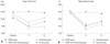
We observed a significant effect of “group” and “time” for the finger-to-floor test (F2,60=47.927; p<0.001 and F3,25=3.760; p=0.023) and for the right popliteal angle (F2,60=52.737; p<0.001 and F3,25=4.075; p=0.017). There was a significant interaction effect between time and type of intervention in the finger to floor test and the right popliteal angle (F6,50=10.79; p<0.001, F6,50=4.20; p=0.002) (Fig. 2). Post-hoc analyses revealed ESWT and ESWT with stretching exercise were significantly different from controls in the finger-to-floor test, and the right popliteal angles, respectively (p<0.05). In addition, the ESWT group, stretching exercise group and ESWT with stretching exercise group had decreased finger-to-floor distances and right popliteal angles immediately after intervention compared with the control group (p<0.05) (Table 1). However, at 4 weeks after the completion of the intervention, finger-to-floor test and right popliteal angles in only the ESWT with stretching exercise group showed a significant improvement compared with the control group (p=0.008 and 0.023, respectively) (Table 1, Fig. 2). These results indicate that ESWT, stretching exercise, and ESWT with stretching for 3 weeks could make the improvement of hamstring tightness immediately after intervention in the healthy participants. In addition, this effect after ESWT with stretching for 3 weeks could last until at least 4 weeks after the completion of intervention.
DISCUSSION
In the ESWT group, stretching group and ESWT with stretching group, hamstring tightness was reduced immediately after treatments, compared with the control group. However, only the ESWT with stretching group showed a significant improvement, compared with the control group, at 4 weeks after intervention. These findings indicate that ESWT or 3-week stretching exercise is effective, but transient, in releasing hamstring tightness, while ESWT combined with stretching has longer effects than ESWT or stretching exercise only.
Mechanotransduction induces biologic responses, including the expression of growth factors, nitric oxide synthesis, and neovascularization.15161718 This mechanism of ESWT is related to regeneration effect in orthopedic injury, which takes about 3 to 4 weeks. In patients with lateral epicondylitis, pain reduction and functional improvement immediately after 3-week ESWT were not prominent, compared to a pre-treatment state. However, there were statistically significant differences in pain and functional scores between the ESWT group and controls in favor of the ESWT at 3 months after treatments. These effects continued and increased up to 12 months.19
The mechanisms of ESWT on muscle tissue are still unknown. Considering the time difference of effects onset, however, the results of the current study suggest a different mechanism of ESWT on hamstring muscles in normal adults from regeneration of soft tissue injury, such as tendinopathy. There are few studies on the effects of ESWT for treatment of muscle problems. In spastic muscles of patients affected by brain lesion, ESWT reduces muscle tone in the short term.1213 In myofascial pain syndrome, ESWT significantly reduces muscle pain immediately after treatments.11 These results support a direct effect of ESWT on muscles.
In addition, reduction of spinal excitability has been proposed as one ESWT mechanism on muscles. However, no significant changes occurred in F wave minimal latency, H-reflex latency, or H-M ratio after ESWT.20 Some authors have proposed that the reduction in muscle tone induced by ESWT can be explained by a mechanism similar to that underlying the effects of ultrasound.21
Ultrasound induces vibrations that act on fibrosis and other intrinsic components of chronically overactivated muscles.21 The vibration and increase in blood flow by ESWT may aid muscle release in a similar manner as ultrasound. Comparison of effectiveness to muscle tightness between ultrasound and ESWT is worthy of future study. Therefore, the most probable theory to explain reduced spasticity or hamstring tightness is a direct effect on the fibrosis of chronic hypertonic or tight muscles including mechanical vibration. These considerations are consistent with recent studies suggesting that the reduced spasticity could be caused by directly acting on fibrosis and rheologic components of chronic hypertonic muscles.22 This study showed that changes in muscle tightness were immediately after ESWT and excludes a major effect of mechanotransduction which is a time consuming and late effect. Therefore, this study regarding normal muscle tightness may help to prove the above theory.
To the best of our knowledge, this is the first study to evaluate ESWT or stretching exercise combined with ESWT on hamstring tightness in normal individuals. Flexibility is an important component of a physical condition that allows the tissue to readily accommodate to stress, to dissipate shock impact, and to improve efficiency of movement, thus minimizing or preventing injury.23 The prevalence of low back pain was found to be increased in patients having tight musculature in the lower spine, as well as the hamstring muscles.24 Hamstring tightness is also a risk factor for the development of patella tendinopathy and patellofemoral pain,2325 hamstring strain injury, and symptoms of muscle damage following eccentric exercise.26
Many mechanisms of the effect of stretching exercise on the enhancement of muscle flexibility have been proposed. However, no clear conclusion on the mechanism of how stretching affects muscle flexibility has been reached. There are two possible mechanisms leading to an increase of the muscle's tolerance to stretching exercise.26 Golgi tendon organs induce muscle-tendon unit relaxation by reducing the effects of motor neuronal discharge. Also, Pacinian corpuscles act as pressure sensors that control pain tolerance. Stretching exercise reduces passive tension and allows greater elongation through small changes in the viscoelastic properties of the muscle-tendon unit. These acute changes in muscle and tendon lengths last only for a short period after stretching exercise. Therefore, these mechanisms support the claim that the effect of stretching exercise does not last long. Although some evidence indicates that stretching exercise increases muscle flexibility and range of motion and reduces spasticity, there is lack of definite evidence.2627 Reportedly, to induce muscle flexibility, one should use stretching exercise as a supplementary method, while receiving other treatment modalities, rather than stretching alone.28 Presently, better results were obtained using ESWT and stretching, compared to stretching alone. By showing longer effectiveness of ESWT combined with stretching exercise, this study is useful, since it indicates the necessity of using ESWT as an adjuvant to stretching exercise.
Hamstring tightness is typically released with common stretching techniques, such as static stretching and proprioceptive neuromuscular facilitation. However, the study results suggest that ESWT may improve hamstring flexibility, especially combined with stretching exercise in healthy participants. Further study with a large number of participants will be needed to clarify the effect of ESWT on the stretching of hamstring tightness. The results also suggested that the mechanism of ESWT on muscle relaxation might be different from tissue regeneration effect wherein a certain amount of time is required.
Limitations of our study include the small number of participants and the short-term period of follow-up. Since there were no previous studies concerning ESWT on muscle tightness, we conducted a pilot study with a small number of participants to prove its efficacy first. Second, the imbalance of sex ratio in this study participant could make it difficult to generalize the present results. Also, the mean age of the subjects was 29.2 years, which means that the subjects of this study did not include many older adults. Further study with a larger number of participants will be needed. Third, there is no definitely unified standard regarding the ESWT equipment currently used in clinical settings (generating focusing, the manner in which the shock wave reaches the target, etc.). Therefore, the most effective doses of ESWT on muscle tightness should be studied in the future. ESWT can also be classified in accordance with level of EFD as low energy with EFD range <0.08 mJ/mm2, intermediate energy with EFD range of 0.08–0.28 mJ/mm2, and high energy with EFD >0.28 mJ/mm2. Usually, EFD applied in clinical practice ranges from 0.01–0.40 mJ/mm.2930 However, it is less likely that a high level of ESWT exceeding 0.28 mJ/mm2 would be more effective on muscle tightness, compared with low or intermediate energy of ESWT, because high-energy ESWT is appropriate for delayed union, but not for muscles. As ESWT has very few side effects, it seems necessary to confirm the effect of regular and elongated application of ESWT and stretching exercise on muscle tightness. Also, further study is needed to compare the effect of ESWT on spastic hamstring muscles of hemiplegic patients with the unaffected normal side.
In conclusion, each ESWT and stretching reduced hamstring tightness immediately after interventions, and only ESWT with stretching exercise maintained the significantly improved relief of hamstring tightness significantly after 4 weeks.

 XML Download