

 PDF
PDF ePub
ePub Citation
 Print
Print


INTRODUCTION
Duchenne muscular dystrophy (DMD) is an X-linked recessive disorder, resulting from mutations at locus Xp21.2 in the DMD gene, that affects approximately one in 3500–5000 live-born males.1 Becker muscular dystrophy (BMD) is also caused by mutations in the same gene locus, but can be distinguished from DMD by its different phenotypic expression, which includes delayed onset, slow progression, and prolonged survival. DMD gene mutations lead to a deficiency of dystrophin, a protein that stabilizes the extracellular matrix and cytoskeleton. The cell membrane destabilization resulting from the loss of dystrophin causes myofiber necrosis and progressive muscle weakness.2 As a consequence, the early symptoms of DMD are usually related to limb muscle weakness, leading to delayed onset of walking, toe walking, and waddling gait. Patients with DMD inevitably experience progressive muscle weakness, becoming non-ambulatory in the second decade of life and eventually requiring respiratory support. Currently, with intensive respiratory care, many patients with DMD can live into their late 20s and beyond.3 Moreover, corticosteroid therapy and better supportive cardiopulmonary care has led to improved survival in patients with DMD, according to a report published by Moxley, et al.4 in 2010.
The DMD gene, which comprises 79 exons, is the largest gene described in humans and encodes a 14-kb messenger RNA that transcribes dystrophin protein.5678 Among all patients with DMD/BMD, DMD gene exon deletion affects 60–65% of patients, whereas exon duplication affects 5–15%. Small mutations, pure intronic deletions, and exonic insertions of repetitive sequences affect the remaining 20–35% of patients.8 Two “hot spot” deletion regions have been identified near the 5'-end of the DMD gene and in the central region around exons 44–55.891011 Conversely, duplications can occur almost anywhere in the gene.121314
Accurate genetic diagnosis of DMD/BMD is important, and the obtained genetic information may help clinicians provide appropriate and reliable genetic counseling, prenatal diagnosis, and gene therapy for those at risk. Among the many methods applied for mutation detection, multiplex ligation-dependent probe amplification (MLPA), which allows multiplex copy number quantification of specific target sequences, has been reported to be a simple, rapid, and reliable tool for the detection of deletions and duplications in the DMD gene.1516 Many studies have used MLPA techniques to define DMD mutation patterns at the exon level in different populations.171819 As DMD/BMD is a genetic disorder, we expected to observe differences in the genetic data with regard to geographic location or race. However, we found that genetic analysis of the DMD gene has rarely been reported in Korea; notably, a single study, based mainly on MLPA data and involving a small number of subjects, was reported by Lee, et al.20 in 2012.
In this report, we describe a retrospective MLPA-based genetic analysis of the DMD gene in 113 Korean patients (86 and 27 patients with DMD and BMD, respectively) and 17 female carriers. We analyzed mutation patterns and hot spot locations of the MLPA-positive results and compared them with results reported from other countries.
MATERIALS AND METHODS
Subjects
We collected retrospective data from 237 subjects with clinically suspected BMD/DMD and from 35 female carriers who had participated in an MLPA study at a university hospital between July 2006 and February 2015. Subjects were primarily diagnosed based on typical clinical presentation (e.g., progressive symmetric muscular weakness, calf hypertrophy, wheelchair dependency before the age of 13 for DMD or 16 for BMD, a creatinine kinase level of more than 10 fold above the normal range, and/or a positive family history compatible with X-linked inheritance).
Subjects with negative results in the MLPA study were tested by other methods (muscle immunohistochemistry or polymerase chain reaction) to confirm the diagnosis of DMD/BMD. Data from subjects with positive results in the MLPA study were included in the analysis of the proportion of deletions/duplications and mutation hot spots. Each proportion was calculated using the total number of patients with DMD/BMD, irrespective of MLPA positivity. This study was approved by the Institutional Review Board (IRB no. 3-2016-0140).
Multiplex ligation-dependent probe amplification
MLPA reactions were performed using the SALSA MLPA probe sets P034 and P035 (MRC-Holland, Amsterdam, the Netherlands). 21 Ligation and amplification were performed on a C1000 Thermal Cycler (Thermo Fisher Scientific, Waltham, MA, USA). All amplified fragments were separated using an ABI 3500DX Genetic Analyzer (Applied Biosystems, Foster City, CA, USA). Using GeneMarker software v.1.8 (SoftGenetics, State College, PA, USA), fragments for each exon from the subjects were depicted as single lines or “peaks.” The peak quantity for each exon was compared with that of normal controls and expressed as a “peak ratio.” Exons with a peak ratio between 0.75 and 1.25 were considered to be in the normal range. Exons with a peak ratio below 0.75 were considered to be deleted, whereas exons with a peak ratio over 1.25 were considered to be duplicated. MLPA results showing either deletion or duplication of specific exons were defined as positive.
Direct sequencing to confirm single-exon deletions
The subjects' MLPA results were initially scanned to identify deletions and duplications. However, probe dysfunction, including ligation problems, can mimic exon deletion, as it presents as reduced or absent peaks. To distinguish true deletions from probe dysfunction, further direct sequencing was performed for all results thought to represent single exon deletion. Where further small mutations were detected from the direct sequencing, although rare, we defined them separately from the positive MLPA results, since the true mutations were not detected by the MLPA analysis.
RESULTS
Clinical characteristics
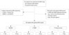
Among the 272 subjects who participated in the MLPA study, 130 showed positive results. Of these, 86 were compatible with DMD, 27 were compatible with BMD, and 17 were female carriers. Two patients were diagnosed with DMD after the detection of point mutations during further DNA sequencing. Among the 140 subjects with negative MLPA results, an additional 9 patients with DMD and four with BMD were diagnosed by muscle immunohistochemistry. In other words, we confirmed 97 patients with DMD and 31 patients with BMD in total. Of the remaining patients, 109 male subjects were later diagnosed with other neuromuscular diseases, and among them, 16 patients had unspecified myopathy. Thirty-five female subjects had taken part in the MLPA study and 17 were found to be MLPA-positive female carriers (Fig. 1). The mean age of the 113 MLPA-positive patients at the time of genetic testing was 18.6±8.6 years for patients with DMD and 23.6±10.1 years for those with BMD. The MLPA-positive female subjects varied in age and were either related to patients with DMD or were symptomatic. Four out of 17 MLPA-positive female carriers were symptomatic.
MLPA mutation pattern and hot spot analysis
Analysis of the DMD gene mutations included both MLPA and direct DNA sequencing results. Among the 128 patients confirmed either by MLPA or other diagnostic tools, MLPA detected deletions in specific exons in 92 patients (71.8%) and duplications in 21 patients (16.4%). Among the 97 subjects diagnosed with DMD, 71 showed deletions (71/97, 73.2%), and 15 showed duplications (15/97, 15.4%) in the DMD gene. Of the 31 subjects diagnosed with BMD, 21 showed deletions (21/31, 67.7%) and six showed duplications (6/31, 19.4%) in the DMD gene. In addition, 11 and six female carriers showed exon deletions and duplications, respectively, in the DMD gene (Fig. 1).
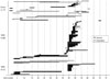
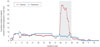
Each deletion or duplication was plotted, as shown in Fig. 2, and the cumulative number of subjects with deletions or duplications within each exon was also plotted, as shown in Fig. 3. Most deletions in patients with DMD/BMD were observed in the central hot spot region between exons 44 and 55 (60.6%) (Figs. 2 and 3). A second previously reported frequent-deletion hotspot region towards the 5'-end was not distinguishable in our results. Duplication events tended to be scattered evenly throughout the entire exon region, and accordingly, no clear central or proximal hot spot region was distinguishable (Fig. 3). Among the 71 DMD patients with exon deletions identified by MLPA, the most common identical deletion was observed in exons 49–50 (7/71, 9.9%), followed by exons 46–47 (5/71, 7.0%), exon 45 (5/71, 7.0%), and exon 51 (5/71, 7.0%). Among 21 BMD patients with exon-deletion MLPA results, the most common deletions were observed in exons 45–47 (7/21, 33.3%), and exons 45–48 (4/21, 19.0%) (Fig. 2).
Further DNA sequencing and point mutations
Among the subjects with MLPA results indicating lower quantity single DMD peaks, who underwent further DNA sequencing, two were found to harbor point mutations (c.4729C>T, p.Arg1577Ter and c.8800G>T, p.Glu2934Ter). The point mutation c.8800G>T, p.Glu2934Ter was found to be a novel nonsense mutation.
DISCUSSION
This study retrospectively evaluated 113 subjects with DMD/BMD and 17 female carriers with positive MLPA results and identified additional patients with DMD/BMD by DNA sequencing or muscle immunohistochemistry. Among 128 patients diagnosed by either MLPA or other diagnostic methods, MLPA detected 92 (71.8%) and 21 (16.4%) subjects with exon deletions and duplications, respectively.
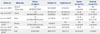
The proportions of deletions and duplications from MLPA studies conducted in other Asian countries are listed in Table 1. Results from this study showed a higher rate of exon deletion and a lower rate of duplication than the study conducted previously in Korea by Lee, et al.20 Instead, our results showed greater similarity with those of the study by Chen, et al.19 involving 119 subjects in China (79.0, 19.8%) and the study by Manjunath, et al.22 involving 83 subjects in India (79.5, 6.5%). We suppose the variation arises from the difference in total population considered in the analysis. In the studies by Chen, et al.19 and Manjunath, et al.,22 the authors calculated deletion/duplication rates only among subjects with a positive MLPA result. Our deletion rates may also have been overestimated, because although we tried to include every DMD/BMD patient, not all patients diagnosed with DMD/BMD proceeded to undergo MLPA analysis during the study period. By contrast, the previous Korean study by Lee, et al.20 tried to consider the total DMD/BMD population in their analysis, showing deletion and duplication rates of 45.5 and 27.2%, respectively. However, the small number of subjects in the previous study cannot be ignored. A study in Taiwan conducted in 2007 by Hwa, et al.17 obtained similar results to the study by Lee, et al.20 from a larger population, with deletion and duplication rates of 36.0 and 24.7%, which were relatively lower and higher, respectively, than the previously published rates in other western countries.81623 Although the authors insisted that these gaps may have resulted from ethnic differences, their deletion rate could have been underestimated, since they included female carriers in the analysis. Overall, our results, which were obtained from a larger number of patients, did not differ greatly from the results of previous studies conducted in other western countries.81623 Therefore, it would be difficult to conclude that a relatively low rate of deletion is characteristic of an Asian or Korean population. The proportion of deletions and duplications in patients with DMD/BMD might not have significant ethnic differences.
As observed, our hot spot study differed slightly from the previous studies and investigated different aspects, compared with previous Korean research. Two deletion hot spot regions were previously described near the 5'-end and the central region around exons 44–55 of the DMD gene.891011 In our research, we could clearly detect the hot spot in the central region, with a distribution rate in this region of approximately 60.6%, similar to rates from other Asian studies. In contrast, a hot spot near the 5'-end was not clearly distinguishable. Further studies will be necessary to explain this discrepancy. Regarding duplication, no level of bias was observed in the central hot spot region, similar to previous studies conducted in China. To date, other studies have failed to identify a duplication hot spot.
This study has limitations, such as failing to define alternative diagnoses for the remaining female carriers and male patients who had unspecified myopathy. These subjects showed negative results for other genetic tests that we conducted. Since we could not conduct whole-gene sequencing for the entire dystrophin gene, there remains a high probability that these subjects may harbor small mutations. This study also has limitations in the manner of computing the deletion/duplication ratio. Only patients with positive results in the MLPA study were analyzed, rather than the entire population of patients diagnosed with DMD/BMD during the corresponding period. However, the rate of deletions/duplications identified in this study was similar to that reported in a recent study conducted in a similar manner in China. In addition, a comparison with deletion/duplication rates of other studies that only included MLPA-positive subjects revealed similarities with our findings. Furthermore, the use of only MLPA-positive subjects did not affect our ability to conduct hot spot analysis, through which we identified a distinct lack of mutation pattern in the previously reported proximal hot spot.
In this study, which included the largest number of patients with DMD/BMD among all similar studies conducted in Korea, slightly different results from previous studies investigating DMD gene mutation patterns were obtained. In addition, our hot spot analysis yielded findings that differed from those previously reported in other Asian countries.
Previously, patients with DMD/BMD had a short life expectancy and experienced relatively similar disease progression. However, because life expectancy has been extended through various interventions, a phenotype-genotype correlation study will be necessary to investigate how disease progression differs among patients in relation to different hot spot traits.
 XML Download