

 PDF
PDF ePub
ePub Citation
 Print
Print


INTRODUCTION
Molecular taxonomy of ovarian cancer suggests several key players in each of its subtypes: p53 in serous type; non-p53 pathways, including K-RAS, in mucinous type; and phosphatase and tensin homolog (PTEN) in endometrioid tumors.1 Recently, another key molecule of ovarian cancer progression has been found, AT-rich DNA-interacting domain 1A (ARID1A), in clear cell carcinoma of ovary (O-CCC).234 ARID1A encodes BAF250a protein, which is one of the largest subunits of the human SWI/SNF chromatin-remodeling complex that functions actively in histone deacetylation and DNA methylation,5 and many of the complex components are identified as tumor suppressors.6 ARID1A participates in cell cycle arrest by regulating c-myc.7 In O-CCC, ARID1A mutation and loss is frequent, which seems to work as a tumor suppressor.2 Loss of this protein has also been found in atypical endometriosis,8 which can transform directly to O-CCC,910 also implying a role in the early stages of carcinogenesis.11
The functional consequences of ARID1A loss in O-CCC are not yet clear. In normal conditions, ARID1A is necessary for SWI/SNF complexes to respond to upstream steroid hormone receptor signaling, such as glucocorticoid, estrogen, and androgen receptors.12 Interestingly, among those steroid hormone receptors, estrogen receptor beta (ERβ) expressional loss is also noted in ovarian cancers,13 which may also be involved in ARID1A loss-dependent O-CCC pathophysiology. Of its downstream signaling, SWI/SNF complex regulates cell adhesion proteins CD44 and E-cadherin.14 Thereby, loss of CD44 and/or E-cadherin could be a sign that ARID1A loss-dependent carcinogenic process is activated.
Overexpression of HNF1β is another recently found O-CCC molecular feature. Positive expression of HNF1β is noted in 90–95% of O-CCCs, contrary to less than 10% of positivity in other epithelium-origin ovarian cancers.15 HNF1β seems to work in the maintenance of E-cadherin expressing epithelial phenotype of O-CCC16 and, thereby, associated with good prognosis.17 However, the functional implications of HNF1β overexpression, particularly in relation to ARID1A loss in O-CCC pathophysiology, have yet to be determined.
O-CCC is often resistant to therapies. If found at an early stage, it can be effectively managed by oophorectomy.18 In contrast, advanced O-CCCs exhibit heterogeneous behavior, being unpredictable with known clinic-pathological information, such as tumor grade, histologic subtype, stage, etc.19 In this situation, the expressional status of ARID1A and other biomarkers, including ER β, E-cadherin, and HNFβ, may provide further prognostic values.1720 In this study, we analyzed the molecular profiles and prognostic factors of O-CCC, focusing on the different clinical and molecular characteristics of ARID1A-positive and ARID1A-negative tumors.
Go to : 
MATERIALS AND METHODS
Patients
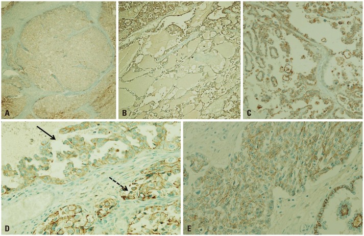
The study subjects were patients diagnosed with O-CCC at Severance Hospital. Medical charts were retrospectively reviewed to compare clinicopathological features including prognosis. For 87 patients from the Yonsei University Health System archived files, O-CCC diagnoses were confirmed in the past 5 years. All of the tumors were examined retrospectively by double-blind test for accuracy of histological subtyping and grading. With exclusion of ambiguous histology or inappropriate immunoprofiles, 70 were eligible for this study (29–67 years of age, median 49 years of age). Four cases with mixed subtypes (endometrioid carcinoma and serous carcinoma), and thirteen cases with an incompatible and distinct immunoprofile were excluded. All of the patients provided written informed consent, and the Research Ethics Board Committee of Yonsei University Health Medicine approved the study. Evaluated parameters included patient age, parity, follow-up periods, tumor size and FIGO stage,21 associated pathology (endometriosis and adenofibroma), histologic grade, and oncologic outcomes (disease recurrence, distant metastasis, and cancer-related death). Histologic grade was based on cellular atypia and mitotic indices of the major parts of the tumors. Low grade was defined as minimal or no atypia in focal areas less than one high-powered field (HPF), a low mitotic index (MI) less than 5/10 HPF, and/or primarily tubular or papillary patterns (Fig. 1A and B), whereas high grade was defined as severe atypia in multifocal areas more than one HPF, a high MI more than 5/10 HPF, and/or a primarily solid pattern (Fig. 1C and D).
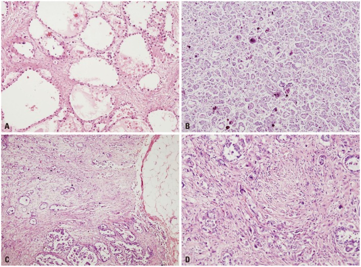 | Fig. 1Histologic grades of O-CCC. (A and B) Low grade O-CCC. (A) tubulocystic or alveolar patterns are the most common patterns seen in low grade tumor. Abortive small tubules are less differentiated (H-E, ×100). (B) Psammomatous calcifications (H-E, ×100). (C and D) High grade O-CCC. (C) Diffuse, solid areas are frequently seen in high grade tumor. Desmoplasia is frequently found in association with infiltrative cells (H-E, ×40). (D) highly cellular spindle cells are parallel with infiltrating tumor cells (H-E, ×200). O-CCC, clear cell carcinoma of ovary; H-E, hematoxylin-eosin.
|
Immunohistochemistry using tissue microarray preparation and interpretation
Macroscopic inspection of whole mount tumor sections was performed to determine whether they were homogeneous or heterogeneous in nature. A core needle was used to make 2-mm holes in recipient blocks. Based on prior inspection, three adjacent areas of ovarian carcinomas from matching donor blocks were transplanted into recipient blocks using a 2-mm core needle. The 4-µm tissue sections were placed on silane-coated slides, deparaffinized, immersed in phosphate-buffered saline containing 0.3% (v/v) hydrogen peroxide, and processed in a microwave oven for 15 min at 700 W in 10 mM sodium citrate buffer (pH 6.5). After blocking for 30 min with 1% (w/v) bovine serum albumin, sections were incubated for 16 h at 4℃ with biotin-labeled rabbit antibodies. The primary antibodies included ARID1A (C-terminal, 1:50, Abcam, Cambridge, MA, USA), HNF1β (SPA002083, 1:400, Sigma-Aldrich, St. Louis, MI, USA), p53 (DO-7, 1:100, Dako, Santa Clara, CA, USA), p38 (Phospho T180+Y182, 1:100, Abcam, Cambridge, MA, USA), E-cadherin (H-108, 1:200, Santa Cruz, Dallas, TX, USA), ERβ (1:50, Proteintech, Rosemonta, IL, USA), and Survivin (EP2880 1:100, Abcam, Cambridge, MA, USA). Streptavidin-conjugated peroxidase was used as the secondary antibody (1:10000). Normal goat serum and subtype-matched normal mouse IgG were used as negative controls. The final reaction product was visualized upon the addition of 0.03% (w/v) of 3, 3'-diaminobenzidine tetrachloride (DAB) for 5 to 20 min.
ARID1A1 expression was interpreted as "loss" if less than 5% of the tumor cells showed nuclear staining. If staining was modest (between 5% and 50% of the tumor cells), expression was interpreted as "focal loss." When more than 50% of the cells were positive, ARID1A was evaluated as "intact." For HNF1β expressions, two-tiered grading was applied: grade 1 in patch staining (focal loss) or grade 2 for diffuse and strong expression. ERβ expressions were graded from 0 to 3+ according to the American Society of Clinical Oncology/College of American Pathologists (ASCO/CAP) guidelines and grade 1, 2, and 3 were considered "positive" (2). E-cadherin membrane staining was graded as grade 0, indicating diffuse loss of more than 10%; grade 1, indicating focal loss of less than 10%; and grade 2, indicating no loss. All of the antibodies were initially applied to whole slides followed by tissue microarray.
Statistical analyses
Demographic data were analyzed using the parametric, independent two-sample t-test and the chi-square (or Fisher's exact test) for continuous and categorical variables, respectively. Categorical variables are reported as frequencies, and continuous variables are reported as means±standard deviations. To compare cancer-specific survival, Kaplan-Meier survival curves were generated for ARID1A-positive and negative tumors. Mean survival times of ARIDA-positive and negative tumors were compared by using log-rank test. All p-values <0.05 were considered to be statistically significant. All analyses were performed with SAS (version 9.2, SAS Institute Inc., Cary, NC, USA) and R statistical software (version 3.1.1, www.R-project.org).
Go to :
RESULTS
Table 1 summarizes patient characteristics. Tumors that were removed were of variable sizes ranging from 2–25 cm (median 8 cm). The majority of tumors were low stage [stage I: 36 (52%); stage II: 5 (7%)] (Table 1). Mean follow-up period were 40±36 months. The numbers of recurrences, metastasis, and deaths due to disease were 4 (3%), 19 (13%), and 10 (14%) cases, respectively.
Table 1
Patient Characteristics
![]()
Immunohistochemical profiles of O-CCCs
We analyzed expressional characteristics of several molecules, including HNF1β, ERβ, and E-cadherin, along with ARID1A. In CCC-associated endometriosis, nuclear staining of ARID1A occurs along the epithelial lining, although CCC shows complete loss of ARID1A (Fig. 2A). Fig. 2B-E shows varying degrees of ARID1A loss in O-CCC, from total expressional loss (Fig. 2B) and focal expression (Fig. 2C) to intact staining (Fig. 2D).
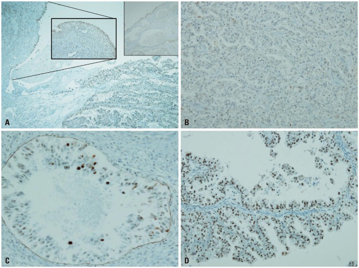 | Fig. 2ARID1A expressions in O-CCC. (A) In endometriosis, nuclear staining occurs along the epithelial lining (inlet), but CCC shows complete loss of ARID1A (DAB, ×40). (B) Total loss of ARID1A in a branched tubular pattern (DAB, ×100). (C) Focal expression (nearly total loss) in an alveolar pattern (DAB, ×200). (D) Moderate preservation of AR1D1A expression in a cystic papillary pattern (DAB, ×200). ARID1A, AT-rich DNA-interacting domain 1A; O-CCC, clear cell carcinoma of ovary; DAB, 3'-3' diaminobenzidine.
|
Complete loss of HNF1β was rarely found (5.7%) in O-CCCs (Fig. 3A), and most of these O-CCCs were negative for p53 or ERβ and loss of ARID1A. In HNF1β-positive cells, HNF1β expression was higher in cells closer to the luminal side (Fig. 3B). Complete loss of ERβ was rare in O-CCCs (Fig. 3C). Luminal protruding hobnail cells were positive for ERβ (Fig. 3D). Loss of E-cadherin or CDH1 was seen in 21.4% of O-CCCs, and E-cadherin loss was variable ranging from complete loss (Fig. 4A and B) to aberrant expression, such as fragmentation and broken linear forms (Fig. 4C and D) or cytoplasmic staining (Fig. 4E).
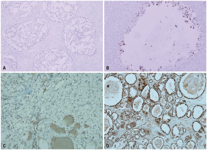 | Fig. 3HNF1β and ERβ expressions in O-CCC. (A and B) HNF1β expression. (A) Complete loss of HNF1-β is rarely found in CCC. However, negative expression of p53 or ER with loss of ARID1A is common (DAB, ×100). (B) Luminal hobnail cells express HNF1-β. More HNF1-β expression occurs in cells closer to the luminal side (DAB, ×200). (C and D) ERβ expression. (C) complete loss of ERβ is common (DAB, ×100). (D) Luminal protruding hobnail cells express ERβ (DAB, ×100). ERβ, estrogen receptor beta; O-CCC, clear cell carcinoma of ovary; ARID1A, AT-rich DNA-interacting domain 1A; DAB, 3'-3' diaminobenzidine.
|
 | Fig. 4E-cadherin expressions in O-CCC. Loss of CDH1, or E-cadherin was manifested variably. (A) Complete loss of CDH1 (DAB, ×10). (B) Partial and weak staining (DAB, ×100). (C) Aberrant CDH1 expression. Aberrant cytoplasmic expression with fragmented or broken linear forms instead of membrane staining is an indicator of E-cadherin disruption (DAB, ×100). (D) Within the same tumor, CDH1 loss is heterogeneous. The solid arrow points to a region of partial CDH1 loss as seen by weak staining along the membrane. The broken arrow points to disrupted fragments in the membrane (DAB, ×200). (E) Images taken at a higher magnification show aberrant staining, such as and non-continuous, fragmented membrane, or cytoplasmic staining (DAB, ×200). O-CCC, clear cell carcinoma of ovary; DAB, 3'-3' diaminobenzidine.
|
Characteristics of ARID1A-negative and ARID1A-positive O-CCCs
The expressions of ARID1A "complete loss," "focal loss," and "intact" were noted in 48 (66%), 12 (17%), and 10 (14%) cases, respectively. We grouped "focal loss" or "intact" expressions in to ARID1A-positive tumors and "complete loss" to ARID1A-negative tumors. Table 2 compares their clinical and pathological characteristics. Patient age, parity, tumor size, and distribution of cancer stage were not statistically different between ARID1A-negative and positive O-CCCs. In contrast, the ratio of histologic high and low grades were significantly different; most (90%) of the ARID1A-negative tumors were low grade, while 41% of ARID1A-positive tumors were high grade (p=0.003) (Table 2).
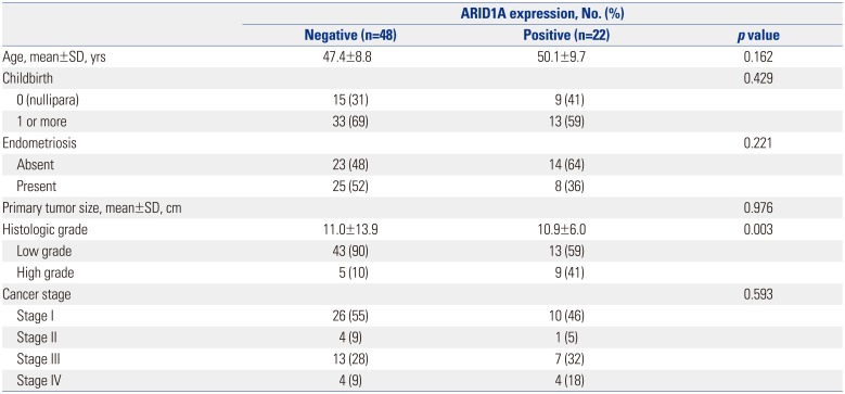
Table 2
Clinico-Pathologic Characteristics of ARID1A-Expressing Ovarian Clear Cell Carcinoma
![]()
Along with this finding, immunohistochemical profiles of ARID1A-positive and negative tumors were further analyzed (Table 3). To simplify comparisons, the expression status of each molecule was categorized as (−) or (+). ARID1A-negative tumors exhibited a mostly homogenous expression profile, HNF1β-positive (96%), ERβ-negative (83%), and E-cadherin positive (83%); whereas ARID1A-positive tumors exhibited rather heterogeneous immunohistochemical profiles, HNF1β-negativity (23%); ERβ-positivity (55%), and E-cadherin loss (41%).
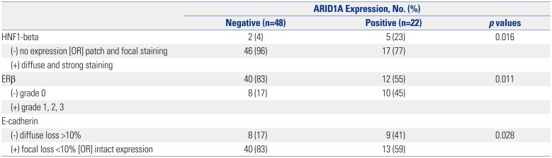
Table 3
Immunohistochemical Profiles of ARID1A-Expressing Ovarian Clear Cell Carcinoma
![]()
Association with oncologic outcomes
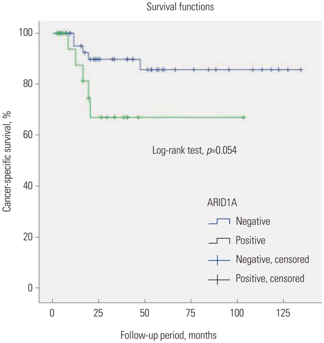
To compare cancer-speficic survival, Kaplan-Meier survival curves were generated for ARID1A-positive and negative tumors (Fig. 5). Mean survival times of ARIDA-positive and negative tumors were 74 months (95% CI 53–95) and 118 months (95% CI 105–131) respectively, and the difference was not statistically significant (p=0.054, log-rank test).
Go to :
DISCUSSION
To date, two significant mutational/expressional molecular characteristics of O-CCC have been found, ARID1A loss and HNF1β-overexpression.215 However, their functional roles in cancer are still largely unknown, and the relationship between those two proteins has not yet been determined. For ARID1A, its loss is suggested as a critical event in endometriosis (EM)-derived O-CCC tumorigenesis.22 Indeed, ARID1A loss has been noted in O-CCC associated atypical EM tissues, as well.3 Since ARID1A is a major component of the human SWI/SNF chromatin-remodeling complex,6 its oncogenic role may be related with remodeling of chromatin and histone re-arrangements4923 or with modulation of estrogenic action.9 We found that ARID1A-loss in O-CCC is linked to a specific immunohistochemical profile: ERβ loss, intact E-cadherin, and HNF1β overexpression. ERβ expressional loss occurs frequently across all epithelial ovarian cancers,24 and ARID1A is important in carrying steroid hormone signaling to the SWI/SNF-induced transcriptional activations.12 Since E-cadherin is one of the downstream activators of the SWI/SNF complex,14 we initially thought that ARID1A loss would accompany E-cadherin loss, which turned out to be not true, as ARID1A-lost tumors were more-likely to have intact E-cadherin expression than ARID1A-expressing tumors. This is in contrast with a previous study that stated that, in gastric cancer, reduced ARID1A expression down-regulates E-cadherin transcription.25 We suspect a hidden significance of HNF1β overexpression here.26 The functional role of HNF1β overexpression in O-CCC has not yet been derived,15 although it may also be mediated by E-cadherin: knock down of HNF1 has been shown to reduce E-cadherin expression and promote epithelial-mesenchymal transition.16 Hypothetically, the uniformly-expressed HNF1β in ARID1A-loss tumors in our study might have served a protective role by maintaining tumor cell E-cadherin expression. Thereafter, we argue that this signaling axis, ERβ/ARID1A/E-cadherin along with HNF1β, requires further molecular functional studies to elucidate the mechanism of O-CCC development.
Using readily available clinical samples and immunohistochemical tools, we sought to characterize ARID1A-negative O-CCCs in comparison with ARID1A-intact (or positive) O-CCCs. While ARID1A-negative tumors exhibited a homogenous immunoprofile, the immunoprofiles of ARID1A-positive tumor were rather heterogeneous: HNF1β-positive rate was slightly low, and the rates of ERβ-negativity and E-cadherin positivity were around 50%. Overall, the frequency of ARID1A loss in our series was slightly higher (69%) than those of previous studies.2320 Nevertheless, this still implies that for about half of O-CCC tumors show intact ARID1A, and their tumorigenic processes require further explanation.26
HNF1β-overexpression and intact E-cadherin are associated with better prognosis in O-CCC,1727 while the prognostic significance of ERβ expression in O-CCC has not been validated. The prognostic effect of ARID1A loss in O-CCC is debatable, some suggesting a poor prognsosis while others reporting no prognostic impacts.172028 In our study, ARID1A expression status did not have a significant impact on survival. This indifference in survival is noticible since most ARID1A-negative tumors in our study were low grade (90%), whereas many of ARID1A-positive tumors were high grade (41%), and histologic grade is directly linked to survival.29
The human SWI/SNF complex regulates gene transcription mainly by modulating DNA methylation status.14 Interestingly, O-CCCs is characterized by wide-spread CpG island promoter hypermethylation.30 In particular, both HNF and ER pathways are frequently methylated in O-CCCs.3132 Moreover, promoter methylation-related expressional changes can lead to disease progression in epithelial ovarian cancers. For instance, methylation status changes result in overexpression of Mucin 13 and carbonic anhydrase 9, both of which contribute to aggressive behavior of affected cancer cells.3334 Therefore, it is probable that epigenetic dysregulations play a significant role in ARID1A-negative O-CCC pathophyology, especially if it depends on HNF and ER pathways.
In this study, we used a modified two-tier histologic grading system based on nuclear atypia and MI. For histological grading of O-CCC, the Shimizu-Silverberg or the International Federation of Gynecology and Obstetrics grading systems can be used. However, a recent study criticized the significances of both systems, which were not effective enough to predict clinical outcomes.35 Along with previous reports, our study may provide a base for development of further molecular subtyping of O-CCC, presumably relying on ARID1A expression status.
In conclusion, ARID1A-positive tumors and negative tumors differed in gross histology and immunoprofiles. This study proposes that ARID1A expression status can be utilized for molecular subtyping of O-CCC. Further study is mandatory to enlighten the molecular events underlying O-CCC, particularly an ARID1A-independent pathway.
Go to :
 XML Download