

 PDF
PDF ePub
ePub Citation
 Print
Print


INTRODUCTION
Pulmonary arterial hypertension (PAH) is a life threatening disease, and is noted for an increase in pulmonary vascular resistance, right ventricular hypertrophy and pulmonary vessel remodeling.1 If it is not treated, it could lead to death. Inflammation and endothelial dysfunction play a significant role in the pathophysiological changes of PAH.234 PAH has been considered a refractory disease to most of the conventional pharmacological therapies.5 At present, alternative pharmacological treatment is being developed, nevertheless, mortality remains still too frequent.
Stem cell therapies have the potential to curesome pulmonary diseases.67 A number of studies demonstrated the efficacy of mesenchymal stem cell administration in many lung diseases including acute lung injury,8 bronchopulmonary dysplasia,910 chronic obstructive pulmonary disease,1112 fibrosing pulmonary injury,1314 pulmonary ischemia reperfusion injury,15 radiation induced lung injury,16 sepsis,17 and PAH.7918 Mesenchymal stem cells have an effect on lung inflammation and injury through paracrine interactions rather than direct interaction. 1920 The administration of human umbilical cord blood derived-mesenchymal stem cells (hUCB-MSCs) has recently been proposed for the treatment of PAH in animal models.21 hUCB-MSCs have advantages in that 1) they can be obtained relatively conveniently and less invasively,22 2) it is possible to differentiate into vascular cell types,23 and 3) they have an ability to secrete paracrine factors regulating inflammatory cytokines.24 We previously reported that hUCB-MSCs improved PAH via suppressing inflammatory cytokine level in lung tissues.6 These molecules have an immunosuppressive effect and lead to the regeneration of injured tissues by the way of paracrine mechanisms.25
In these earlier studies, diverse dosage, treatment timing and frequency were used, and the results of these studies showed variable rates of improvement. However, there is little information on the effects of a low-dose of and the administration timing of hUCB-MSCs, especially early timing. The purpose of this study was to examine the effect of low dose early treatment of hUCB-MSCs on PAH and compare it with the reversal therapy.
MATERIALS AND METHODS
Animals
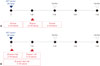
Study protocols were approved by the Institutional Animal Care and Use Committee of Ewha Womans University School of Medicine. Six-week old male Sprague-Dawley rats (180–220 g; Orient, Seongnam, Korea) were induced to PAH by subcutaneous injection of 60 mg/kg monocrotaline (MCT). We performed this study in two-parts (Fig. 1). The first part of the experiment was to determine the effective dose range because low-dose transplantation has an advantage from a clinical standpoint (Fig. 1A). We tested three different doses of hUCB-MSCs; 3×10⁶ cells/rat (UA group), 1.5×10⁶ cells/rat (UB group) and 3×10⁵ cells/rat (UC group) in MCT-induced PAH rats. Thus, the hUCB-MSCs were diluted in 500 µL of Dulbecco's modified Eagle's medium and were directly administered into the external jugular vein of rats placed in a supine position. The transfusion of hUCB-MSCs was performed randomly .The second part of the experiment was to optimize the timing and frequency of hUCB-MSCs transplantation (Fig. 1B). The experimental groups were divided into three groups; UD group (early treatment group of low dose hUCB-MSCs at day 1), UE group (dual transfusion group of low dose hUCB-MSCs at day 1 and week 1), UF group (reversal treatment group of low dose hUCB-MSC at week 1).
Preparation of hUCB-MSCs
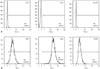
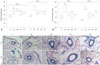
hUCB-MSCs were obtained from Medipost Inc., and isolated human mesenchymal stem cells were expanded in culture according to the method previously described.6 The cells were positive for the cell surface markers such as CD73, CD90, and CD105, but negative for the hematopoietic cell-specific surface markers such asCD34, CD45, as well as for the major histocompatibility complex class II marker HLA-DR (Fig. 2).
Flow cytometry
After the mesenchymal cells were isolated from human umbilical cord blood, hUCB-MSCs were cultured at passage 4. The hUCB-MSCs were trypsinized and suspended in Dulbecco's modified Eagles medium at a concentration of 5×10⁶ cells/mL. The cells were then incubated in various non-labeled primary antibodies, followed by fluorescein isothiocyanate-conjugated or phycoerythrin-conjugated immunoglobulin G antibodies. Subsequently, hUCB-MSCs were washed, fixed and analyzed.
Induction of differentiation
Cultured cells were incubated in osteogenic, adipogenic or chondrogenic differentiation medium. The medium was changed every 2 days. After completion of differentiation, differentiation was confirmed by morphology and accumulation of alkaline phosphatase, oil red-O-staining or safranin-O-staining sulfated proteoglycans.
Hemodynamics
A hemodynamic study was performed at weeks 2 and 4 after MCT injection. The rats were anesthetized with intraperitoneal Zoletil (Virbac, Carros, France) and Rompun (Bayer Korea, Seoul, Korea). A catheter connecting a pressure electricity transducer was inserted in the external jugular vein and then advanced into the right ventricle (RV) for assessment of RV pressure and the carotid artery for arterial pressure.
Body and organ weight measurement
Body weights were weighed and tissues were harvested. Rats were excised, and RV, left ventricle (LV)+septum (S), lung, kidney and liver were removed, and the tissues were then weighed. RV to LV+S ratio was used as an index of right ventricular hypertrophy.
Pathological observation
For determination of pulmonary arteriole medial wall hypertrophy, thickness between the internal and external elastic lamina of two sides of arterioles was measured.6 The percentage of medial wall thickness was calculated as follows; % medial wall thickness=(medial wall thickness 1+medial wall thickness 2)/(vessel diameter)×100. For assessment of neomuscularization, the number of muscularized vessels was counted in a ×200 magnification field. Twenty or more fields were examined in each rat for analysis.
Western blot anaylysis
Protein expression levels of endothelin receptor A (ERA, SantaCruz biotechnology, Santacruz, CA, USA), B-cell lymphoma 2 (Bcl-2, SantaCruz biotechnology, Santacruz, CA, USA) in the lung tissues, collagen 1 (SantaCruz biotechnology, Santacruz, CA, USA) and collagen 3 (Abcam, Cambridge, UK) in the heart tissues and glyceraldehyde 3-phosphate dehydrogenase (GAPDH, Santa Cruz Biotechnology, Inc., Santa Cruz, CA, USA) were determined by western blot analysis. Thus, frozen tissues were homogenized in the lysis buffer (Proprep; iNtRON, Seongnam, Korea), and western blot analysis was done as previously described.6
Statistics
All data are expressed as median (q1, q3). Data were analyzed using SPSS (SPSS v22.0; SPSS Inc., Chicago, IL, USA) for windows. Statistical comparisons were performed as follows. The Mann-Whitney test was used for significant differences between the control (C) and monocrotaline (M) groups. The Kruskal-Wallis test was used for significant differences between M group and U groups. If significant, we further conducted Pairwise by Mann-Whitney test among the U groups. Raw p-value<0.05 was considered statistically significant.
RESULTS
Characterization of human umbilical cord blood
Flurescence-activated cell-sorting (FACS) analyses revealed that the cells used were MSCs. The evidences are as follows. First, the hUCB-MSCs were negative for CD14, CD45, and HLA-DR, implying that we excluded CD14+ cells for macrophages or granulocytes, and CD45+ cells for hematopoietic stem cells. Second, the hUCB-MSCs were positive for CD73, CD90, and CD105 (Fig. 2). According to the International Society for Cellular Therapy, human MSCs are defined as the adherent cells derived from human tissues that are positive for CD73, CD90, and CD105 but negative for CD11b, CD14, CD34, CD79, and HLA-DR surface marker. Therefore, in terms of immunophenotype, the cells used in the present study were considered to be MSCs.
Differentiation of hUCB-MSCs into type II alveolar epithelial cells
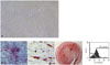
We investigated the immunophenotype of hUCB-MSCs to confirm differentiation potential. hUCB-MSCs have the potential to differentiate into type II alveolar epithelial cells, and are known to have multi-lineage differentiation potential to form bone, adipose and cartilage. As shown in Fig. 3, FACS data indicated that hUCB-MSCs indeed have differentiation potential into type II alveolar epithelial cells. Surfactant protein-C (SP-C), a type II alveolar epithelial cell marker, is not normally expressed in undifferentiated hUCB-MSCs, but it is expressed in hUCB-MSCs after differentiation. SP-C is an extremely hydrophobic 4 kda peptide produced by type II alveolar epithelial cells and it is critical for maintenance of respiratory structure and function. The above results, therefore, had us carefully speculate that hUCB-MSCs can differentiate into type II alveolar epithelial cells.
Effect on MCT-induced PAH according to dose of hUCB-MSCs
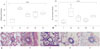
The optimal dose of hUCB-MSCs has not been established for PAH to assess the feasibility of low-dose hUCB-MSCs transfusion via the jugular vein. Therefore, we investigated the dosedependent effects on PAH in MCT-induced PAH rat. RV pressure was significantly increased in M group compared with C group at weeks 2 [C vs. M; 8.0 mmHg (6.0, 10.0) vs. 29 mmHg (28.0, 42.5)] and 4 [C vs. M; 11.0 mmHg (8.0, 12,0) vs. 49.5 mmHg (37.3, 69.3)]. Decreased RV pressure was observed in all three groups (UA, UB, and UC), but there was no significant difference among the three groups at week 2 [UA vs. UB vs. UC; 14.5 mmHg (13.3, 18.0) vs. 13.0 mmHg (12.5, 16.5) vs. 16.0 mmHg (13.5, 20.0)] and 4 [UA vs. UB vs. UC; 20.5 mmHg (18.5, 21.8) vs. 15.5 mmHg (14.0, 20.0) vs. 14.0 mmHg (13.5, 16.5)] (Fig. 4A). RV hypertrophy showed tendency to decrease in all three groups compared with M group, although the low-dose group (UC) showed slightly higher RV/LV+S ratio at week 2 [C vs. M vs. UA vs. UB vs. UC; 0.26 (0.25, 0.26) vs. 0.36 (0.36, 0.37) vs. 0.29 (0.28, 0.30) vs. 0.29 (0.14, 0.31) vs. 0.31 (0.28, 0.36)] and 4 [C vs. M vs. UA vs. UB vs. UC; 0.28 (0.25, 0.29) vs. 0.80 (0.65, 0.97) vs. 0.44 (0.24, 0.52) vs. 0.43 (0.24, 0.51) vs. 0.69 (0.55, 0.71)]. However, the differences between M and U groups were not statistically significant (Fig. 4B). Medial wall thickening of the pulmonary arterioles was improved, however, there was no significant difference among the three U groups at week 4 [C vs. M vs. UA vs. UB vs. UC; 21.24 (15.82, 22.75) vs. 42.31 (40.58, 44.17) vs. 31.63 (27.19, 32.25) vs. 34.78 (27.01, 36.05) vs. 32.52(31.9, 35.53)] (Fig. 5A). The number of intra-acinar arteries was improved in UA and UB groups, but there was no significant difference among the three groups at week 4 [C vs. M vs. UA vs. UB vs. UC; 0.80 (0.78, 1.06) vs. 1.91(1.43, 2.25) vs. 1.31 (0.99, 1.48) vs. 1.07 (0.94, 1.21) vs. 1.45 (1.35, 1.55] (Fig. 5B). The representative data of Victoria blue staining are shown in Fig. 5C. The above results suggest that the lowest-dose (3×10⁵ cells/rat) of hUCB-MSCs can also be effective in treating PAH rats. Because the low dose therapy has potential clinical significance in cost effectiveness, we carried out second experiment of low-dose hUCB-MSCs with added factor of treatment timing.
Therapeutic effect according to different timing using low dose hUCB-MSCs in MCT-induced PAH rats
To verify the therapeutic effect on MCT-induced PAH rats according to different timings using low dose hUCB-MSCs, we compared mean RV pressure among three groups (UD, UE, UF). The result showed that hUCB-MSCs decreased the mean RV pressure in all three groups at weeks 2 [C vs. M vs. UD vs. UE vs. UF; 11.0 mmHg (11.0, 12.0) vs. 27.0 mmHg (25.0, 33.0) vs. 16.0 mmHg (15.0, 16.0) vs. 14.0 mmHg (13.0, 16.0) vs. 16.0 mmHg (16.0, 18.0)] and 4 [C vs. M vs. UD vs. UE vs. UF; 11.0 mmHg (10.0, 11.0) vs. 44.0 mmHg (38.0, 45.0) vs. 15.0 (11.0, 23.0) vs. 13.0 mmHg (11.0, 18.0) vs. 13.5 mmHg (13.0, 14.0), not significant]. The difference among U groups was not statically significant (Fig. 6A). Therefore, not only reversal therapy but also prophylaxis of hUCB-MSCs improved PAH in terms of RV pressure.
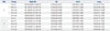
To explore whether these therapeutic strategies have an effect on RV hypertrophy, we compared RV/LV+S ratio (Table 1). As shown in Fig. 6B, RV/LV+S ratio was increased in M group compared with C group but there was no significant difference between M and U groups at weeks 2 [C vs. M vs. UD vs. UE vs. UF; 0.29 (0.29, 0.33) vs. 0.40 (0.40, 0.44) vs. 0.40 (0.34, 0.57) vs. 0.32 (0.27, 0.37) vs. 0.34 (0.32, 0.40)] and 4 [C vs. M vs. UD vs. UE vs. UF; 0.31 (0.31, 0.32) vs. 0.81 (0.67, 0.92) vs. 0.71 (0.66, 0.78) vs. 0.62 (0.58, 0.65) vs. 0.68 (0.54, 0.69)].
Next, we investigated pathological changes to confirm the preventive effects of hUCB-MSCs on PAH. As seen in Fig. 7A and C, medial wall thickness of pulmonary arteriole was slightly decreased in the UD, UE, and UF groups but not significant at weeks 2 [C vs. M vs. UD vs. UE vs. UF; 26.78 (23.86, 29.70) vs. 38.22 (32.23, 44.63) vs. 40.43 (38.04, 43.77) vs. 34.76 (29.11, 39.93) vs. 35.40 (31.98, 36.86)] and 4 [C vs. M vs. UD vs. UE vs. UF; 26.89 (25.82, 29.33) vs. 40.90 (40.89, 49.22) vs. 33.62 (31.55, 38.21) vs. 36.16 (36.02, 39.73) vs. 36.67 (30.47, 39.11)]. Furthermore, the number of intra-acinar arteries was significantly decreased in all three groups at week 4 [C vs. M vs. UD vs. UE vs. UF; 0.71 (0.70, 0.79) vs. 1.80 (1.78, 2.05) vs. 1.10 (0.86, 1.38) vs. 1.13 (1.12, 1.24) vs. 1.57 (1.41, 1.65)] (Fig. 7B).
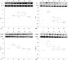
We examined the protein expression levels of ERA, Bcl-2 in the lung tissues and collagen 1, collagen 3 in the heart tissues after hUCB-MSCs transfusion (Fig. 8). In the lung tissues, Bcl-2 was significantly decreased in the UF groups compared with the M group at week 4 [M vs. UD vs. UE vs. UF; 2.33 (2.26, 2.41) vs. 1.08 (0.91, 1.51) vs. 1.09 (0.74, 1.44) vs. 0.76 (0.65, 0.80)], whereas collagen 3 in the heart tissues was significantly decreased in the UD and UF groups at week 4 [M vs. UD vs. UE vs. UF; 1.81 (1.46, 2.16) vs. 1.17 (1.03, 1.49) vs. 1.21 (1.10, 1.39) vs. 1.09 (1.09, 1.17)]. There were no significant changes between M and U groups in ERA and collagen 1.
DISCUSSION
The results in the present study suggested that low-dose (3×10⁵ cells/rat) reversal treatment of hUCB-MSCs via jugular vein did improve PAH and low dose early treatment also partially prevented MCT-induced PAH in our rat model. The ability to use low-dose stem cells has great potential to help commercialize hUCB-MSCs because low dose transfusion has an advantage of competitive price compared with other therapeutic strategies, and the cost for stem cell therapy can cause a significant economic burden to households. Furthermore, early transfusion of hUCB-MSCs has a significant implication because progression of the disease is relentless and refractory and early treatment was as effective as reversal treatment on RV pressure in this study.
This research is meaningful because low dose treatment reduces the costs of therapy and is the first to investigate the early transfusion effect of hUCB-MSCs on PAH. Early treatment attenuated expression levels of collagen 3 protein in the heart tissues: abnormally high expression levels caused by MCT were suppressed by an early transfusion of hUCB-MSCs, and therefore, early treatment may improve RV systolic function because increased deposition of collagen protein contributes to the deterioration of cardiac systolic function. These results led us to conclude that dual or reversal treatment of hUCB-MSCs is more effective than early treatment.
In the present study, we used hUCB-MSCs that could be differentiated into type II alveolar epithelial cells. Because the specific dose range of hUCB-MSCs for PAH has not yet been determined, we performed a dose escalation test. Dose raged from 3×10⁶ cells/rat to 3×10⁵ cells/rat showed similar levels of improvement, demonstrating that low-dose cell therapy has effects on MCT-induced PAH rats.
The main effects of the early transfusion of hUCB-MSCs on PAH are as follows. First, early stage treatment decreased mean RV pressure. Effects of reversal treatment have been previously confirmed by changes of several protein expression levels related to PAH and immunomodulation in MCT-induced PAH rats.67 However, the present study is a first report on early stage treatment. Second, early treatment improved pulmonary pathology. The number of neomuscularized intra-acinar arteries were decreased. Third, early treatment attenuated protein expression levels of collagen 3 in the heart tissues. Therefore, the effects of prevention therapy are comparable to that of reversal therapy.
These findings may have important implications for early stage therapy of PAH. PAH is progressively worse and occasionally fatal despite various medical interventions. PAH ordinarily leads to death within 2-3 years after first diagnosis, therefore, hUCB-MSCs injection at the initial phase is critical for prognosis because it is difficult to get back the lung structure and function once it becomes worsen. There is a report that early intervention may improve the prognosis of PAH. In 2002, Sitbon, et al.26 reported that the survival of patients with medical treatment was determined by the severity at baseline which was determined by clinical outcomes and hemodynamics, and suggested that early medical care can bring a favorable prognosis. Our data suggested that hUCB-MSCs in a PAH rat model are capable of differentiate into type II alveolar epithelial cells: we confirmed that differentiation induced hUCB-MSCs were possible to be specialized into type II alveolar epithelial. In 2003, Ortiz, et al.27 studied that MSCs may limit the injurious effect of bleomycin by replacing type II alveolar epithelial cells. Therefore, the present data indicate that early transfusion of hUCB-MSCs has beneficial effects on the prevention of PAH.
Low-dose hUCB-MSCs may improve PAH by regulating protein expression levels in lung and heart. The exact mechanism of low-dose hUCB-MSCs' effect is not currently known. Nevertheless, it has been suggested that hUCB-MSCs control the cardiac collagen deposition via paracrine mechanism. In this study, hUCB-MSCs decreased collagen 3 protein expression in the heart tissues, suggesting that low-dose hUCB-MSCs may improve RV function because increased collagen contents contribute to cardiac malfunction. Another possibility is that hUCB-MSCs downregulated Bcl-2 protein expression in the lung tissues, which is related with apoptosis. We earlier showed that high dose hUCB-MSCs have similar effects although slightly better. Therefore, it is likely that low-dose hUCB-MSCs act similarly as high-dose transfusions, resulting in the recovery of protein expression levels.
MSCs not only have reversal therapeutic effects on diverse diseases but also have prophylactic effects on several diseases. MSCs have been used in preclinical and clinical trials in treatments for graft-versus-host disease28 and autoimmune disease such as systemic sclerosis and type 1 diabetes mellitus.2930 In 2007, Mei, et al.31 that MSCs injection into the jugular vein significantly prevented lipopolysaccharide-induced acute pulmonary inflammation in mice. In 2008, Polchert, et al.32 suggested that activated MSCs by IFN-γ could prevent graft-versus-host disease. MSCs also have prophylactic effects on lung disease. In 2005, Zhao, et al.33 showed that bone marrow derived endothelial-like progenitor cells could prevent the MCT-damaged lung and microvasculature structure, and in 2013, Pierro, et al.34 reported that hUCB-MSCs prevented bronchopulmonary dysplasia by paracrine effect without adverse lung effect.
The effect of the early transfusion of MSCs in several diseases might be due to immune regulation ability by paracrine effect. MSCs have been shown to have a preventive effect on graft versus host disease through prophylactic immunosuppressive pathway by T cell regulation. Prophylactic treatment of bone marrow-derived MSCs by airway delivery improves arrested alveolar and vascular growth in part through paracrine activity in a bronchopulmonary dysplasia model.35 These findings suggest that early stage administration of hUCB-MSCs has immunomodulatory ability and recover injured alveolar and vascular cells as well.
The exact mechanism of hUCB-MSCs' prevention effect in PAH is not clearly understood. Whether it is an anti-apoptotic effect, inhibition of neo-muscularization of pulmonary arterioles or inhibition of cardiac fibrosis remains unknown. Various factors may influence these therapeutic actions on PAH. One probable mechanism of the prevention effect is controlling cardiac remodeling via paracrine mechanism. It has been known that MSCs are capable of cardiac repair in an infarcted rat heart,36 and we showed in the present study, that a preventive administration of hUCB-MSCs attenuated cardiac hypertrophy and increased cardiac protein expression levels of collagen 3, thus suggesting that hUCB-MSCs can possibly control cardiac hypertrophy and fibrosis by regulating a number of molecules such as collagen. The administration of hUCB-MSCs via the external jugular vein has shown an ability to control the collagen deposition and anti-apoptotic mechanisms by both preventive and reversal treatment. It is, therefore, highly likely that this identical mechanism may be involved in both therapeutic actions. However, the exact mechanism of preventive effect in PAH rat models is still unclear.
Limitations of this study are as follows. First of all, preventive intervention on PAH may not have important clinical meaning because the prevalence rate of PAH is low and early diagnosis is difficult. Second, the mechanism has not yet been elucidated although there are several reports that MSCS improved MCT-induced PAH rats. Third, it has not been confirmed that hUCB-MSCs have a potential to differentiate into type II alveolar epithelial cells in animal models. Fourth, this study was performed using small number of animals.
In conclusion, prophylactic early application of hUCB-MSCs on PAH may provide a basis for the development of a new approach to the treatment of PAH. The present study demonstrated that low-dose hUCB-MSCs treatment is effective in MCT-induced PAH rats. Also, low-dose early treatment improved PAH as well, although dual or reversal treatment is more effective.


 XML Download