

 PDF
PDF ePub
ePub Citation
 Print
Print


INTRODUCTION
Stenosis of the subclavian artery (SCA), although not common, occurrs in a number of patients. Most etiologies are atherosclerotic, and patients may present with vertebrobasilar insufficiency (subclavian steal syndrome), angina due to reversal of flow from an internal mammary arterial graft (coronary subclavian steal syndrome), and arm ischemia or weakness due to distal embolization or claudication. Treatment of these lesions has traditionally been through surgical bypass. Because of the lower complication rate, high technical success rate and good long-term patency rate (>80% at 5 years), catheter-based treatment has become more and more popular.1 We present a case of patient with total situs inversus and stenosis of the left SCA, who was treated with catheter-based revascularization with stent that was complicated with SCA perforation with hemothorax. Percutaneous stenting with a graft stent was used as a rescue, but this procedure was complicated with occlusion of the common carotid artery. The occlusion of the common carotid artery was finally re-opened by carotid stenting. We present a way to rescue perforation of large artery during percutaneous intervention.
CASE REPORT
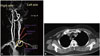
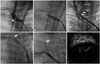
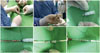
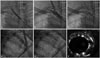
This 73-year-old female, with a history of coronary artery disease and situs inversus, had recently noticed left arm exercise weakness. The magnetic resonance angiogram and computer tomography of the upper limb vessels showed critical stenosis of the left-side SCA with calcification near the orifice (Fig. 1). Percutaneous revascularization was done via right femoral artery approach, and severe left SCA stenosis was confirmed (Fig. 2A) (Supplementary Video 1, only online). Direct stenting in the left SCA was carried out with a balloon-expansible Express LD 10×25 mm stent (Boston Scientific Corporation, Natick, MA, USA) (Fig. 2B) up to 8 atmospheres for 13 seconds. However, severe shortness of breath and hypotension developed after stenting. The blood pressure was down to 88/56, and the heart rate was up to 90. The oxygen saturation was down to 74%. Emergent intubation, fluid infusion, inotropic agent with norepinephrine were given. The immediate angiography showed vascular perforation in the stented segment of left SCA (Fig. 2C) (Supplementary Video 2, only online). Emergency balloon inflation within the stent was done with an 8×40 mm balloon. The cardiovascular surgeon was also consulted. However, due to the high surgical risk of bypass, endovascular treatment was suggested. We decided to do a retrograde approach through the left brachial artery by surgical cut-down. Since there was no suitable graft stent in our cath lab, we modified and cut the iliac extension of self-expanding Endurant II graft stent 10×82 mm (Medtronic, Inc., Minneapolis, MN, USA), which was originally designed to treat abdominal aortic aneurysms (AAA), to a suitable length (around 30 mm) (Fig. 3). After retrograde wiring, an operator-modified Endurant II graft stent was deployed in the stent segment (Fig. 2D) and the perforation was sealed successfully. After stenting of the graft stent, however, slow flow of left common carotid artery (LCCA) was noted, and angiography confirmed the near total occlusion of the LCCA, occluded by the SCA graft stent (Fig. 2E) (Supplementary Video 3, only online). A firm wire (Conquest pro, Asahi Inc, Seto, Japan) was successfully advanced outside the graft stent and into the LCCA. Despite sequent balloon dilations at the bifurcation site, the flow to the LCCA was still poor. Intravascular ultrasound also confirmed the severe compromise of the LCCA ostium which was caused by the graft stent (Fig. 2F). Therefore, we put a balloon-expansible carotid stent, Express 7×37 mm (Boston Scientific Corporation), from the LCCA to the left brachiocephalic artery (Fig. 4A). TIMI III flow of the LCCA was restored after stenting (Fig. 4B-E) (Supplementary Video 4, only online). Intravascular ultrasound also confirmed proper expansion of the LCCA stent (Fig. 4F). The follow-up chest radiography revealed left-side hemothorax. After thoracocentesis with drainage and proper care, the patient was discharged. Till now, the patient has been free from symptoms for six months.
DISCUSSION
Percutaneous angioplasty has become an increasingly popular therapy for treating symptomatic SCA stenosis. The technical success rate of percutaneous therapy for SCA stenosis with stent is up to 91% to 100% and ranges from 83% to 94% in totally occluded SCA.2 The complication rate has also been low. No peri-procedural deaths have been reported. Stenting of SCA stenosis is also characterized by its proven efficacy, long-term patency, lack of a need for general anesthesia, and shorter healing hospital stay.3 Therefore, when an intervention is indicated, a percutaneous approach is currently favored over surgical intervention.
There are several instructional points in our present case. First, what kind of mechanism is involved in the SCA perforation after stenting? Despite many advantages, there are still some possible procedural complications, including stroke, dissection, distal embolization, arterial thrombosis, and access site complication. SCA perforation caused by stenting has rarely been reported; however, as in our case, it may cause severe complications, such as hemothorax and hypotension. Therefore, it is highly possible that perforation might be caused by an over-sized stent or/and too rapid inflation of the stent, especially in a calcified lesion. It would be better off if we had inflated the balloon slowly and correlated with the patient's discomfort. In addition to angiography, further image modality, such as intravascular ultrasound, could help choose more precise stent size.
Second, how to deal with an iatrogenic SCA perforation? Due to massive blood loss at the time, immediate balloon inflation was mandatory to seal the perforation. In our case, the cardiovascular surgeon suggested endovascular intervention, rather than surgical bypass, because of higher surgical risk and complication rate. Due to the lack of any usual cover stent with such big size, we had to modify the iliac extension of Endurant II stent graft, which was originally designed to treat AAA. Furthermore, because this graft stent was designed for AAA, the delivery system was too short to approach SCA lesion by femoral approach. This made us to use the retrograde approach through the brachial artery by surgical cut-down. However, it was not easy to place this modified stent in a precise position, making the stent to accidentally occlude the LCCA. Because of the strong and firm force of the graft stent, the balloon could not dilate the occlusion well, and we had to put a balloon-expanded stent in the LCCA to keep the ostium open. In our opinion, before stenting a graft stent, a protective wire should be put in every important branch that might be impinged by the stenting. In this way, locating the stent position would be more precise, and total occlusion of the side branches could be avoided.
We described herein our valuable experience in dealing with a rare complication of SCA perforation while stenting the SCA. Choosing an SCA stent of a suitable size or even an under-sized SCA stent should be considered, and gradual slow inflation of the stent may also help lessen the risk of perforation. Except for open surgery, modified graft stent described in the present study is an alternative choice. While using this therapy, protective side branches wiring in addition to precise locating, should also be considered if the risk of jailing is high.
 XML Download