

 PDF
PDF ePub
ePub Citation
 Print
Print


INTRODUCTION
Large to giant aneurysms in the distal internal carotid artery (ICA) are challenging to manage because it is difficult to secure the complete topographic isolation of aneurysms while maintaining the collateral circulation through the anterior cerebral artery, middle cerebral artery, anterior choroidal artery (AchA), and posterior communicating artery (PcomA). The feasibility of achieving satisfactory clip placement is largely dependent upon proximal control of the parent vessels and obtaining complete visualization of the aneurysm including its surrounding structures. Retrograde suction decompression (RSD) is a useful technique for the treatment of large or giant distal ICA aneurysms. We describe a modified RSD technique using a commercialized carotid shunt device.
SURGICAL TECHNIQUE
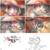
As shown in Fig. 1, cerebral angiography displays fusiform dilation of the right distal ICA and eccentric posterior aneurysm formation measuring 1.2×1.1×0.7 cm. The right M1 and A1 portions are incorporated, and the aneurysm is located just distal to the orifice of the PcomA. The AchA emerges from the side wall of the aneurysm.
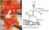
To secure this aneurysm, a standard pterional craniotomy was performed with removal of the sphenoid ridge. The sylvian fissure was widely split. The ipsilateral common carotid artery (CCA) was surgically exposed and isolated with a 4-cm horizontal incision along the skin crease at the level of C5–6. Vessel loops were applied at the proximal and distal edges of the exposed CCA. A purse string suture (diameter 5 mm) with a 6-0 Prolene was performed at the CCA midway between the two loops. A linear incision was performed with a No. 11 blade in the center of the suture. A Pruitt-Inahara® carotid shunt (LeMaitre Vascular Inc., Burlington, MA, USA) was prepared with heparin solution. The ICA balloon and safety balloon of the Inahara carotid shunt were tested. The insertion length of the Inahara carotid shunt within the ICA was determined preoperatively via head and neck CT angiography. The external carotid artery (ECA) of the patient was hypoplastic and there was no possibility of catheter malposition into the ECA. Only the ICA portion of the Inahara carotid shunt was inserted and used to purse the suture (Fig. 2).
Attention was now transferred to the aneurysm. The M1 proximal and A1 proximal portions were temporarily clipped. Trapping was achieved through the ICA balloon inflation of the Inahara carotid shunt catheter and tightening of the proximal vessel loop. Retrograde suction was performed with a 50 mL syringe. The aneurysm was effectively deflated (Supplementary Video 1, only online). Definitive clipping was performed with five ring clips with preservation of AchA (Fig. 1). We performed suction decompressions twice and discarded the aspirated blood. During balloon-deflation, we maintained the tightening of the proximal vessel loop, which moderately reduced blood flow and decreased aneurysmal wall tension. Consequently, we were able to adjust the clips easily without an additional RSD. Blood flow through the parent ICA, A1, M1, and AchA was verified by transcranial Doppler. Postoperative angiography revealed control of the aneurysm (Fig. 3).
DISCUSSION
Retrograde suction aspiration was first tried by Batjer and Samson,1 who inserted an angiocatheter into the cervical ICA and treated over 40 cases with giant paraclinoid aneurysms using this technique. Carotid artery dissection developed in a case in their series, which required emergency endarterectomy. Tamaki, et al.2 reported the use of the RSD technique under the name of ‘trapping-evacuation’. They deflated aneurysms by aspirating intra-aneurysmal blood through the ICA via the superior thyroid artery. To avoid carotid dissection, RSD was also introduced through the ECA3 or CCA.4 Such RSD techniques require temporary clips on the cervical carotid arteries to trap the aneurysm. Recently, an RSD technique using endovascular balloons has become an important adjunct to facilitate the dissection and clipping of large and giant aneurysms.5 The advantages of this modification are that it eliminates the need to expose the carotid artery in the neck, and that it allows simultaneous intraoperative angiography. However, even with the use of the endovascular RSD technique, it is difficult to complete the dissection and clipping of the aneurysm within only a few minutes in most cases.6789 The introducing catheter is placed in the artery and the balloon remains seated in the ICA throughout the surgery, which lasts for several hours. As in any endovascular procedure, the principal pitfall of suction decompression is the risk of embolism during the procedure, particularly when repeated inflations of the balloon are necessary.910
The Pruitt-Inahara® carotid shunt device was originally used to maintain cerebral perfusion during carotid endarterectomy.11 It is easily available, applicable, and inexpensive. It does not require experience or special skills to manipulate. The balloon is optimally sized for ICA placement, and temporary clips are not applied extracranially to the carotid artery. The carotid arteriotomy is small and sized to fit just the carotid shunt catheter, which measures 4 mm and is placed in the CCA. Therefore, its use may minimize ICA trauma. We used heparin (3000 units) to prepare the carotid shunt catheter and systemic heparinization was not required. The balloon occlusion was limited to 5 min at a time and was repeated after sufficient time intervals. Simply fastening the vascular loops could relieve tension in the aneurysmal wall without additional aspiration. The use of purse string sutures permits stable catheter sustention. This device features a safety balloon on the safety sheath to avoid ICA overinflation-induced arterial damage.

 XML Download