

 PDF
PDF ePub
ePub Citation
 Print
Print


INTRODUCTION
Cataract and glaucoma are common eye diseases, especially in older patients. Many older individuals with glaucoma also have cataracts, decreasing visual acuity, contrast sensitivity, and the accuracy of glaucoma examinations. For these reasons, many patients with glaucoma undergo cataract surgery.12
Prior to cataract surgery, multiple biometric analyses are performed, including measurement of axial length, keratometry, and anterior chamber depth. It is expected that minimum refractive error will remain after surgery, and the ultimate degree of postoperative error will be similar to the predicted value determined preoperatively. For this purpose, many surgeons use various formulas to calculate the intraocular lens (IOL) power, conduct multiple biometric examinations, and perform surgery using a small incision technique of phacoemulsification.3 However, some refractive errors inevitably develop, which can be disturbing to patients.45
Many previous studies have reported that IOL power predictions for cataract surgery in patients with angle-closure glaucoma (ACG) can be inaccurate. ACG may be associated with an abnormally shallow anterior chamber depth, short axial length, and high crystalline lens vault. This is in contrast to open angle glaucoma (OAG), which is usually associated with relatively normal anatomy.4 However, individuals with OAG may also develop refractive errors post-trabeculectomy because of surgery-related changes in anterior chamber depth and axial length. Furthermore, both ACG and OAG may be accompanied by con-nective tissue abnormalities that can influence the sclera, angle, zonules, and capsular bag orientation.67 In patients with glaucoma, refractive errors can be bothersome and lower vision quality.89 Refractive errors may also interfere with accurate visual field testing and sensitive detection of progressive nerve fiber defects, which are necessary for optimal management of patients with glaucoma.910
Changes in angle configurations may occur after cataract surgery; however, subtle changes in angle morphology cannot be detected by traditional methods. Recently, anterior segment swept-source optical coherence tomography (SS-OCT) has been used to evaluate anterior segment configurations. Anterior segment OCT is a non-contact method that can provide quantitative, objective data. When only the scleral spur is marked, the built-in software can automatically calculate many anterior segment parameters. Furthermore, the analytical properties of anterior segment SS-OCT are being continuously refined.1112
The objective of the current study was to compare refractive errors after cataract surgery in patients with OAG and an intraocular pressure (IOP) under 21 mm Hg to postoperative refractive errors in patients with cataracts but not glaucoma. We also sought to outline correlations between postoperative refractive errors and angle parameters obtained by SS-OCT.
MATERIALS AND METHODS
The study protocol followed the tenets of the Declaration of Helsinki and was approved by the Institutional Review Board of Gangnam Severance Hospital, Yonsei University College of Medicine. Informed consent was obtained from all subjects.
Patients
Between January 2015 and December 2015, patients from Ga-ngnam Severance Hospital Eye Center (Seoul, South Korea) were recruited. This prospective, comparative, observational study classified the patients into two groups: OAG group and CAT group. The OAG group included patients with glaucomatous optic nerve changes and visual field defects. To minimize the effects of IOP on IOL positioning, only patients with low-tension OAG were enrolled. Low-tension OAG was diagnosed by a glaucoma specialist according to the presence of the following: 1) glaucomatous visual field defect confirmed by two reliable visual field tests; 2) typical appearance of a glaucomatous optic nerve head, including a cup/disc ratio >0.7 and cup/disc ratio asymmetry >0.2, with diffuse or focal neuroretinal rim thinning, disc hemorrhage, or vertical elongation of the optic cup; 3) maximum untreated IOP <21 mm Hg on all three measurements obtained at different times on separate visits during follow-up; and 4) a normal and open-angled anterior chamber during slit-lamp and gonioscopic examination. The CAT group was defined as patients with no other ocular disease, except the cataract.
No patient had any other ocular disorder affecting aqueous outflow or angle morphology except the cataract and glaucoma. Clinical exclusion criteria included ACG, exfoliative syndrome, neovascular glaucoma, age-related macular degeneration, and proliferative diabetic retinopathy. Patients with prior corneal surgery, trabeculoplasty, cycloablation, or any incisional glaucoma procedure (such as trabeculectomy, tube shunt, or deep sclerectomy) were also excluded.
Preoperative and postoperative assessments
All patients underwent visual acuity determination, axial length measurements, and evaluation by tonometry, keratometry, sp-ecular microscopy, and indirect ophthalmoscopy. The power of the inserted IOL was calculated using the SRK-T formula by non-contact biometry (IOL-Master500®; Carl Zeiss Meditec, Dublin, CA, USA). Non-contact biometry can measure the axial length, keratometry values, anterior chamber depth, and anterior chamber width. Using these procedures, the expected postoperative refractive error was determined. The IOP measurement was performed using Goldmann applanation tonometry (AT 900®; Haag-Streit, Koeniz, Switzerland) after applying topical anesthesia (proparacaine HCl, Alcaine®; Alcon, Fort Worth, TX, USA). Biometric evaluations were performed three times for more accurate values, and we selected the modal value for analysis. If all of the values were different in repeated biometry, we selected the median value.
Postoperative refractive error values were obtained using an auto-refractometer (TONOREF®, Nidek Co., Ltd., Gamagori, Japan) 2 months postoperatively. Comparisons between the expected refractive errors and postoperative real refractive errors were performed. The postoperative refractive error was calculated by the equation of spherical equivalents: spherical errors+ 1/2 cylindrical error. ΔSE was calculated as the expected refractive error minus the postoperative real refractive error. Absolute ΔSE was the absolute value of the difference between the expected and postoperative real refractive error.
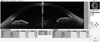
SS-OCT was performed before surgery. Anterior segment parameters were obtained by anterior OCT (Casia SS-1000, Nagoya, Japan). One operator obtained all angle images. The patient's eyes were undilated, and the testing was performed in dark, identical room conditions. To generate entire angle images, the upper eyelids were gently raised by the examiner with a long cotton tip. Using the “angle analysis mode” of the Casia SS-1000, images were recorded in the nasal, temporal, superior, and inferior angle quadrants by moving the arrow bar. The best images were selected thereafter using the automatic calculating software in the SS-OCT to obtain several anterior segment parameters. After marking the position of the scleral spur, the following measurements were automatically generated: angle op-ening distance (AOD) at 500 µm and 750 µm from the scleral spur (AOD 500 and AOD 750), trabecular-iris space (TISA) area at 500 µm and 750 µm (TISA 500 and TISA 750), area of the angle recess (ARA) at 500 µm and 750 µm (ARA 500 and ARA 750), and trabecular-iris angle (TIA) at 500 µm and 750 µm (TIA 500 and TIA 750) (Fig. 1). Scleral spur marking was therefore very important in this study for reliability of anterior segment parameter determinations. One investigator (WL) first marked the scleral spur site, and after 1 week, a second investigator (SHL) confirmed the site.
Surgical management
All patients were administered mydriatic drops [5 mg phenylephrine HCl plus 5 mg tropicamide (Mydrin-P®; Taejoon Pharmaceutical, Seoul, Korea)] before surgery. One surgeon (SGJ) performed all cataract operations under topical anesthesia (proparacaine HCl, Alcaine®; Alcon). A 2.75-mm clear corneal incision was made at the temporal side of the cornea, and the anterior chamber was filled with an ophthalmic viscoelastic substance (Healon®; Abbott Laboratories, Chicago, IL, USA). An approximately 5.5–6.0-mm continuous curvilinear capsulorrhexis was performed. Lens extraction was performed by phacoemulsification (INFINITI®; Alcon), and a foldable IOL (Hoya iSert®, Hoya, Tokyo, Japan) was inserted into the capsular bag. Postoperatively, the patients were treated with gatifloxacin eye drops (Handok, Seoul, Korea) and prednisolone acetate eye drops (Allergan, Irvine, CA, USA) four times per day for 2 weeks. No complications were noted during or after surgery.
Statistical analysis
The data were analyzed using SPSS, version 20, software for Windows (SPSS Inc., Chicago, IL, USA). Parametric paired t-tests, chi-square tests, and multiple regression tests were performed. All patients were included. Results were considered statistically significant if the p-value was <0.05. Data are presented as means±standard deviations, unless indicated otherwise.
RESULTS
Baseline characteristics and changes in intraocular pressure
The CAT group included 63 eyes of 63 patients, and the OAG group included 45 eyes of 45 patients (Table 1). The sex distribution and mean ages were similar in both groups. The axial length was also not significantly different between the two gr-oups. Anterior angle parameters also were not significant different between two groups (Supplementary Table 1, only online). Before surgery, the mean IOP was significantly higher in the OAG group (13.19±2.82 mm Hg) than in the CAT group (12.37±2.81 mm Hg). At 2 months after surgery, the mean IOP was not significantly different between the OAG group (11.26±2.95 mm Hg) and CAT group (11.54±2.39 mm Hg). Thus, there was a significant reduction in IOP after surgery in the OAG gr-oup (p=0.013).
Preoperative and postoperative refractive error
Preoperative predicted spherical equivalents did not differ between groups: -0.50±0.85 diopters (D) in the OAG group and -0.41±0.72 D in the CAT group (p=0.602) (Table 2). Postoperative spherical equivalents were -1.18±1.37 D in the OAG group and -0.80±0.85 D in the CAT group, reflecting ΔSE values of -0.68±0.53 and -0.40±0.38, respectively. ΔSE was significantly greater (reflecting a more myopic value) in the OAG group than in the CAT group (p=0.023). Preoperative cylindrical errors (as-tigmatism) were -0.89±0.72 in CAT group and -1.15±0.87 in OAG group. Postoperative cylindrical errors were -0.92±0.77 in CAT group and -1.05±0.82 in OAG group. There was no significant astigmatic changes between that before and after surgery (p=0.522, 0.630, respectively). Thus, unpredicted myopic refractive error increased in OAG patients after cataract surgery. Absolute ΔSE, reflecting the absolute value of the unpredicted postoperative refractive error, was greater in the OAG group than in the CAT group (0.76±0.46 vs. 0.49±0.32, respectively, p=0.011).
Among the 45 OAG patients, 23 (51.11%) had an absolute ΔSE of more than 0.5 D and 11 (24.45%) had an absolute ΔSE more than 1.0 D (Table 3). Among the 63 CAT patients, 22 (34.92%) had an absolute ΔSE more than 0.5 D and 4 (6.34%) had an absolute ΔSE more than 1.0 D. The percentages of patients with an absolute ΔSE more than 0.5 D or more than 1.0 D were higher in the OAG group than in the CAT group (p<0.001 for both comparisons).
Correlations with unpredicted postoperative refractive errors
In the OAG group, preoperative angle parameters obtained by SS-OCT were correlated with unpredicted postoperative refractive error. The correlations were most prominent for parameters at the superior and inferior quadrants, but not significant at temporal and nasal quadrants. The AOD 500, ARA 500 and 750, TISA 500 and 750, and TIA 500 in the superior and inferior quadrants were all significantly correlated with ΔSE (Table 4) (Supplementary Table 2, only online). Anterior chamber depth, lens vault, and anterior chamber width, however, were not correlated with ΔSE. In the CAT group, there were no correlations between any preoperative angle parameters and ΔSE (Table 5) (Supplementary Table 3, only online).
In univariate linear regression analysis, several angle parameters at the superior and inferior quadrants were negatively correlated with ΔSE in the OAG group. In multivariate linear regression analysis, AOD 500 at the superior quadrant was the only parameter significantly correlated with the ΔSE, after adjusting for age, sex, anterior chamber depth, axial length, and IOP (Table 6). It was negatively correlated with ΔSE.
Neither preoperative IOP nor postoperative IOP was correlated with postoperative unpredicted refractive error. OAG patients used a mean of 1.53±0.61 species of anti-glaucoma eye drops. At 2 months after cataract surgery, they used a mean of 0.53±0.93 species of anti-glaucoma eye drops. Among patients with OAG, 33 used prostaglandin analogues, eight used beta blockers, 11 used fixed combination of timolol and dorzolamide, and six used alpha agonist. Twenty-one subjects used two species of anti-glaucoma eye drops and three used three species. There was no significant correlation between prostaglandin analogue use and preoperative angle parameters (Supplementary Table 4, only online). There was also no significant correlation between prostaglandin use and ΔSE in the OAG group (β=0.137, p=0.369).
DISCUSSION
This is the first published study to examine unexpected refractive errors after cataract surgery in patients with low-tension OAG. We explored the possibility that preoperative angle morphologic characteristics may lead to an increased incidence of unexpected postoperative refractive errors in our patient population. We found that specific preoperative angle parameters in the superior and inferior quadrants were correlated with postoperative unpredicted refractive errors causing myopia. Our results suggest that patients with low-tension OAG who have a wide angle in the superior and inferior quadrants may be more likely to experience unexpected myopic errors after cataract surgery than patients without glaucoma.
Patients with ACG usually have a narrow anterior chamber depth. After cataract surgery, these patients may develop a wider anterior chamber. Because of this anterior chamber deepening, an inserted IOL could become tilted and positioned posteriorly, resulting in postoperative unexpected refractive errors. The angle morphology of patients with ACG usually differs from that of patients with normal eyes.4 After cataract removal and IOL insertion, older patients with OAG may likewise have resolution of a crowded angle caused by the cataract, as extraction of a thick lens and implantation of a thin IOL deepens the anterior chamber and moves the anterior capsule posterior to Schlemm's canal.113 These angle configuration changes could also cause the IOL to tilt or become decentered in patients with OAG.
The composition of the extracellular matrix of the trabecular meshwork is a key to the pathogenesis of primary OAG.14 Extracellular matrix is regulated by specific degradation of extracellular matrix components and selective deposition of new matrix material produced by trabecular meshwork cells, including collagens, glycosaminoglycans, proteoglycans, fibronectin, and elastin.15 The degradation involves matrix metalloproteinases (MMPs), a family of secreted zinc-dependent pro-teinases with selective substrate specificity. Based on reported changes in extracellular matrix composition in the trabecular meshwork of patients with glaucoma, it has been suggested that an imbalance of the MMP to tissue inhibitor metalloproteinase (TIMP) ratio could be involved in the development of glaucoma.151617
According to Wang, et al.,7 patients with OAG usually have low ocular rigidity. The anterior chamber of patients with OAG also has higher levels of MMPs and TIMPs, compared with the anterior chamber of normal eyes.1819 Variable levels of MMPs and TIMPs can modify the trabecular meshwork and angle structure, including the zonules, lens capsule, iris, and ciliary body.1617 Nga, et al.18 reported altered levels of MMPs and TIMPs, as well as an imbalance of the MMP/TIMP ratio, in the aqueous humor of primary OAG eyes, compared to non-glaucomatous control eyes. Total MMP and TIMP levels were significantly higher than in controls. Increased MMP/TIMP ratios were also noted in OAG eyes. Fountoulakis, et al.17 reported MMP-2/TIMP-2 ratios of 4.04 in patients with primary OAG and 2.07 in control patients, and the ratios of MMP-2/TIMP-2 and MMP-2/TIMP-1 were higher in primary OAG patients than in controls. Especially in glaucoma patients, an imbalance of MMP/TIMP ratios might contribute to changes in angle structure after cataract surgery.
Prostaglandin analogues, which are often prescribed to patients with glaucoma, can modulate intra- and extraocular extracellular matrix via MMPs.20 The mechanism by which prostaglandin analogues reduce IOP has been well studied and is believed to occur by enhancement of the uveoscleral pathway through regulation of MMPs and resultant remodeling of the extracellular matrix.21 It has been shown experimentally that prostaglandin analogues induce a dose-dependent increase in MMPs in aqueous humor and human ciliary smooth muscle cells, leading to remodeling of the extracellular matrix and increasing the space between bundles of smooth muscle cells.22 Although use of prostaglandin analogues was not significantly correlated with postoperative refractive errors in our study, the possibility remains that prostaglandin analogues are modulators of angle structure through their action on MMPs.
Among the four quadrants (superior, inferior, nasal, and temporal), the angle parameters of the superior quadrant were de-emed to be important determinants of postoperative refractive errors. In the sitting or lying position, the superior quadrant is the area most influenced by gravity. In patients with OAG and a wide preoperative angle (greater AOD 500) in the superior qu-adrant, a greater unpredicted myopic refractive error was observed after cataract surgery. With widening of the superior angle, posterior movement or tilting of the IOL is more likely to be influenced by gravity. Even a slightly tilted IOL can lead to myopic refractive error at the IOL center (view axis). Kang, et al.4 investigated the inaccuracy of IOL prediction for cataract surgery due to IOL shift or tilt in patients with ACG. Posterior shifting of a large capsular bag after cataract removal results in deepening of the anterior chamber. IOL shifting or tilting could produce refractive errors due to anatomical differences in ACG eyes, compared to non-glaucomatous eyes.4 In our study involving detailed evaluation of preoperative parameters using SS-OCT, a preoperative wide AOD 500 was particularly likely to result in greater unpredicted refractive error after cataract surgery in patients with OAG.
This study has some limitations. A relatively small number of subjects were enrolled, and the duration of the study was 2 months. For more reliable conclusions, more subjects and a longer follow-up time are needed. Furthermore, the difference in refractive error was relatively small. Approximately 0.25 D to 0.50 D differences can occur with typical cataract surgery. Nevertheless, this study is the first to specifically examine perioperative refractive errors in patients with OAG and to identify correlations between postoperative unpredicted errors and pre-operative angle parameters; these parameters did not include anterior chamber depth, but they did include subtle parameter changes that could only be detected by high resolution SS-OCT. Even relatively small diopter differences can interfere with the visual acuity and quality of patients with glaucoma.


 XML Download