

 PDF
PDF ePub
ePub Citation
 Print
Print


INTRODUCTION
Nephrin, a podocyte-related protein, is a protein to form a major component of slit diaphragm that tightly restricts the passing of large proteins such as albumin.1 Since the discovery of nephrin mutation in congenital nephrotic syndrome of the Finnish type,2 the role of nephrin in the pathogenesis of diabetic glomerulopathy and proteinuria has been suggested by several data.345678910
Preeclampsia (PE) is characterized by proteinuria associated with renal pathology delineated as endothelial cell swelling, loss of endothelia fenestration, widening of subendothelial space, and deposition of hyaline material.1112 Although the disruption of basement membrane and podocyte was not evident in electron microscopic examination of kidney of preeclamptic patient,13 urinary excretion of podocyte and nephrin suggesting podocytopathy has been recently observed in patients with PE.1415161718 Furthermore, in vitro demonstration of nephrin shedding from the podocyte, induced by endothelin-1 secreted from the endothelial cells which were exposed to the sera of PE patients, may be the underlying mechanism of proteinuria in PE.19 Thus, detecting urinary excretion of podocyte-related protein may serve as a potent predictive or early diagnostic tool for PE. In light of this, a prospective longitudinal study was conducted to evaluate the nephrin levels in serum and urine of normal pregnancy to establish a standard reference value and to compare them with patients who subsequently developed PE.
MATERIALS AND METHODS
Participants
From October 2010 to August 2012, 117 healthy women with singleton pregnancies with confirmed gestational age with means of ultrasonography in the first trimester were enrolled between 6 to 20 weeks of gestation at 2 participating medical centers (Yonsei University Healthcare Center and MizMedi Women's Hospital, Seoul, Korea) adhering to a common protocol. Women with uncertain dating of the pregnancy, multi-order pregnancy, spontaneous abortion, intrauterine fetal death, fetal anomaly, diabetes, hypertensive disease, renal disease, gestational proteinuria, urinary tract infection, vascular or connective tissue disease, under medication other than multivitamin supplement, language barrier, and smoker were considered ineligible for the study. Patients complicated with hypertension, diabetes, renal disease, or connective tissue disease were also excluded from the study. PE was defined as newly onset hypertension (either diastolic blood pressure of at least 90 mm Hg or systolic blood pressure of at least 140 mm Hg on two occasions) accompanied by clinically significant proteinuria, defined as one of the followings: random urine dipstick results of at least 1+ on two occasions or results of at least 2+; or 24-hour urine protein of at least 300 mg. This study was reviewed and approved by the institutional review board and written informed consent was obtained from patients prior to the enrollment.
Procedures
Technicians performing the assays were blinded to the patient's information and pregnancy outcome. Eligible patients were requested to submit urine and serum samples at the time of enrollment, at 11 to 15 weeks, 16 to 20 weeks, 21 to 28 weeks, 29 to 40 weeks of gestation, and at 4 to 6 weeks postpartum. Serum and urine samples were stored at -70℃ in aliquots until analysis. Enzyme-linked immunosorbent assay (ELISA) for nephrin was performed in duplicate with the use of commercial kit (USCN Life Science Inc., Wuhan, China). The minimal detectable dose was 0.0156 ng/mL for nephrin.
Statistics
Statistical analysis was performed using SAS 9.2 version (SAS Institute Inc., Cary, NC, USA). Comparison of basic characteristics between pregnancies with normal outcome and women with subsequent PE was conducted using Mann-Whitney U-test and Fisher's exact test. Comparison of serum and urine nephrin level according to the sampling points was performed using Linear mixed model. Comparison of serum and urine nephrin levels between normal and patients with subsequent PE was conducted using Mann-Whitney U-test. For all analyses, a p-value of <0.05 was considered statistically significant.
RESULTS
Patients' characteristics
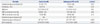
Of 117 uncomplicated pregnant women enrolled, 9 pregnancies terminated prior to 20 gestational weeks, and 9 delivered elsewhere. Among 99 patients (84.6%) who delivered at the study centers, 12 patients (12.1%) later developed PE at a median gestational age of 34+4 weeks (range 29+5–36+6 weeks). At postpartum, 16 were loss to follow up following delivery, 2 withdrew from the study at postpartum, and 1 did not submit urine sample. A total of 80 patients were eligible for final analysis. Basic characteristics of eligible cases and those who developed PE are shown in Table 1.
Subanalysis of 12 patients who subsequently developed PE
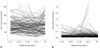
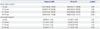
The median interval between the specimen collection and diagnosis of PE was 9 days (range 3–19 days). The nephrin levels in specimens obtained prior to the onset of PE from women who subsequently developed PE were significantly higher, compared to that of the normal pregnant controls (Table 3). Increase in serum and urinary nephrin preceded the clinical manifestation of PE. However, as delineated in the longitudinal plots (Fig. 2), third trimester alteration was more pronounced and discriminative in the urine nephrin while the serum level overlapped with the normal control.
DISCUSSION
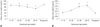
The major findings of this prospective study are: 1) serum nephrin level showed a decreasing trend while urine nephrin increased in the latter half of the pregnancy, but both values returned to the 1st trimester level at postpartum; and 2) a prominent increase in the serum and urine nephrin level antedated the onset of PE.
Garovic, et al.15 were the first to make an important observation about a significant excretion of podocyte in the preeclamptic patient's urine, suggesting podocyturia as a novel marker for the diagnosis of PE. Similar to Garovic's finding, Aita, et al.14 demonstrated that acute but transient podocyturia in PE is closely related to heavy proteinuria. However, when it comes to clinical application, determining podocyturia by overnight incubation and staining the urinary cells with podocyte-related proteins such as podocin, podocalyxin, neprhin, and synaptopodin is time-consuming. To better utilize the significant finding of Garovic, et al., subsequent studies pertinent to urinary podocyte-specific proteins in PE followed.1720 In the recent studies, soluble podocyte-specific proteins were measured in the urine samples, using ELISA rather than cell per se.1720 The result not only demonstrated that the urine concentration of nephrin is significantly increased in PE, compared with normal pregnancy, but also compared with pregnancy complicated with chronic hypertension.20 These data are expected to provide a future perspective on the use of urinary podocyte-related proteins, resulting from the podocyte injury and glomerular barrier dysfunction occurring in the PE for early diagnosis for PE, or differential diagnosis of hypertensive or proteinuric disease during pregnancy.
In this context, normative data of serum and urine nephrin level during pregnancy should be available to first understand the gestational pattern. To our best knowledge, the present study is the first to illustrate the gestational changes in serum and urine nephrin level in a prospective manner. The reason for the decrease and increase of nephrin in serum and urine, respectively, during latter half of pregnancy is unclear. Nevertheless, we speculate that the physiologic expansion of plasma volume occurring in the latter half of pregnancy may contribute to the decrease of serum nephrin level. And as vascular endothelial growth factor (VEGF) blockade is known to induce podocyte injury,19 late increase in the soluble fms-like tyrosine kinase 1 (sFlt-1) that normally occurs in pregnancy21 may lead to subclinical podocyte injury resulting in increased urinary shedding of nephrin. Furthermore, slight podocyturia in the absence of clinically significant proteinuria observed in a normal pregnancy substantiates our speculation.14
One concern raised in the present study is that the urine nephrin concentration of normal pregnant women at 3rd trimester was much lower than that measured previously by Wang, et al.20 (0.26 ng/mL vs. 86 ng/mL). However, this may possibly due to difference in commercial immunoassay kit used (USCN Life Science vs. Exocell) or difference in subject ethnicity (Korean vs. African).3
Of note, women in whom PE later developed had significantly higher levels of urine nephrin at 32–35 gestational weeks when compared to those with normal pregnancy outcome, whereas this difference was not evident in the serum nephrin level. This observation, albeit small sample size, indicates that the nephrin in the urine is not a result of increased filtration of serum nephrin, but rather caused by a direct shedding from the renal podocytes injured by circulating factors.
Moreover, the increase in urine nephrin level preceded the onset of clinical manifestation of PE by 9 days (range 3–19 d). Although the statistical significance cannot be determined due to small sample number, the sensitivity and specificity of urine nephrin at 3rd trimester at a cutoff value of 0.85 ng/mL were 100% and 97%, respectively. In comparison, the time interval was much greater in serum sFlt-1 and soluble endoglin (sEng) with 5 weeks and 2 to 3 months, respectively.2122 Considering the fact that preventive strategy for PE is unavailable at this time, use of sFlt-1 and sEng for predicting PE would only make patients and clinicians suffer the fear of disease occurrence for substantial period of time. Thus, the use of urine nephrin that represents relatively shorter time interval between the rise and the onset of PE, compared with serum sFlt-1 and sEng, may clinically be more feasible in the prediction of subsequent development of PE or in making decision at a timely basis. Furthermore, urine nephrin may be incorporated together with other placenta-derived biomarkers in an effort to improve the predictability and/or accuracy of PE.2324252627282930
Our study has several limitations. The main weakness of this study was a small study population in PE. Some of the analyses may not have enough power to demonstrate the statistical significance. Second, there was a lack of information on some potential confounders such as circadian variation of urine protein. Patients with glomerular proteinuria and fairly well-preserved renal function have circadian rhythm for proteinuria. Therefore, we were unable to adjust for this factors and to exclude a confounding effect on the result. The difference in the median gestational age at sampling in the 3rd trimester between the control and the patient who subsequently developed PE may affect the result (36+1 and 33+1 wks, respectively, p=0.011). This may be the case, since serum nephrin tends to decrease with advancement in gestation. However, the level of urine neprhin remained consistent with narrow standard error, and the degree of increment was far greater than the control. Furthermore, when urine nephrin level of the PE group was compared to that of earlier gestational age control, the value was still significantly higher in PE group. Lastly, due to a small sample size of our study, feasibility of urine nephrin as a predictive factor in PE remains to be assessed by prospective longitudinal study involving large number of pregnancies with subsequent PE.
In conclusion, as the onset of PE was preceded by the rise in the serum and urine nephrin in comparison to normal pregnancy, serum and urine nephrin may be useful as a predictive marker of PE.

 XML Download