

 PDF
PDF ePub
ePub Citation
 Print
Print


INTRODUCTION
Fracture is a serious complication that often occurs in advanced chronic kidney disease (CKD) patients, causing patients to often become bed-ridden (or have restricted ambulation) for a long time, and it also has detrimental effects on clinical outcomes and quality of life. The effect of mild to moderate renal dysfunction on increased fracture risk is considered controversial, although it has been verified in a few studies recently.1234 The risks of fracture are known to be influenced by both quantity and quality of bone.5 Bone biopsy is a standard method in order to diagnose bone quality and type of CKD-mineral bone disorder (MBD). However, due to its invasiveness, bone biopsy is not used frequently in clinical settings. Even though bone mineral density (BMD) is a useful tool for evaluating bone quantity and fracture risk in the general population, its application in CKD patients has been limited, as it has been shown that low BMD cannot predict risk of fracture in renal failure patients.6 Despite controversy surrounding BMD, recent studies have shown that CKD patients who experienced fracture had low bone mass in meta-analysis7 and osteoporosis assessed by dual-energy X-ray absorptiometry (DXA) can predict fracture risk in patients with CKD.89 These results suggest that BMD can serve as an alternative surrogate marker for fracture risk in CKD patients.
Renal clearance tests using inulin or inulin analogs are the gold standard for measuring glomerular filtration rate (GFR). However, difficulties during the infusion procedure and risks of anaphylactic allergic reactions limit its routine clinical application. In addition, creatinine clearance rate calculated from 24-hour urine collection can estimate renal function. However, it is not convenient to collect urine specimens throughout a day. Clinically, serum creatinine levels are used to estimate renal function, although the accuracy of this approach is easily affected by individual muscle mass variations and renal tubular secretion levels. On the other hand, a recent study has presented cystatin C as a more valid marker for renal function estimation, overcoming the limitations of serum creatinine.10 Several equations have been formulated to calculate estimated GFR (eGFR): Modification of Diet in Renal Disease (MDRD),11 CKD-Epidemiology Collaboration (CKD-EPI) creatinine (CKD-EPI-Cr),12 and CKD-EPI cystatin C (CKD-EPI-Cys).10
Cystatin C is known to be an effective marker of adverse outcomes in CKD patients, such as coronary artery disease, heart failure, and stroke.131415 In addition, previous research has revealed that cystatin C acts as an inhibitor of bone resorption in vitro16 and that it possibly reflects hip fracture risks in older women.17 In light of this, we hypothesized that cystatin C-based eGFR might be more precisely correlated with loss of bone mass than creatinine-based eGFR. Among the eGFR methods described above, we aimed to identify the formula that would be optimal for correlating osteopenia in CKD patients by measuring BMD by DXA.
MATERIALS AND METHODS
Ethics
This study was performed in accordance with the Declaration of Helsinki. It was approved by the Institutional Review Board at each participating hospital: Gachon University Gil Medical Center (GIRBA2553), The Catholic University of Korea Seoul St. Mary's Hospital (KC11OIMI0441), Kangbuk Samsung Hospital (2011-01-076), Seoul National University Bundang Hospital (B-1106/129-008), Seoul National University Hospital (1104-089-359), Seoul Eulji Hospital (201105-01), Severance Hospital (4-2011-0163), Inje University Busan Paik Hospital (11-091), and Chonnam National University Hospital (CNUH-2011-092). Written informed consent was provided by every participant in the study.
Study design and population
This study was cross-sectional in design, using patient-based cohort data from the KoreaN cohort study for Outcome in patients With Chronic Kidney Disease (KNOW-CKD) study.18 KNOW-CKD study was performed over 5 years, from 2011 to 2015, and enrolled 2450 CKD patients at nine university hospitals in Korea. In the present study, we included only 1529 CKD patients who were enrolled until December 2013. In this study, we could not obtain Z-score data from BMD. Male patients aged <50 years and pre-menopausal female patients were not suitable to diagnose osteopenia with only T-score data. The mean age of menopause in Korean women is 49.7 years old;19 therefore, we excluded patients aged <50 years (n=527). The patients who had missing data on cystatin C levels (n=169) or BMD T-scores (n=53) were also excluded. Finally, 780 patients were included in the study.
Data collection
All data analyzed in this study were collected at study enrollment. Baseline demographic data included age, gender, body mass index (BMI), smoking status, blood pressure, and medical histories, such as diabetes mellitus, hypertension, coronary artery disease, cerebrovascular disease, and congestive heart failure. Laboratory data included complete blood cell counts, blood urea nitrogen levels, serum creatinine and cystatin C levels, calcium and phosphate concentrations, lipid profiles, albumin levels, intact parathyroid hormone levels, 25-hydroxyvitamin D and 1,25-dihydroxyvitamin D concentrations, and C-reactive protein levels. We collected 24-hour urine specimens to analyze daily urine protein and creatinine levels. Other methodological issues were described in our previous study.18
Creatinine and cystatin C measurement and eGFR formula
Serum creatinine levels were measured using an isotope dilution mass spectrometry (IDMS)-traceable method at a central laboratory, and cystatin C concentration was assessed by immunonephelometry calibrated against the reference. We compared three eGFR methods in this study: four-variable MDRD by IDMS-traceable method (MDRD), creatinine-based CKD-EPI (CKD-EPI-Cr), and cystatin C-based CKD-EPI (CKD-EPI-Cys). The formulas were taken from previous studies101112 and are described in Supplementary Table 1 (only online). Additionally, we calculated eGFR using Korean-specific equations to compare with CKD-EPI-Cys formula (Supplementary Table 1, only online).20
BMD measurement
BMD was measured at baseline by DXA, and total hip and femur neck T-scores were used to diagnose osteopenia. Spine BMD measurement can be misleading if there are anatomical abnormalities in the bone, extensive osteophyte formation, or aortic calcifications, thus we excluded L-spine T-score data in the analysis. The T-score was expressed as the number of standard deviations of the BMD based on the mean value of a healthy, 30-year-old individual of the same gender and ethnicity as the patient. Osteopenia was defined as a T-score under -1.0 at two pre-defined points (total hip and femur neck). According to the definition of osteopenia, we divided the patients into two groups: the normal bone mass group (T-score >-1.0) and the osteopenia group (T-score ≤-1.0).
Statistical analyses
We performed statistical analyses using IBM SPSS for Windows, version 21 (IBM Corporation, Armonk, NY, USA). Numerical variables were expressed either as means±standard deviations for data with standard normal distributions or as medians (interquartile range) for skewed data. Categorical variables were expressed as numbers (percentages). We analyzed the differences between the groups using Student's t-test or the Mann-Whitney U test for continuous variables and the chi-squared test for categorical variables. Multiple logistic regression analyses were performed to verify independent factors related to osteopenia after adjusting for confounding variables. Receivers operating characteristic (ROC) curves were created for the eGFR calculation methods, and area under ROC curves (AUCs) were compared to identify the eGFR equation best correlated with osteopenia according to the method of DeLong, et al.21 Analyses related to ROC curves were performed using MedCalc® for Windows, version 14.8.1 (MedCalc Software, Ostend, Belgium). To make up for the difference of baseline renal function between normal bone mass group and osteopenia group, we conducted 1:1 propensity score matching (PSM) analysis according to the baseline CKD stage using IBM SPSS for Windows, version 23 (IBM Corporation).
RESULTS
Characteristics of the study subjects
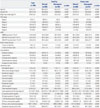
Baseline demographic and laboratory data of all patients are presented in Table 1. Among a total of 780 patients, the mean age was 61.0±7.0 years and 291 (37.3%) were female. When the patients were classified into groups according to CKD stage, the CKD stage 3 (42.7%) and stage 4 (29.4%) groups were the largest. In the entire cohort, 185 (23.7%) patients had osteopenia at the total hip and 312 (40.0%) at the femur neck. At both pre-defined points, the proportion of female patients was higher among the osteopenia patients, and the osteopenia patients were generally older and had lower mean BMIs. The patients in the normal bone mass group mainly had CKD stages 1 to 3, although the osteopenia group predominantly included CKD stage 4 and 5 patients. Co-morbid conditions were not statistically different between the two groups. With respect to laboratory data, the osteopenia group had higher blood urea nitrogen and cystatin C concentrations and lower mean eGFR levels than the normal bone mass group. However, serum creatinine levels in the osteopenia group were not higher, and the total hip osteopenia group showed statistically lower mean serum creatinine concentrations than the normal bone mass group for the total hip. In addition, serum calcium, 25-hydroxyvitamin D, and 1,25-dihydroxyvitamin D levels were lower in the osteopenia group; however, serum phosphate and intact parathyroid hormone levels were higher in the osteopenia group. Furthermore, 24-hour urine creatinine levels were lower in the osteopenia group for both total hip and femur neck osteopenia groups, and 24-hour urine protein concentrations were not significantly different between the two groups.
eGFR as an independent factor associated with osteopenia
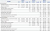
To identify the relationship between eGFR and osteopenia, multiple logistic regression analysis was performed using three models. All three models showed that eGFR calculated by CKD-EPI-Cys was independently associated with osteopenia at the total hip after adjusting for confounding variables [odds ratio (OR) 0.98, 95% confidence interval (CI) 0.97–0.99, p=0.004] (Table 2). Similarly, eGFR calculated by CKD-EPI-Cys showed comparable results for osteopenia at the femur neck (OR 0.98, 95% CI 0.98–0.99, p=0.001). However, the models using creatinine-based eGFR data showed discordant results between total hip and femur neck assessments. Osteopenia at the femur neck correlated with results determined using creatinine-based eGFR methods (MDRD, CKD-EPI-Cr); results at the total hip, however, were not related to those determined using creatinine-based eGFR formulas (MDRD, CKD-EPI-Cr) (Supplementary Table 2 and 3, only online).
Comparison of osteopenia AUCs depending on eGFR formulas
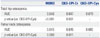
We plotted three ROC curves to illustrate the relationship between osteopenia and eGFR and compared the AUCs to determine which eGFR calculation was best correlated with osteopenia in CKD patients. Among the methods, CKD-EPI-Cys had the largest AUC at the total hip (AUC 0.678, vs. MDRD, p<0.001; vs. CKD-EPI-Cr, p=0.001) (Table 3) (Supplementary Fig. 1, only online). At the femur neck, CKD-EPI-Cys also showed similar trends when we compared the AUC thereof with those of the other eGFR formulas (AUC 0.665, vs. MDRD, p=0.008; vs. CKD-EPI-Cr, p=0.011) (Table 3) (Supplementary Fig. 2, only online). The data comparing AUCs of Korean-specific eGFR equation and the CKD-EPI formula at two pre-defined points are provided in Supplementary Table 4 (only online). The AUC of the CKD-EPI equation was larger than that of the Korean-specific and creatinine based eGFR equation, and it was also statistically significant.
Propensity score matching analysis data
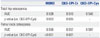
As shown in Table 1, baseline kidney function was different between the normal bone mass group and osteopenia group. This could be a confounding factor to solidify the effect of cystatin C-based equation on osteopenia. For this reason, we conducted 1:1 PSM analysis according to baseline CKD stage. After PSM, 185 patients of each group were selected in regard to total hip osteopenia, and 312 patients of each group were chosen for femur neck osteopenia. Table 4 lists the result after PSM analysis, and the AUC of CKD-EPI-Cys was the significantly largest among the AUCs of all three equations.
Subgroup analysis according to the gender
A previous study reported gender differences in BMD in the elderly.22 Therefore, we performed subgroup analysis to find differences according to gender (Table 5). As expected, the AUCs of the CKD-EPI-Cys were the biggest among the three methods irrespective of gender at both pre-defined points. In terms of osteopenia at the total hip, CKD-EPI-Cys was superior to the other formulas in men (vs. MDRD, p=0.003; vs. CKD-EPI-Cr, p=0.007), but not in women (vs. MDRD, p=0.054; vs. CKD-EPI-Cr, p=0.111). However, at the femur neck, CKD-EPI-Cys was not superior to the other formulas when assessing osteopenia in both male and female patients, except when compared to MDRD in males (vs. MDRD, p=0.039).
DISCUSSION
We compared three eGFR formulas used to estimate renal function and osteopenia using BMD as a standard, and found that CKD-EPI-Cys was the best formula in CKD patients. For the first time, we used ROC curve analysis to demonstrate that the cystatin C-based eGFR formula was superior to conventional creatinine-based eGFR calculation methods to detect osteopenia in CKD patients.
CKD patients experience loss of bone mass accompanied by a decline in renal function, and the prevalence of fractures has been reported as 18–47% in CKD stage 3 and 423 and 30–52% in ESRD patients.623 Bone mass decline has been revealed as one of the factors associated with increased fracture risks in CKD patients recently, although current guidelines recommend not testing BMD in CKD patients. Therefore, there is an unmet clinical need for effective markers for predicting bone mass decline in CKD patients. Cystatin C recently came into the spotlight as a valid marker for estimating renal function, and some studies have reported that cystatin C-based eGFR equation is superior to creatinine-based eGFR ones in estimating renal function and predicting cardiovascular events.131415 Furthermore, a recent study also proposed cystatin C as a predictor of hip fracture risk in elderly female patients,17 and several previous studies regarding the association between mild decreased renal function assessed by cystatin C in the elderly and loss of bone mass have also been reported.242526 However, little is known about the relationship between cystatin C and decreased bone mass determined by BMD in CKD patients, and further research is needed on this patient group.
We measured BMD using DXA in all patients at enrollment, and the results were analyzed by three eGFR formulas. As mentioned earlier, the Kidney Disease Improving Global Outcomes CKD-MBD guidelines recommend that BMD testing should not be performed routinely, because BMD cannot predict fracture risks and the type of renal osteodystrophy in CKD patients.5 However, several reports on general populations with mild renal function decline and bone loss,12426 and recent studies with CKD patients have demonstrated contrasting results: BMD can predict fracture risk in patients with CKD.8927 Considering the invasiveness of bone biopsy, BMD assessment can be an effective method to predict fracture risk in CKD patients.
In this study, CKD-EPI-Cys had the largest AUC among the three eGFR calculation methods for reflecting osteopenia in CKD patients. In contrast to CKD-EPI-Cys, eGFR calculated using creatinine (MDRD and CKD-EPI-Cr) showed discrepancies between the two BMD measurement sites in reflecting osteopenia. All of these results consistently support the superiority of cystatin C-based eGFR formula over creatinine-based eGFR formulas for detecting osteopenia in CKD patients. A previous study on women without clinical CKD (eGFR ≥60 mL/min/1.73 m2) demonstrated a relationship between increased risk of hip fracture and higher cystatin C levels, but not higher serum creatinine levels.17 We suggest that early changes in bone metabolism due to mild deterioration of renal function, including changes in calcium, phosphorus, and vitamin D homeostasis, cannot be easily detected by serum creatinine changes and that cystatin C could reflect these changes earlier than serum creatinine. We also suggest that individual muscle mass variations can hinder the accuracy of creatinine-based eGFR calculation methods. In patients with low muscle mass, creatinine-based eGFR might overestimate renal function, compared to measured GFR. Therefore, creatinine-based eGFR formulas cannot accurately reflect renal function in patients with low muscle mass. Finally, cystatin C is not only implicated in renal function, but also has considerable extrarenal effects that can impact outcomes.1528 Therefore, other underlying mechanisms of cystatin C, such as interaction with inflammatory factors and direct effects on bone resorption,162930 might explain why cystatin C is particularly effective in predicting bone mass changes in CKD patients.
In addition, gender subgroup analysis showed that CKD-EPI-Cys was not statistically significantly associated with osteopenia in female patients. The most plausible explanation for this is the relatively small number of the female patients included in the study. Nevertheless, we also speculate that the effect of menopause in females aged >50 years may play an important role in BMD changes, making it hard to diagnose osteopenia by CKD-EPI-Cys alone. Therefore, additional investigations combining the effects of menopause and renal function decline are needed to predict bone mass changes in elderly female CKD patients.
Bone quantity and quality are the main factors contributing to bone force, and bone quantity decline has an additive effect on increasing fracture risk in CKD-MBD patients. The evaluation of bone quality by bone biopsy is a proven-diagnostic method to predict fracture risks in CKD-MBD patients.5 Therefore, high-risk patients need to be assessed for fracture earlier with respect to decreased bone mass, and using cystatin C to accurately assess renal function and predict skeletal events could be a useful tool in CKD patients.
This study has several limitations. First, we did not directly measure GFR, and thus, we could not compare eGFR with accurately measured GFR. Second, we could not obtain data about menopause, fracture history, and medication history, such as steroid use, as well as data about Z-score and measured BMD values, muscle and lean body mass, and bone turnover markers. Due to these shortcomings, exclusion of the patients aged less than 50 years old may affect the results of this study. Third, this is a cross sectional study, and we did not check the incidence of fracture or serial BMD changes. Although our findings demonstrated an important aspect of deterioration of renal function and low BMD, future investigations taking into account fracture incidence data are needed. Despite these caveats, this study emphasizes, for the first time, the importance of cystatin C-based eGFR in the detection of bone mass decline in CKD patients, and may provide clinical evidence that calls for early detection strategies to prevent loss of bone mass and fracture risks in CKD patients with cystatin C measurement.
In conclusion, deterioration of renal function assessed by CKD-EPI-Cys correlates with osteopenia better than eGFR calculated using creatinine-based methods in CKD patients. CKD-EPI-Cys is the most powerful method for detecting osteopenia among three eGFR formulas tested, suggesting it might be a useful tool for assessing the risk of skeletal events in CKD patients.

 XML Download