

 PDF
PDF ePub
ePub Citation
 Print
Print


INTRODUCTION
Takotsubo cardiomyopathy, also known as stress-induced cardiomyopathy, is transient left ventricular dysfunction with chest pain, normal coronary angiography, and electrocardiographic changes that mimic acute myocardial infarction.1 Although the long-term prognosis of patients with takotsubo cardiomyopathy is generally excellent with a complete recovery of left ventricular function without complications, recent studies have revealed that one-fourth to one-half of patients suffer acute phase complications, such as cardiogenic shock, ventricular fibrillation, intracavitary thrombus formation, and stroke.23 Furthermore, cardiac rupture leading to death has been reported, where this rupture occurred mostly in the left ventricle.4 We describe a case of a woman with takotsubo cardiomyopathy with right ventricular involvement who developed right ventricular free wall rupture following ventricular septal rupture.
CASE REPORT
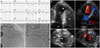
A 73-year-old woman with long-standing ankylosing spondylitis treated with adalimumab presented to an emergency department complaining of chest pain and dyspnea for 5 days. Her husband had died 10 days previously, causing her severe emotional stress. Her heart rate was 112 beats/min, and blood pressure was 102/68 mm Hg. A grade 4/6 holosystolic murmur was audible at the apex. An electrocardiography showed prominent ST-segment elevation in leads V2–5 (Fig. 1A). Troponin I was 1.3 ng/mL (reference range, <0.12 ng/mL) and creatine kinase was 141 U/L (reference range, 55–170 U/L). Bedside echocardiography showed an akinesis from the mid to apical left ventricle, but preserved systolic function within the basal segment (Fig. 1B). Left ventricular ejection fraction was 58% and flow acceleration in left ventricular outflow tract was not noted by color Doppler. Right ventricular systolic function was also reduced, particularly within the mid to apical segments. Estimated pulmonary artery systolic pressure was 59 mm Hg. In addition, a ventricular septal defect resulting from apical septal rupture with a left-to-right shunt was noted (Fig. 1B). Pericardial effusion was not present in the initial echocardiographic assessment. Angiography demonstrated no significant stenosis of the epicardial coronary arteries (Fig. 1C), suggesting the diagnosis of ventricular septal rupture complicated by takotsubo cardiomyopathy with right ventricular involvement.
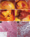
The patient was taken to the operating room 3 hours after admission for emergency repair of the ventricular septal rupture. However, immediately before induction of anesthesia, her blood pressure suddenly collapsed with pulseless electrical activity. After 5 minutes of resuscitation, cardiac activity was restored. Sternotomy was done, and the pericardium full of fresh blood was found. Both left and right ventricles had necrotic changes with hemorrhage (Fig. 2A, B, and C). Cardiopulmonary bypass was established and the apex was incised. Ventricular septal wall rupture was confirmed (Fig. 2D). Also, another rupture within the right ventricular free wall measuring 20×18 mm was noted (Fig. 2C), which had led to the cardiac tamponade that presented in the operating room. Patch closure of the ventricular septal rupture with right ventriculoplasty was performed. Pathological examination of the ruptured ventricular septum revealed disrupted myocytic integrity in multiple foci with lymphohistiocytic infiltrate and myocytic necrosis (Fig. 2E and F). Although she was sent to the intensive care unit after weaning of the cardiopulmonary bypass, extracorporeal membrane oxygenation support was required because of refractory shock 4 hours after surgery. The patient died 5 days after admission.
DISCUSSION
Takotsubo cardiomyopathy is a reversible disease often triggered by acute emotional or physical stress.5 Most takotsubo cardiomyopathies are expected to almost completely resolve within several days to weeks following the acute event, and the prognosis is generally good.6 However, recent studies have revealed that up to half of the patients with takotsubo cardiomyopathy may have acute phase complications.234 Similar to previously reported cases of cardiac rupture in takotsubo cardiomyopathy, our patient had prominent ST-segment elevation in leads V2–5, suggesting that the observed ST-segment elevation on the precordial leads may be a high risk of cardiac rupture.4 Also, Kumar, et al.4 reported that patients with cardiac rupture in takotsubo cardiomyopathy were more frequently Asian females with older age and had higher blood pressure with higher left ventricular ejection fraction than those without cardiac rupture. Therefore, our patient could be considered as a high risk of cardiac rupture according to previous reports. In takotsubo cardiomyopathy, hypercontraction and possibly functional basal obstruction of the left ventricular outflow can increase afterload in the left ventricle. This can lead to left ventricular rupture. Although our patient had no flow acceleration in left ventricular outflow tract contrary to previous cases of increased left ventricular outflow tract gradient,78 these can be attributed to our echocardiographic assessment after the development of cardiac rupture.
Although takotsubo cardiomyopathy commonly involves the left ventricle, recent studies have reported right ventricular involvement in 28% to 50%.91011 Furthermore, right ventricular involvement was associated with poor clinical outcomes.91011 In the present case, concomitant right ventricular involvement of takotsubo cardiomyopathy resulted in a highly friable right ventricle susceptible to rupture by mechanical wall stress, acute increase of volume and pressure overload worsened by left to right shunt. Before the patient was taken to the operating room, pericardial effusion was not present on echocardiography and she was hemodynamically stable. Although several left ventricular ruptures complicated by takotsubo cardiomyopathy have been reported, rupture of right ventricle is exceedingly rare.
Our case report illustrates that takotsubo cardiomyopathy can be life threatening in the acute phase. Ventricular septal rupture can be complicated by takotsubo cardiomyopathy. Furthermore, an acute increase of right ventricular afterload by left-to-right shunt can lead to right ventricular rupture, particularly when right ventricular involvement of takotsubo cardiomyopathy is present. Thus, ventricular septal rupture in biventricular takotsubo cardiomyopathy may be a harbinger of cardiac tamponade by right ventricular rupture. Clinical suspicion and early diagnosis of potentially life threatening complications such as ventricular rupture is crucial even in patients with takotsubo cardiomyopathy.
 XML Download